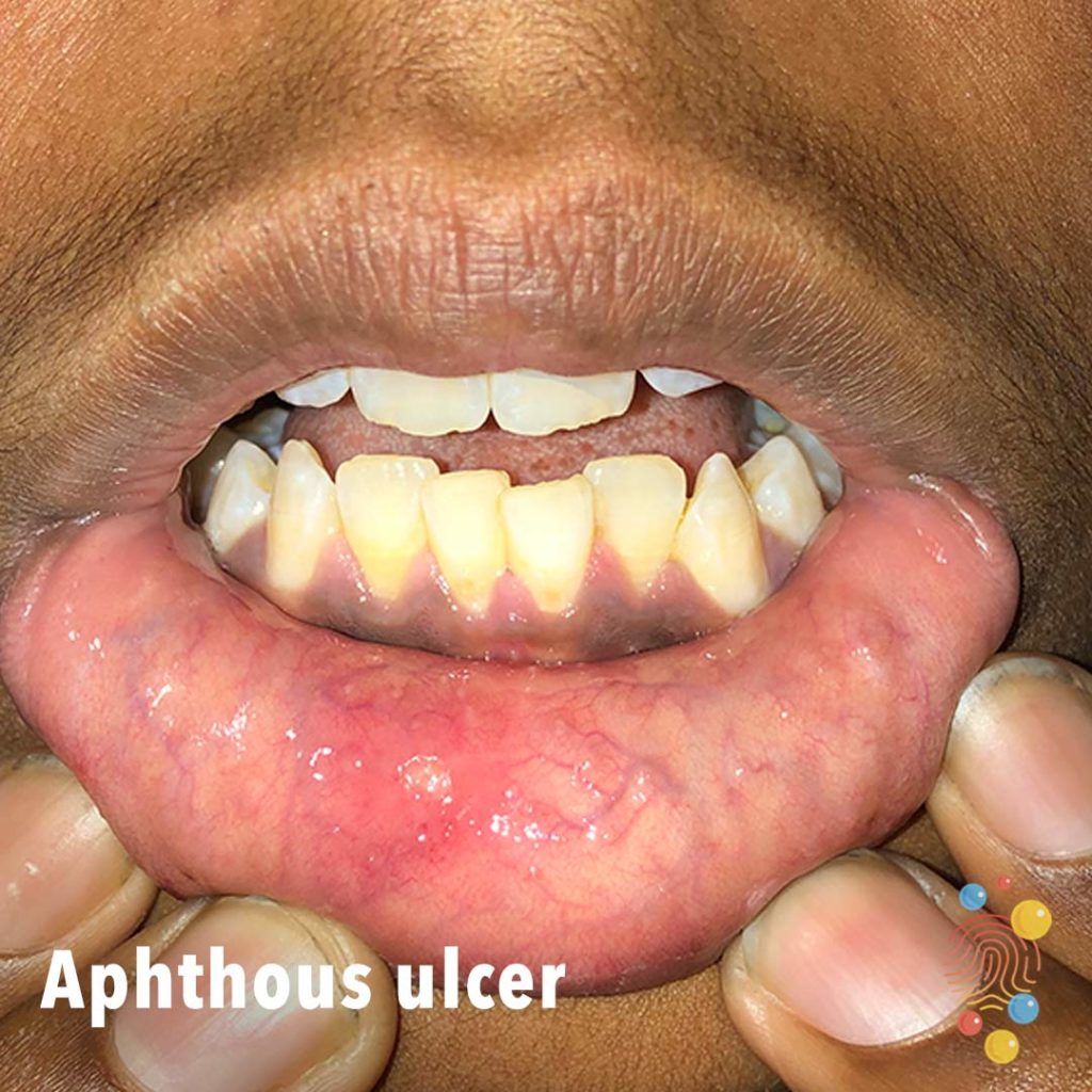
Aphthous Ulcer
Learn more about aphthous ulcers
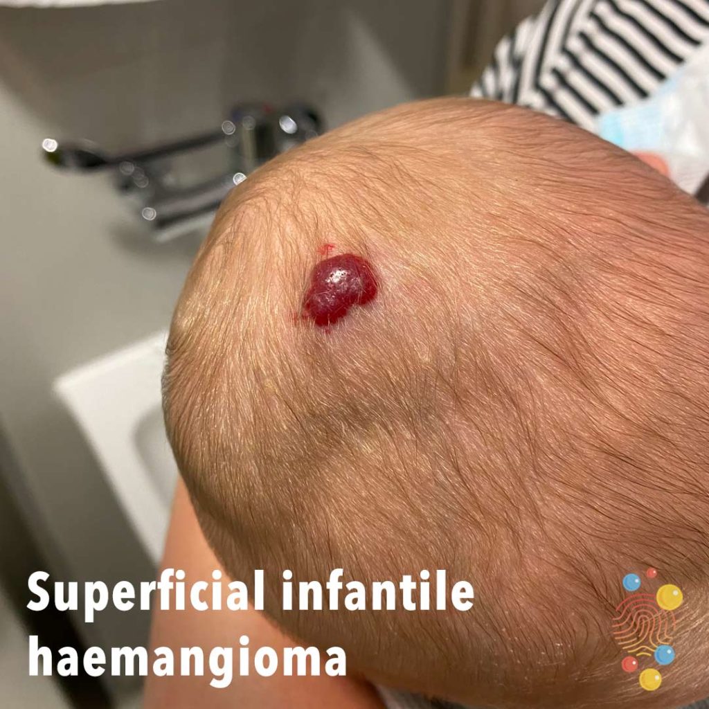
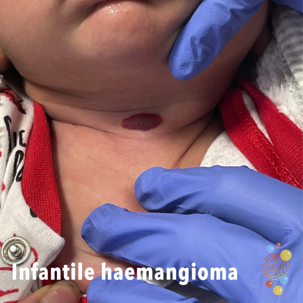
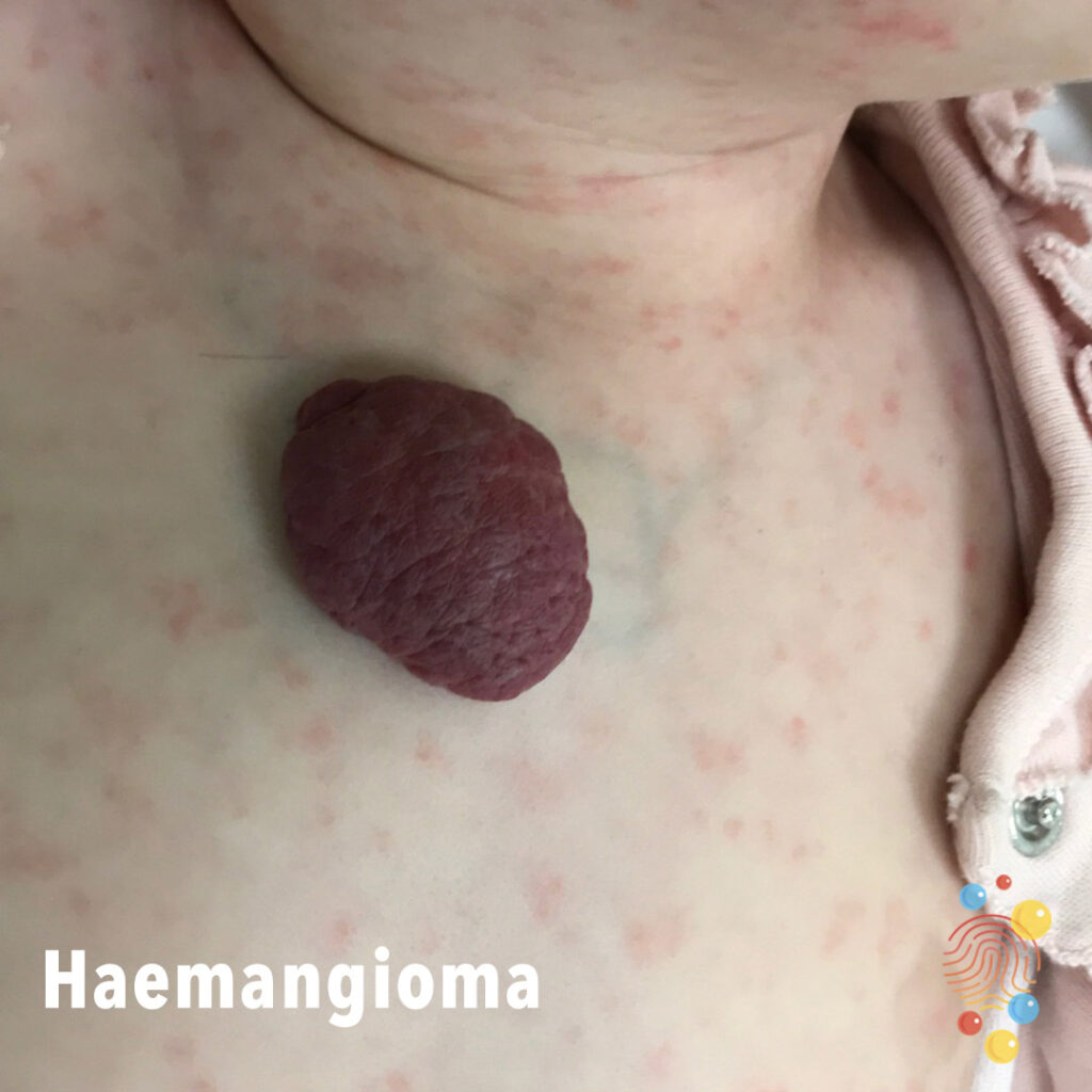
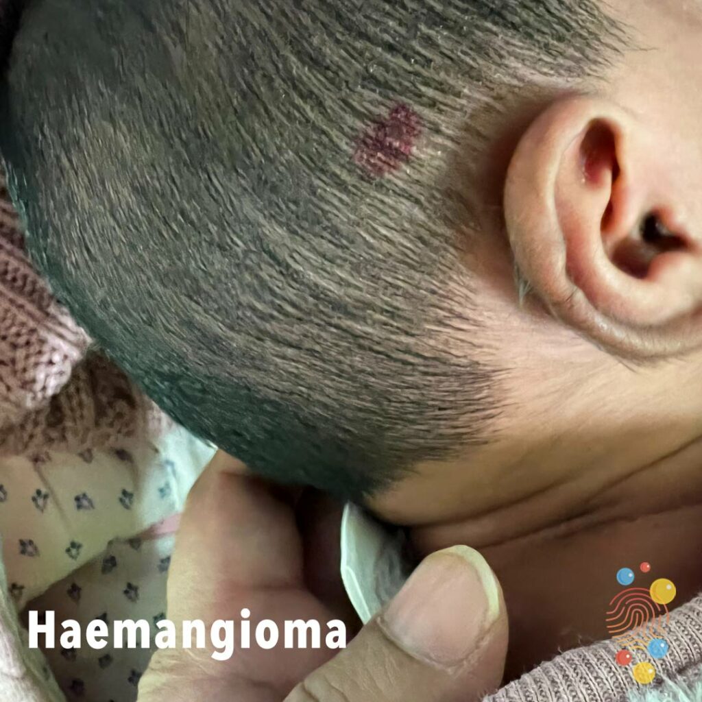
Superficial Infantile Haemangioma
Learn more about haemangiomas

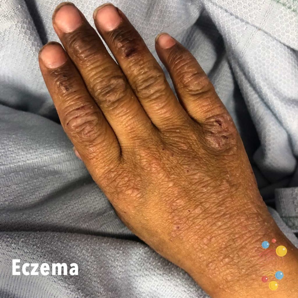
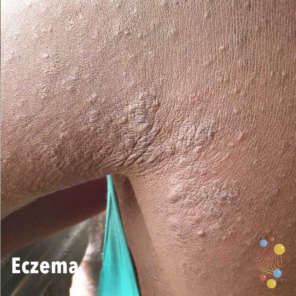
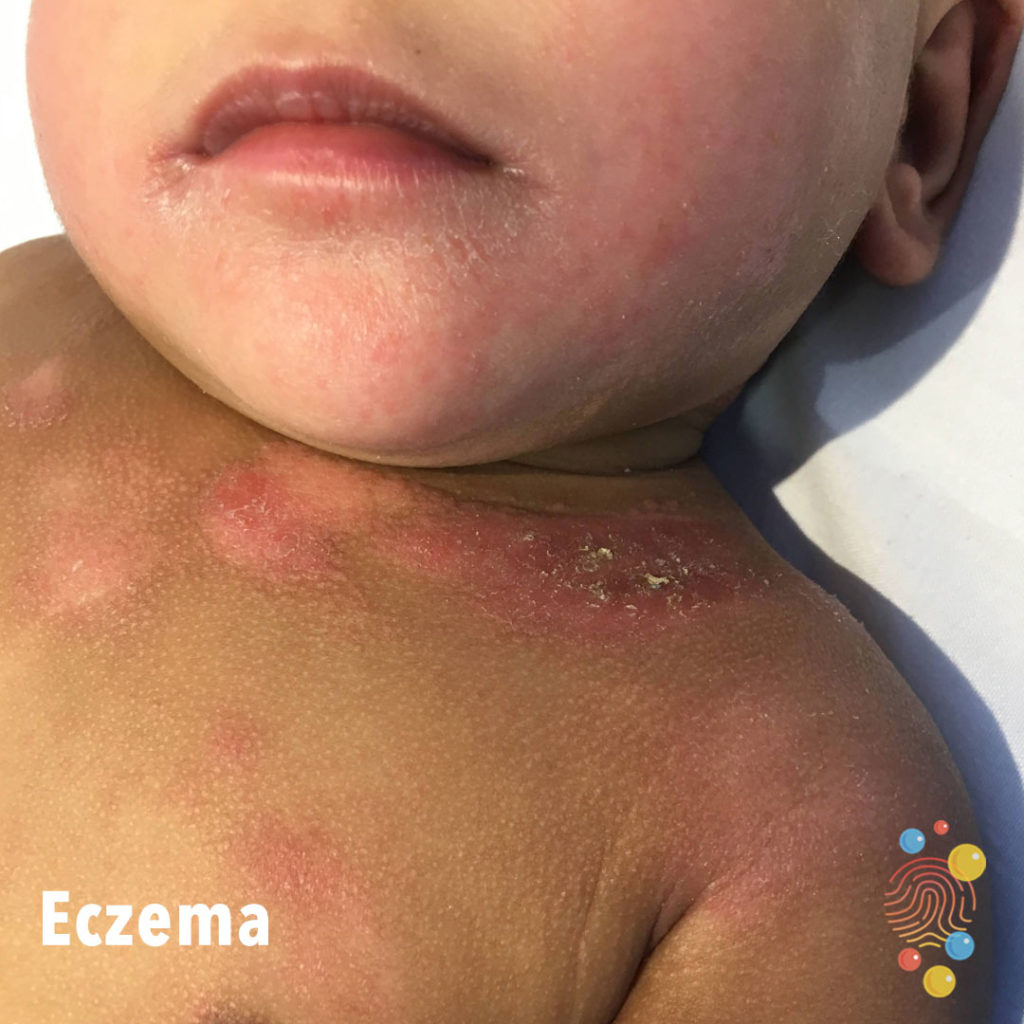
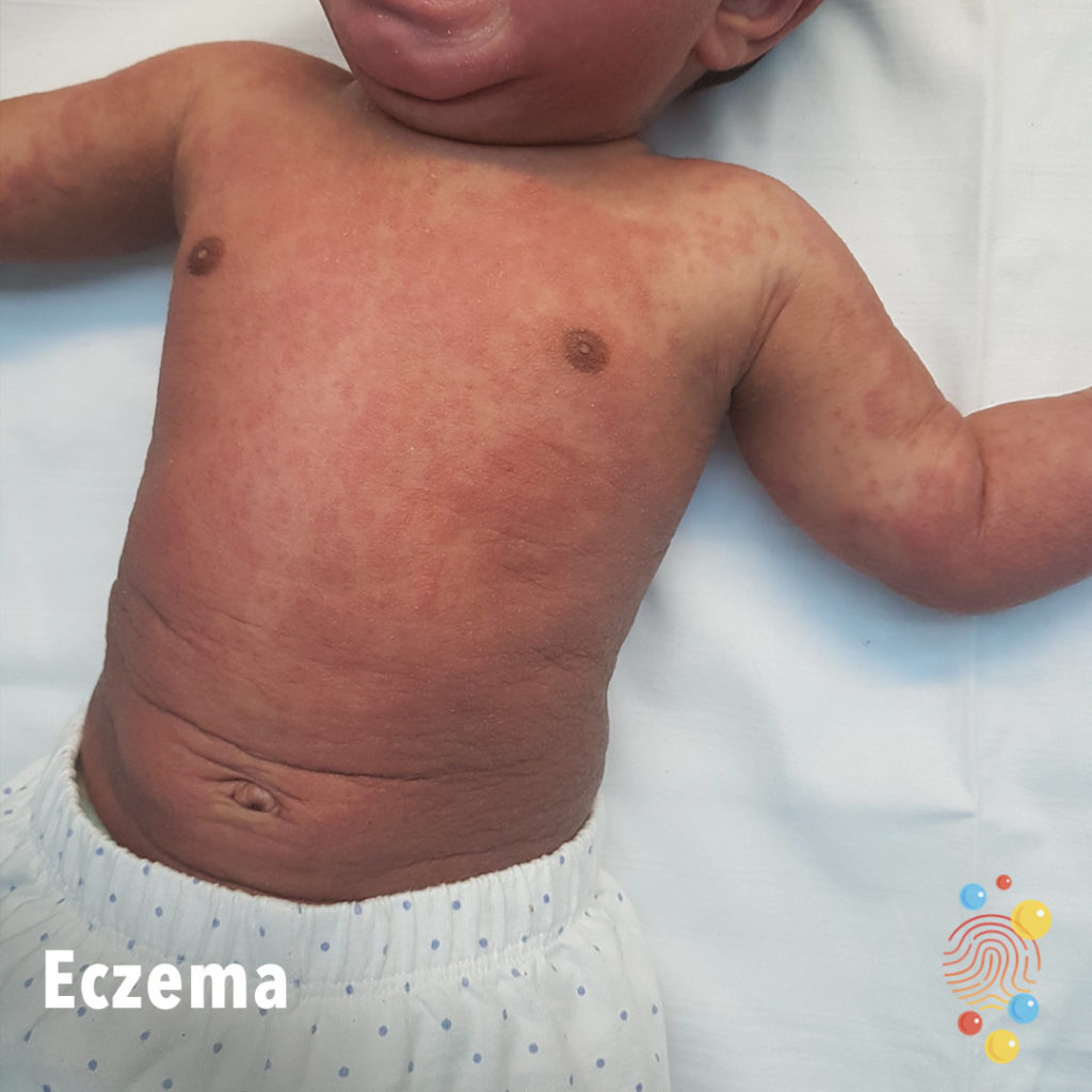
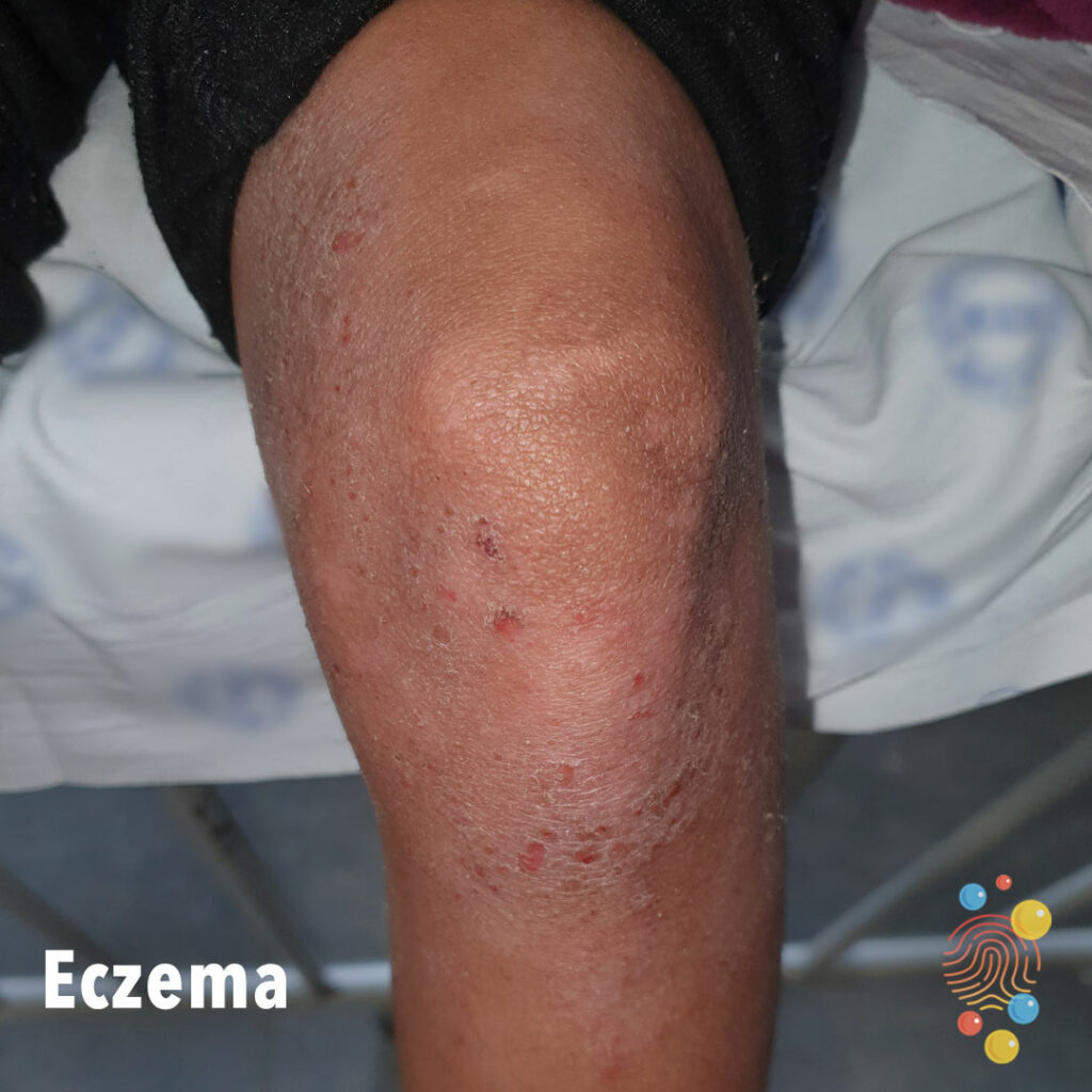
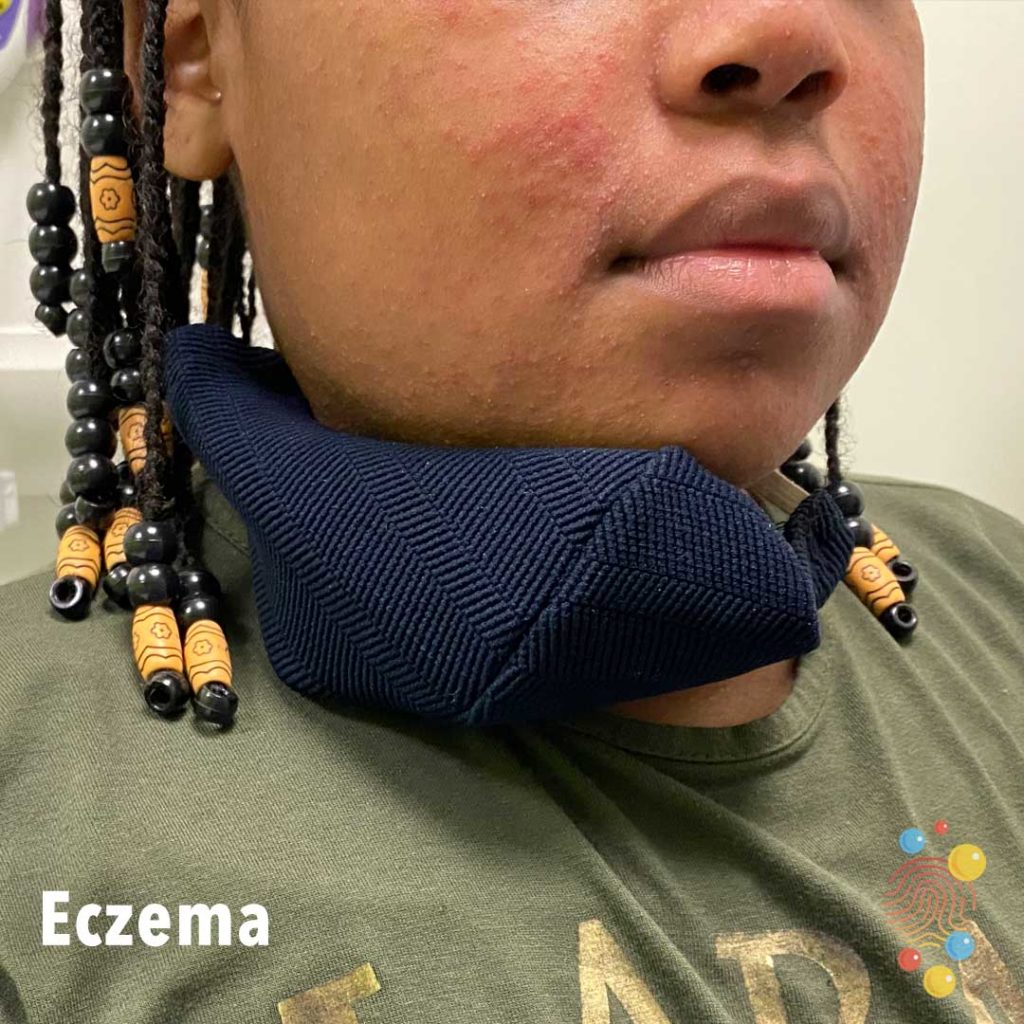
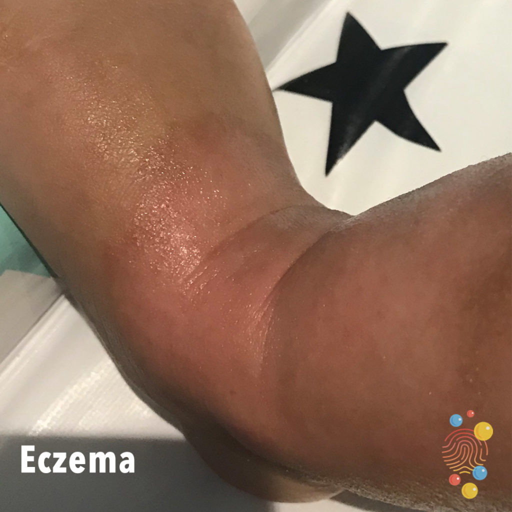
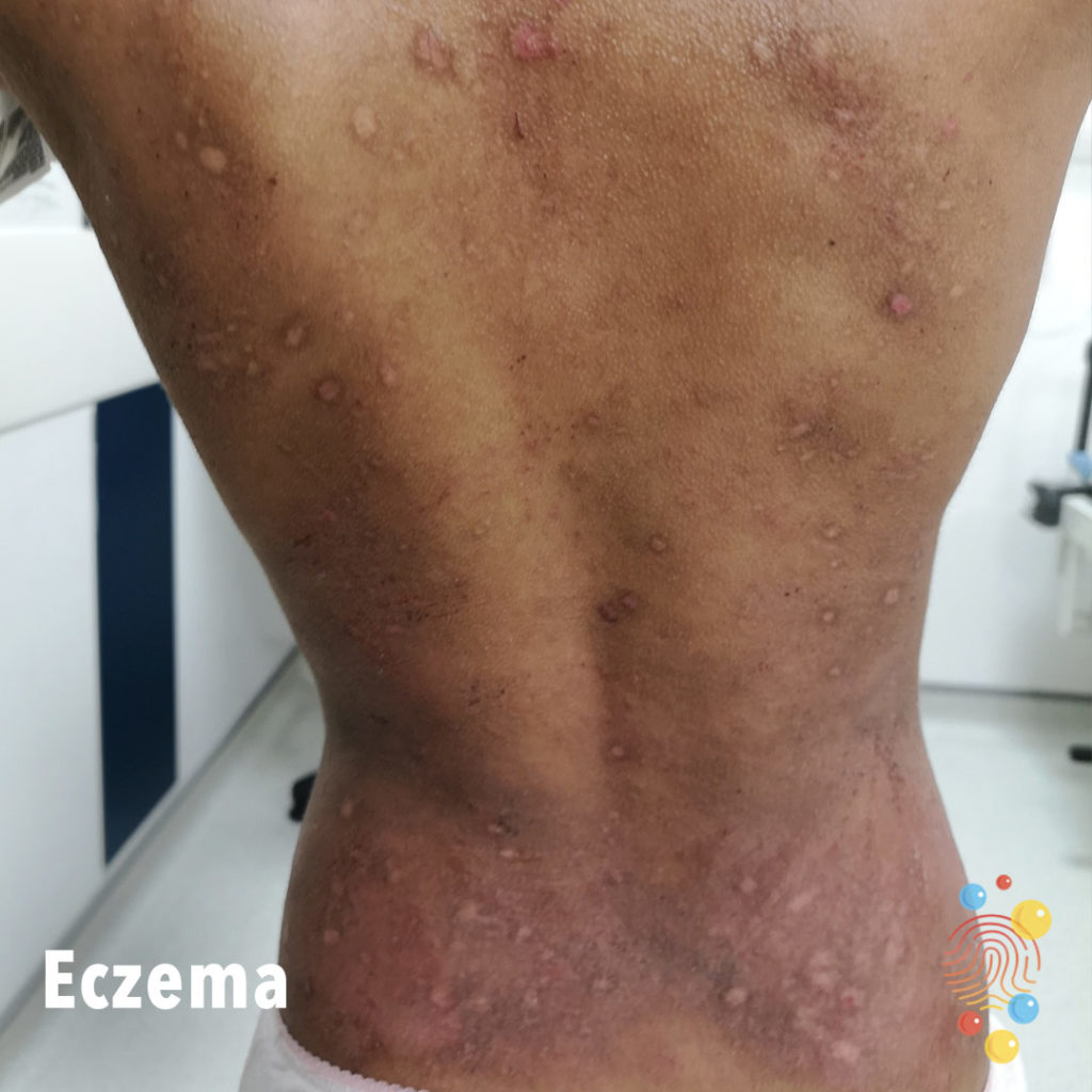
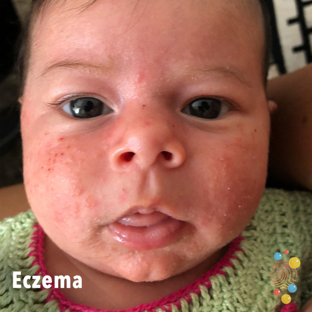
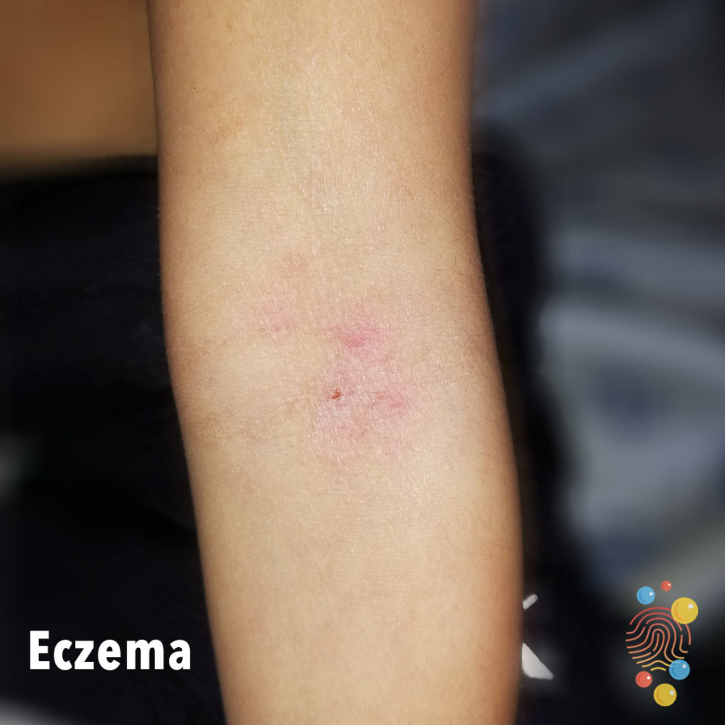
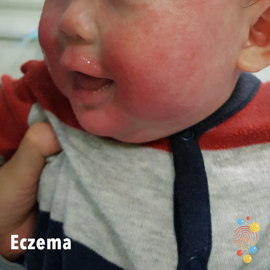
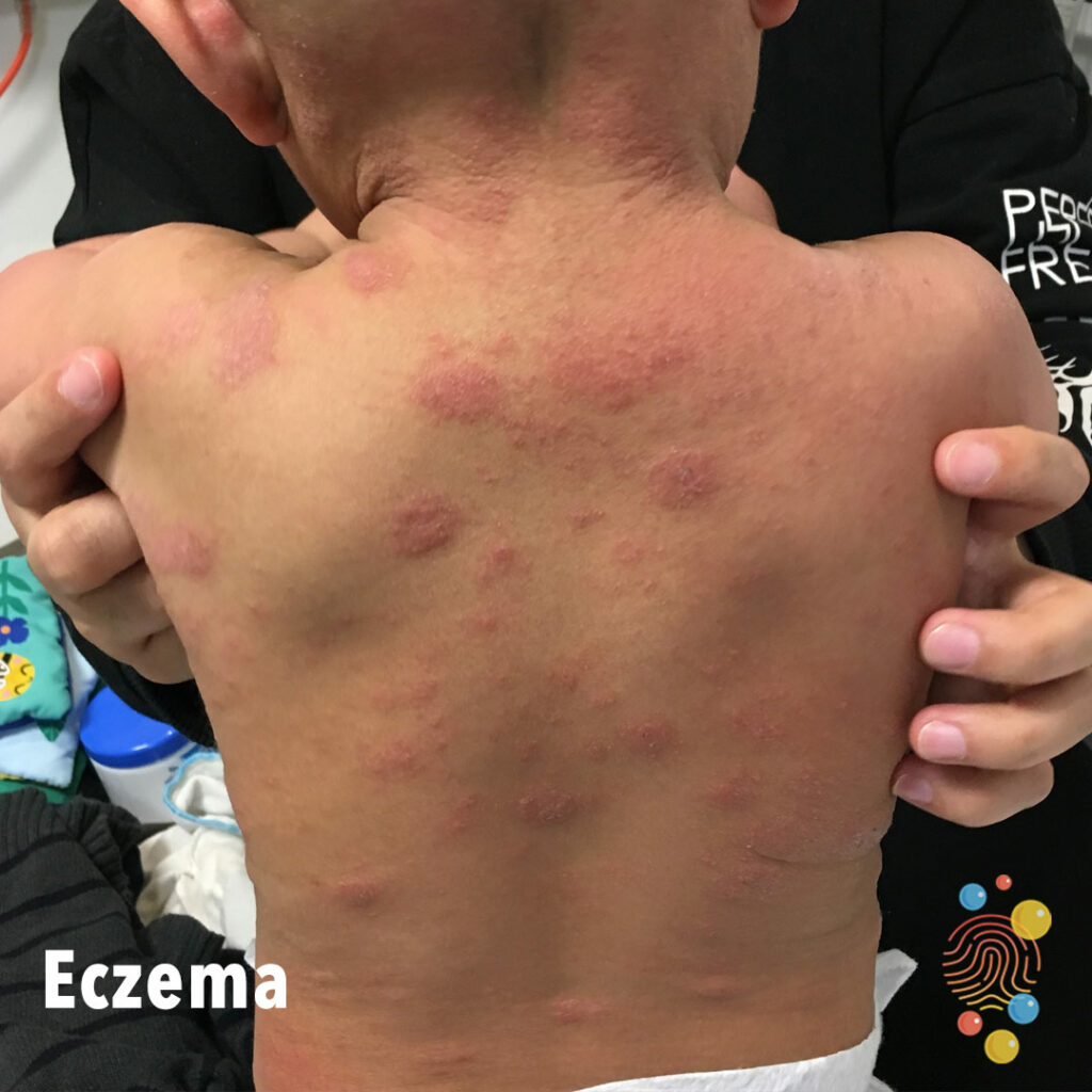
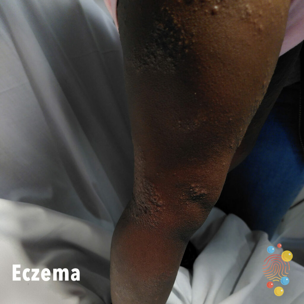
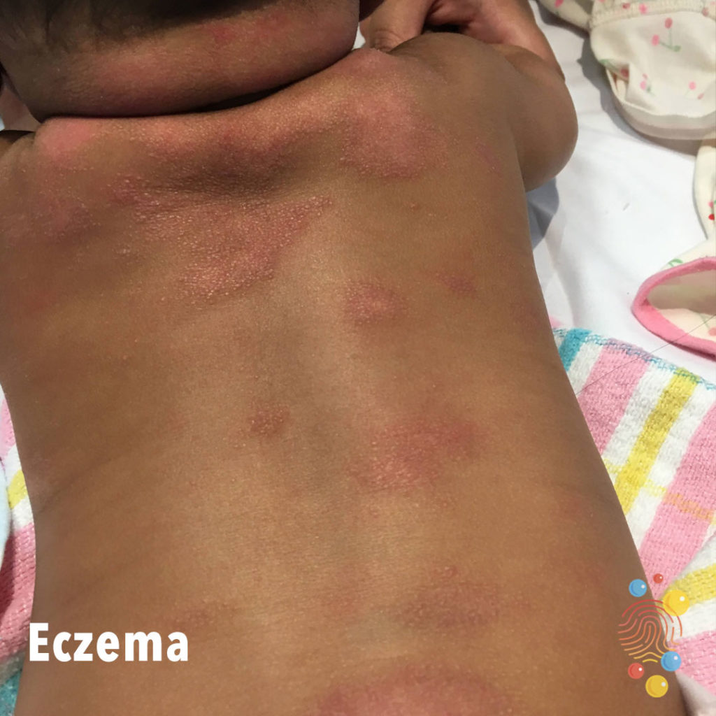
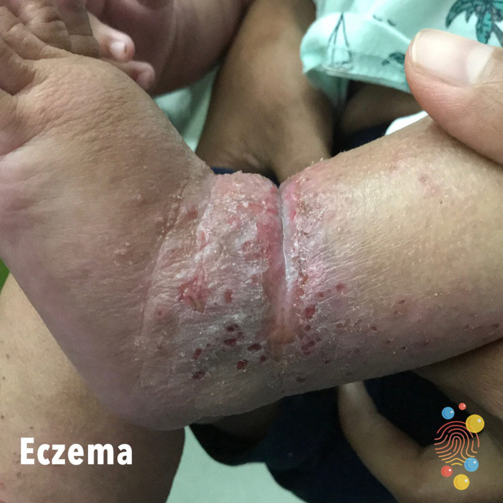
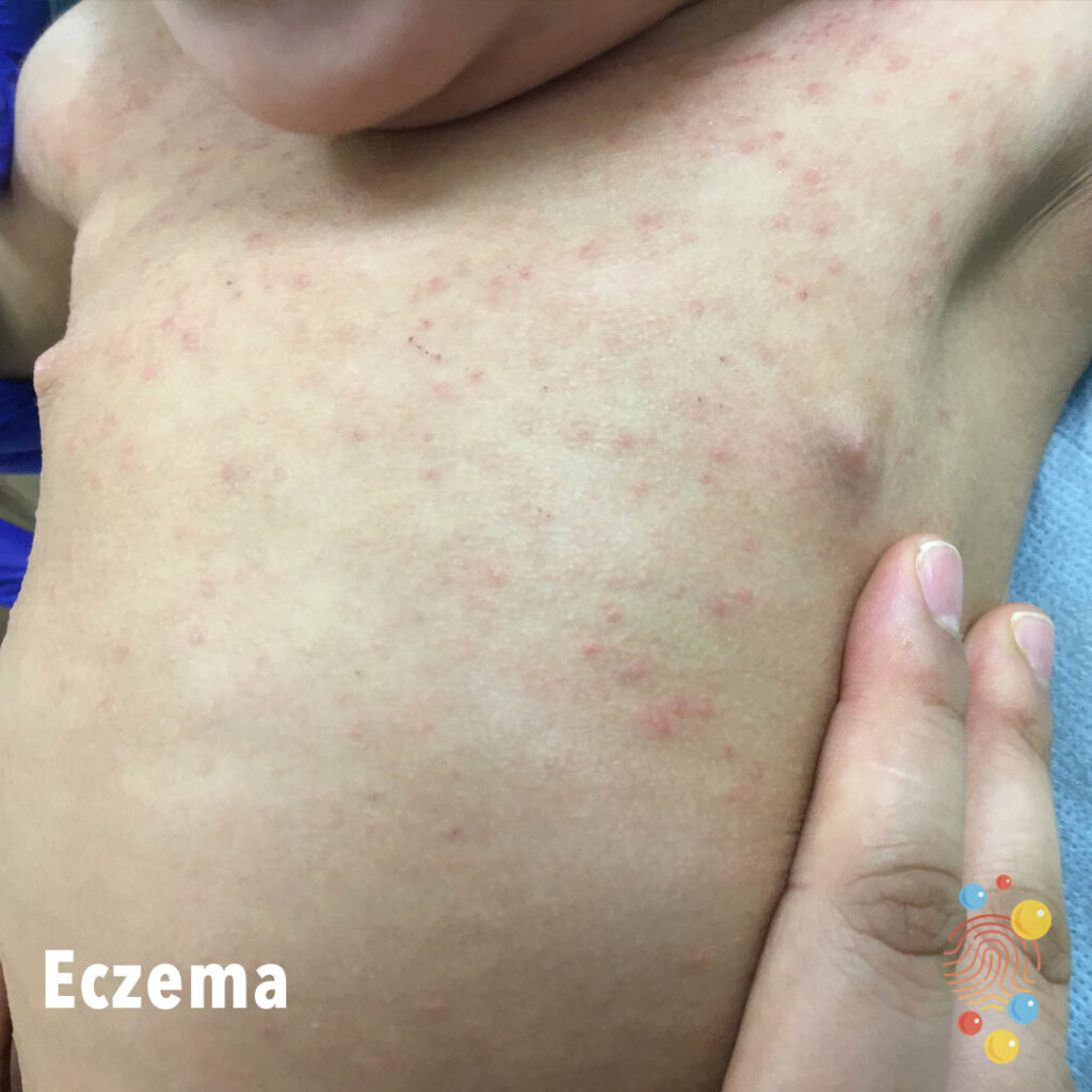
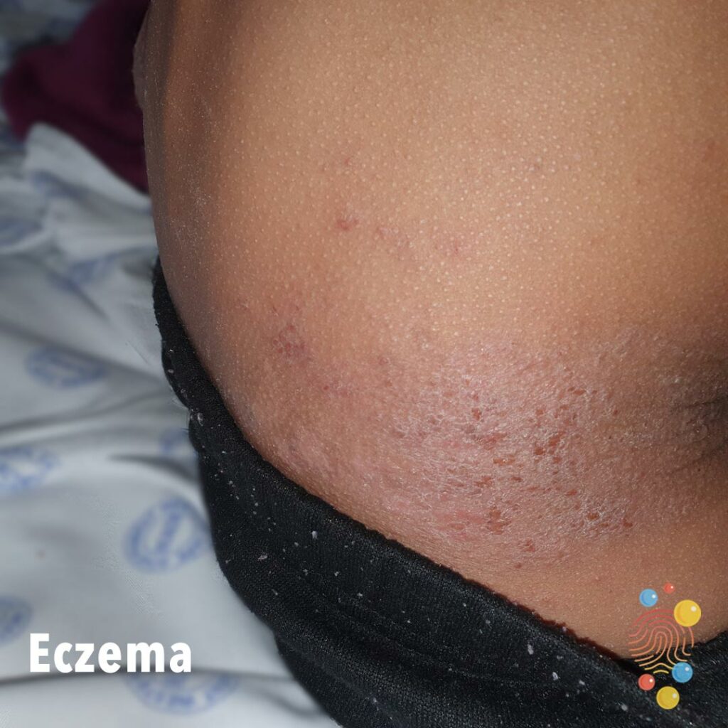
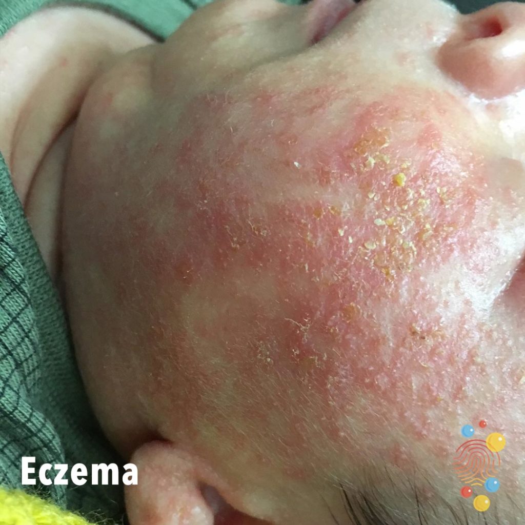
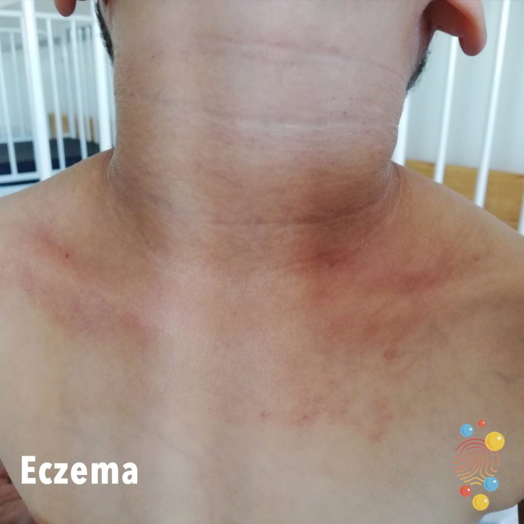
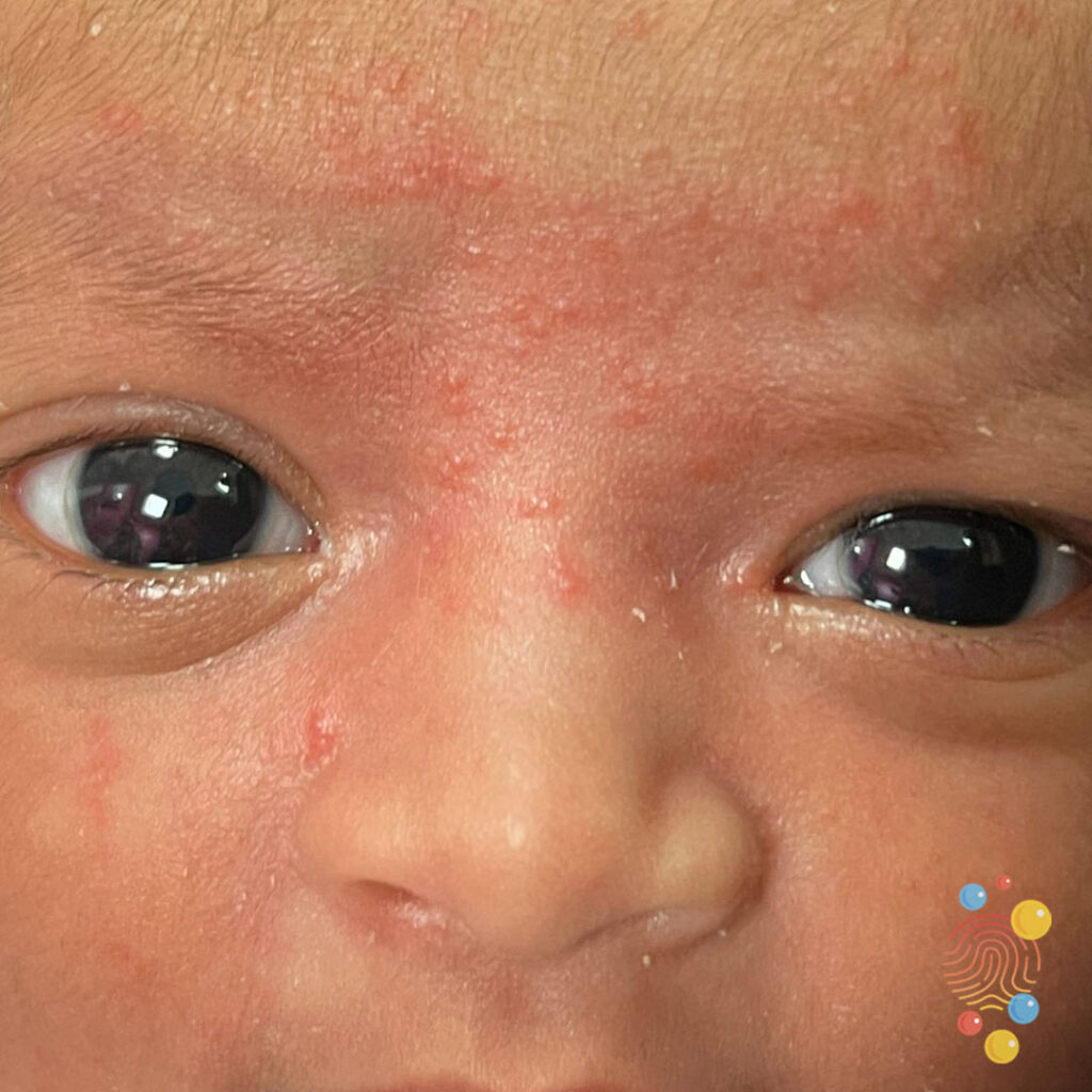
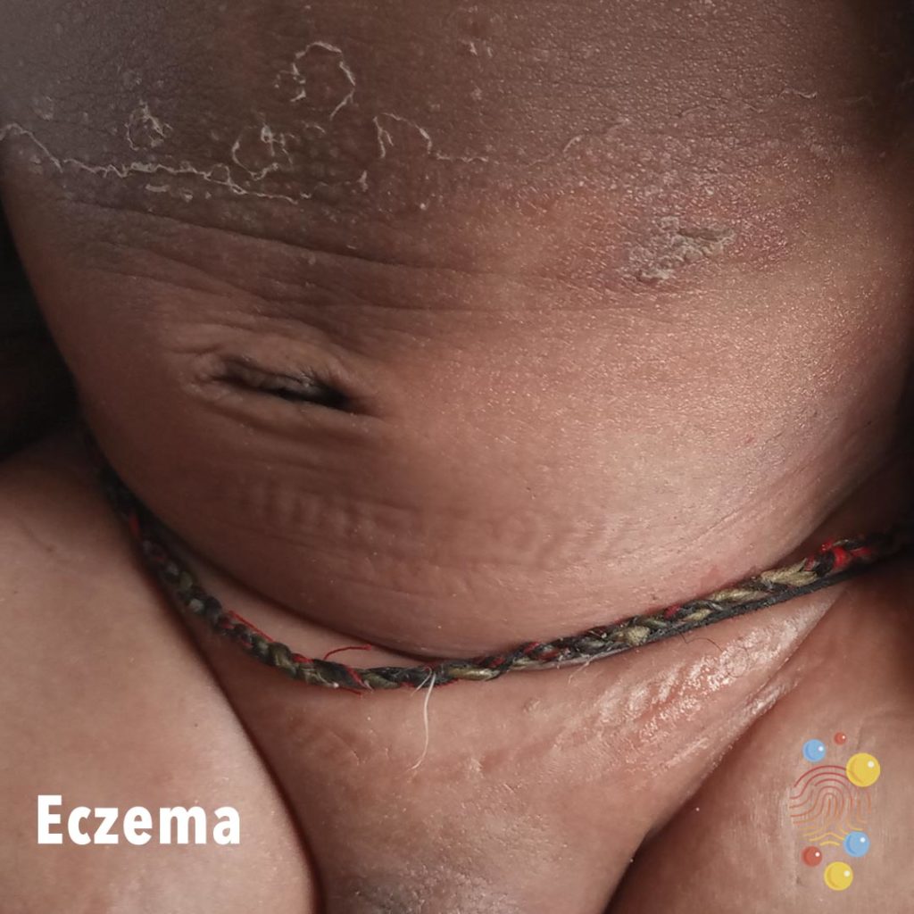
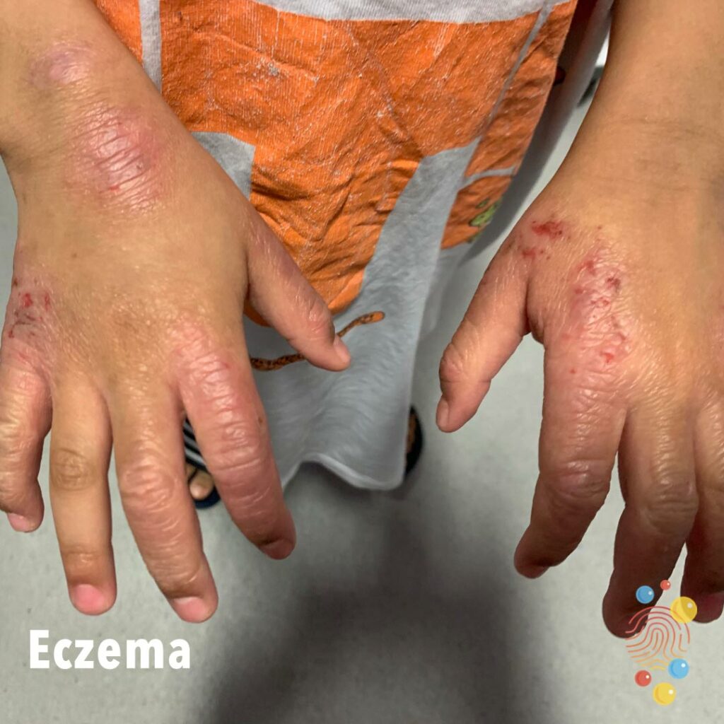
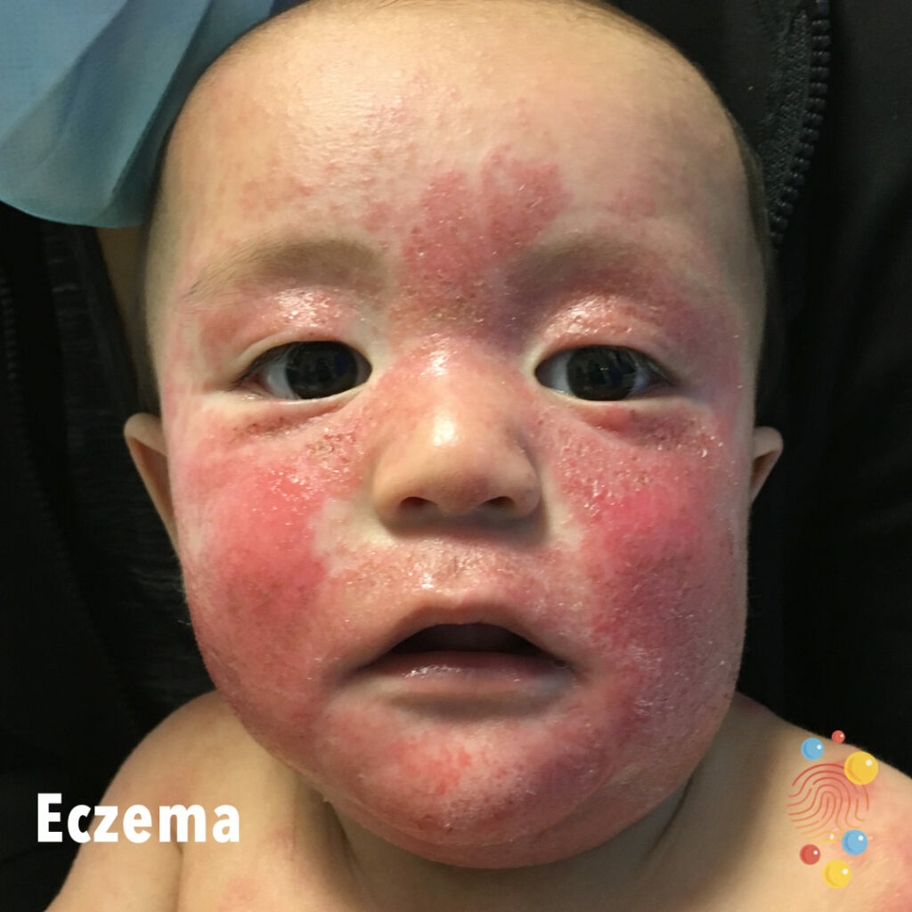
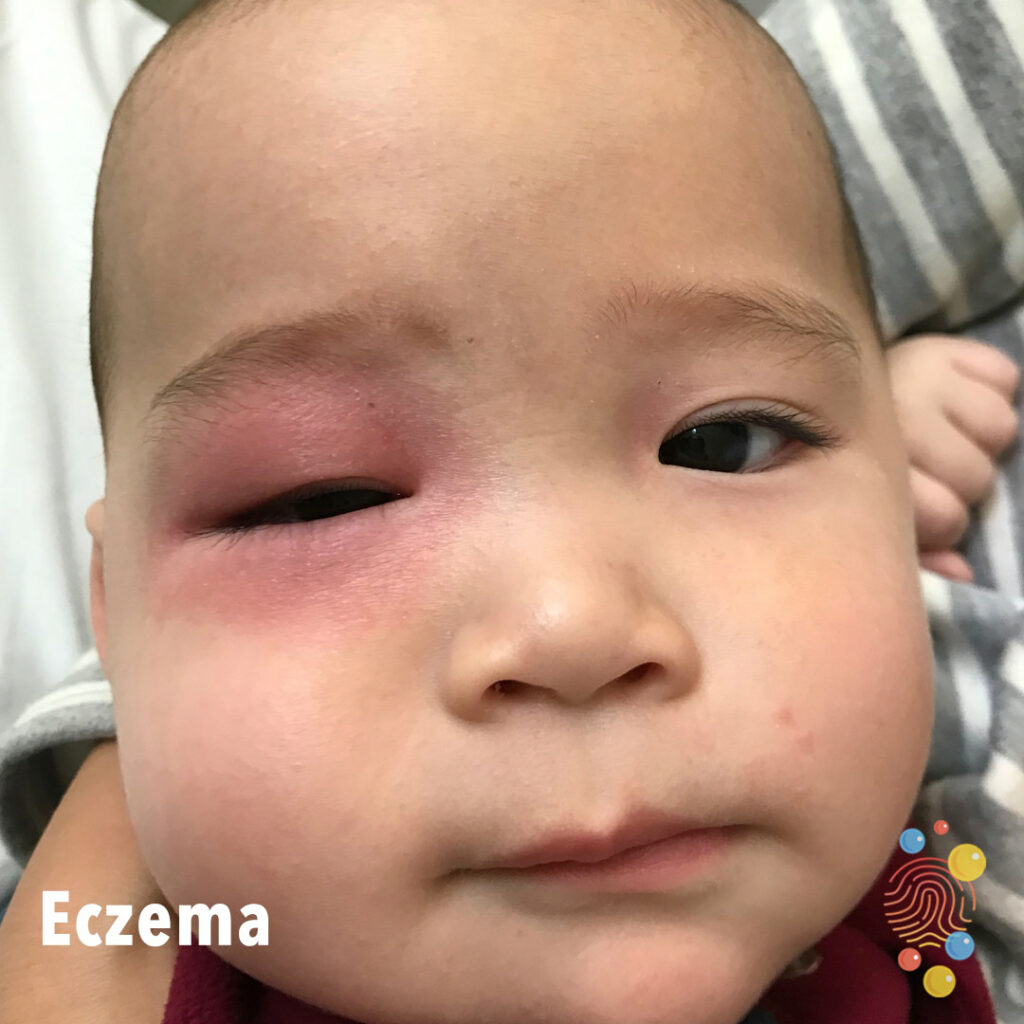
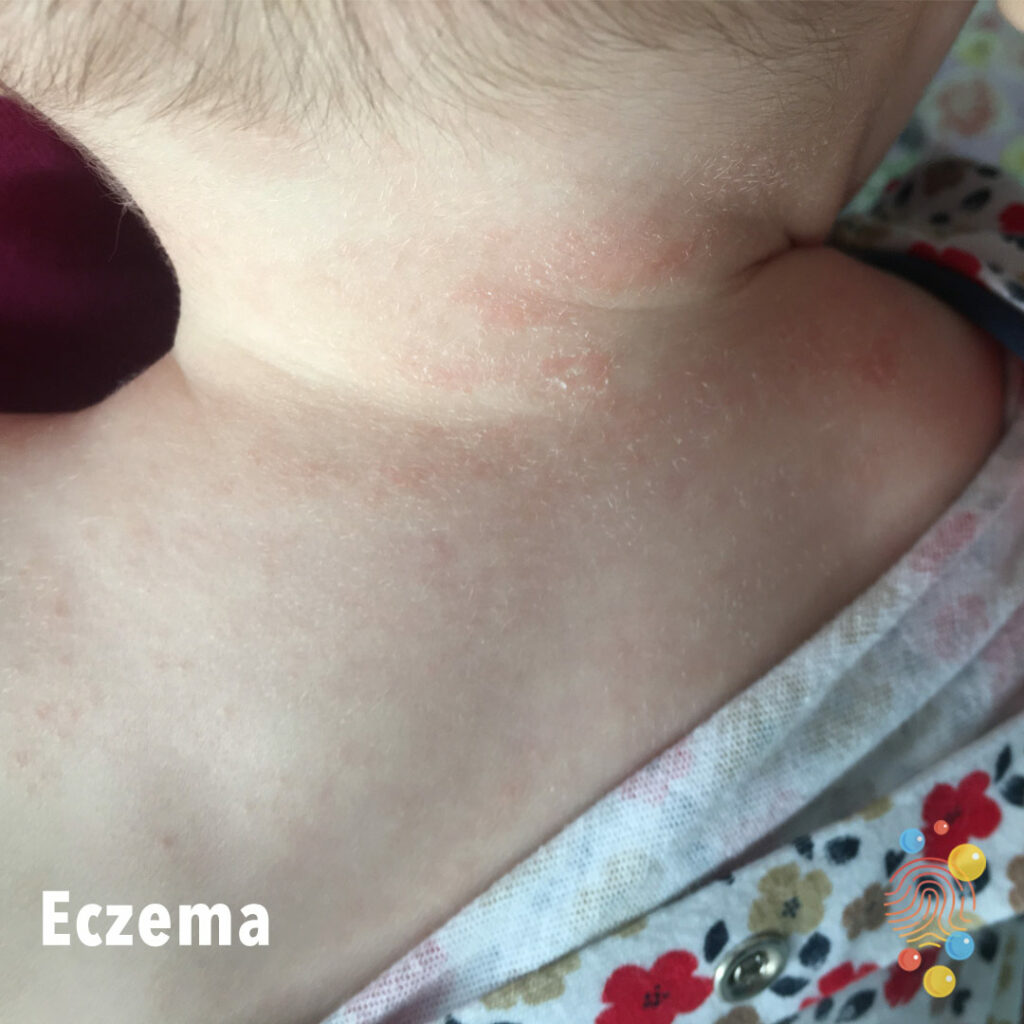
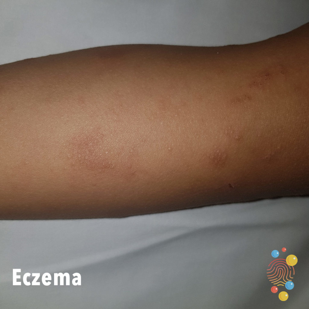
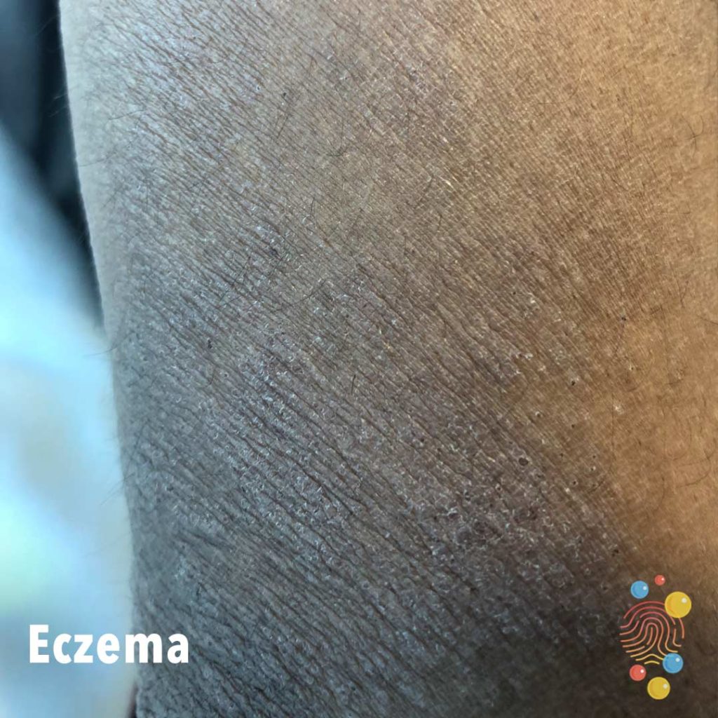
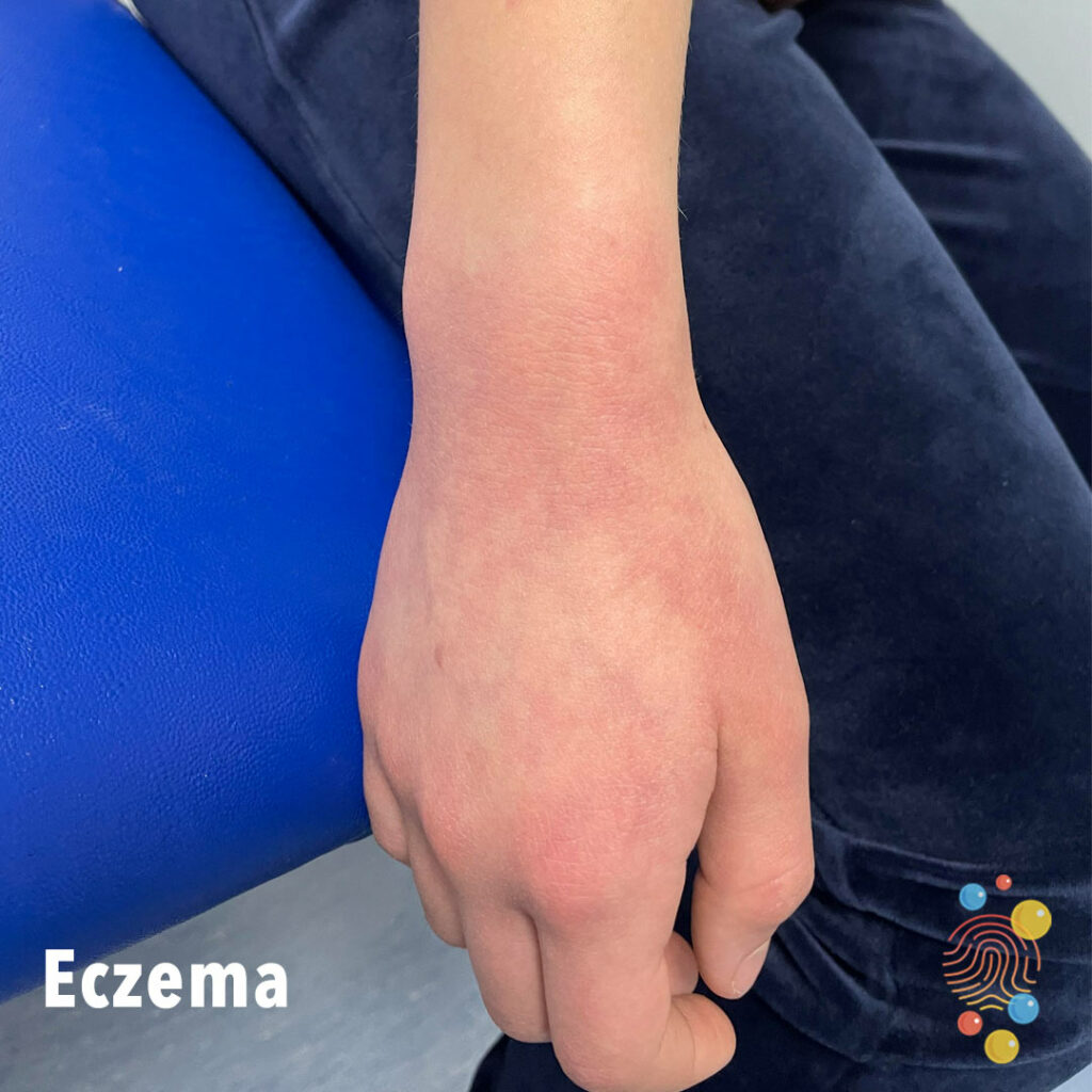
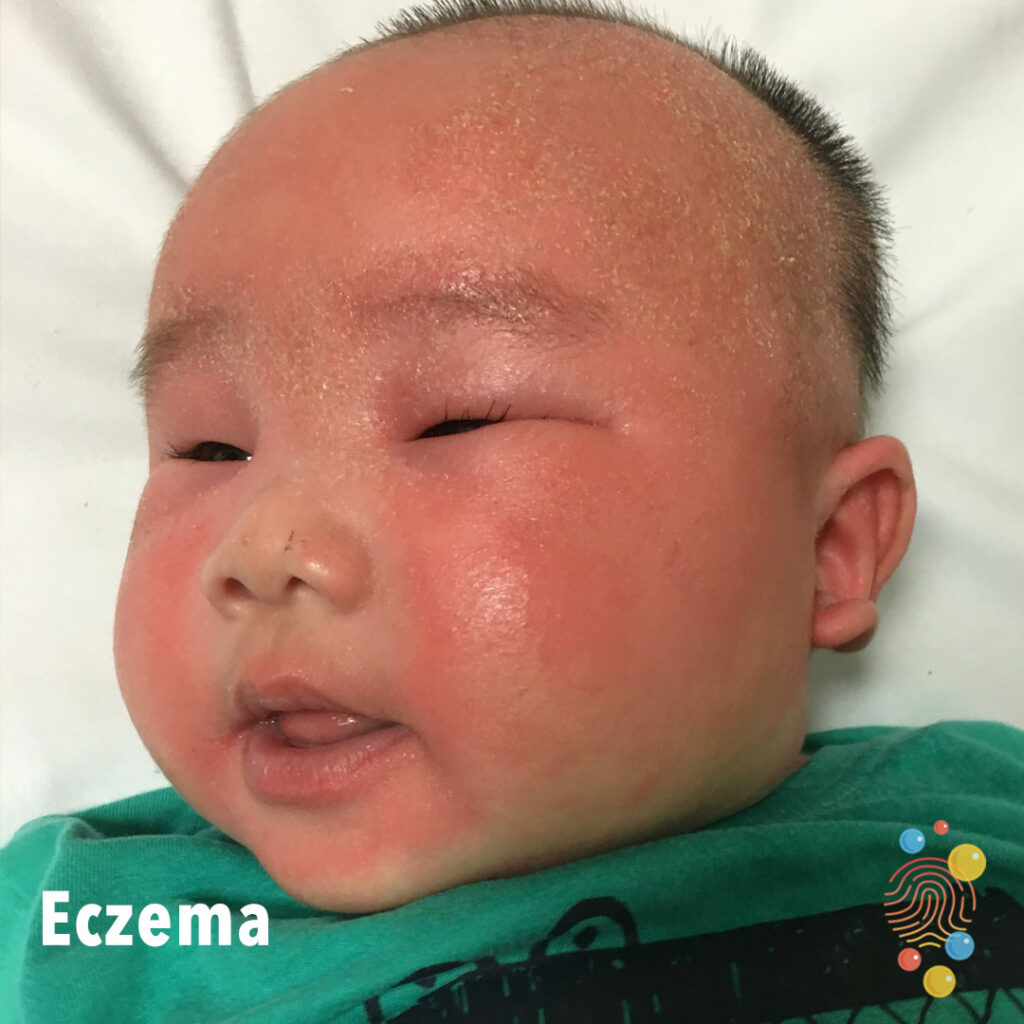
Eczema
Learn more about eczema
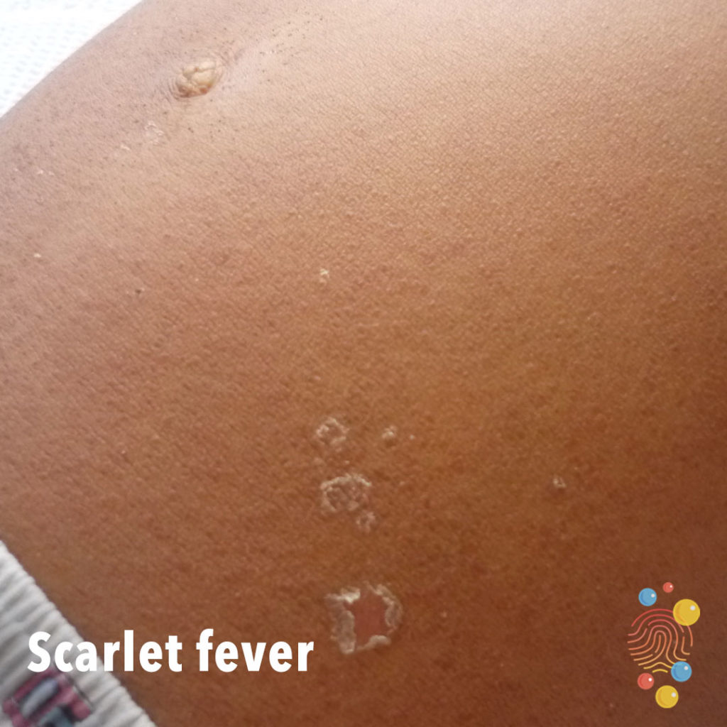
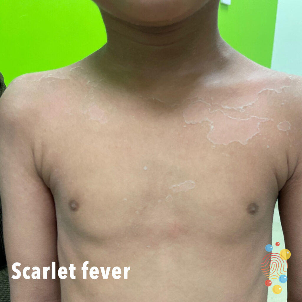
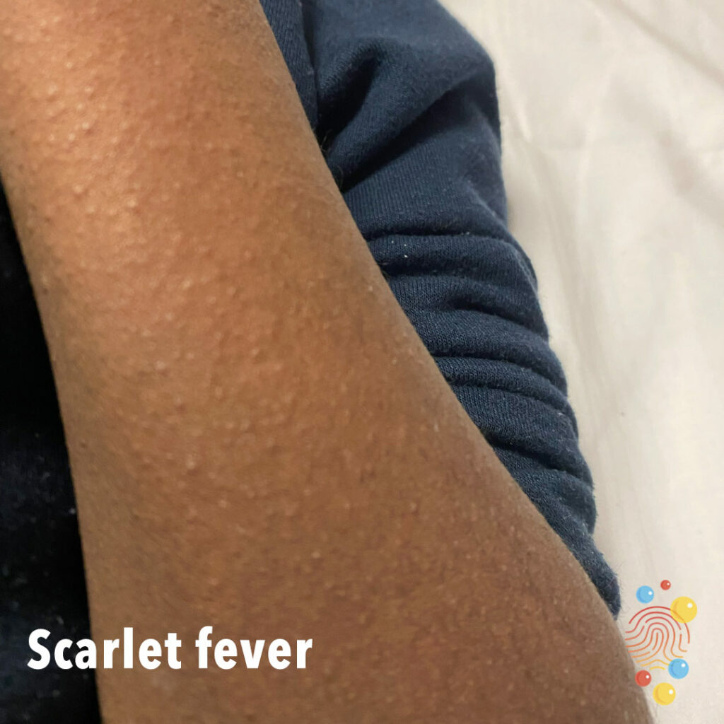
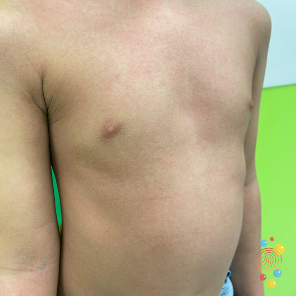
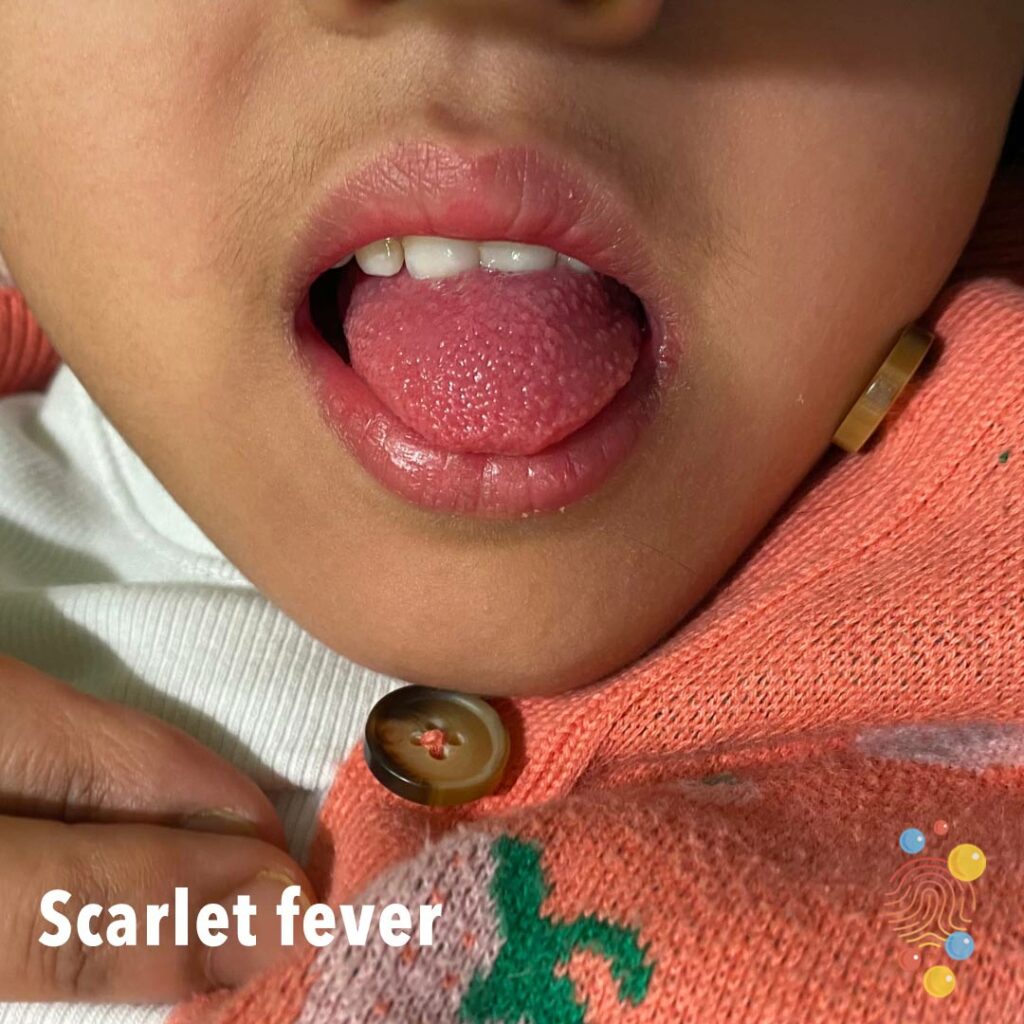
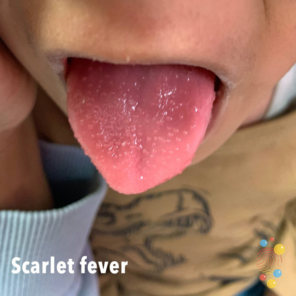
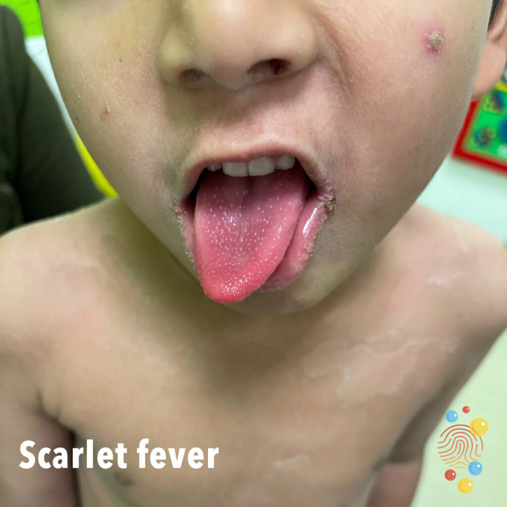
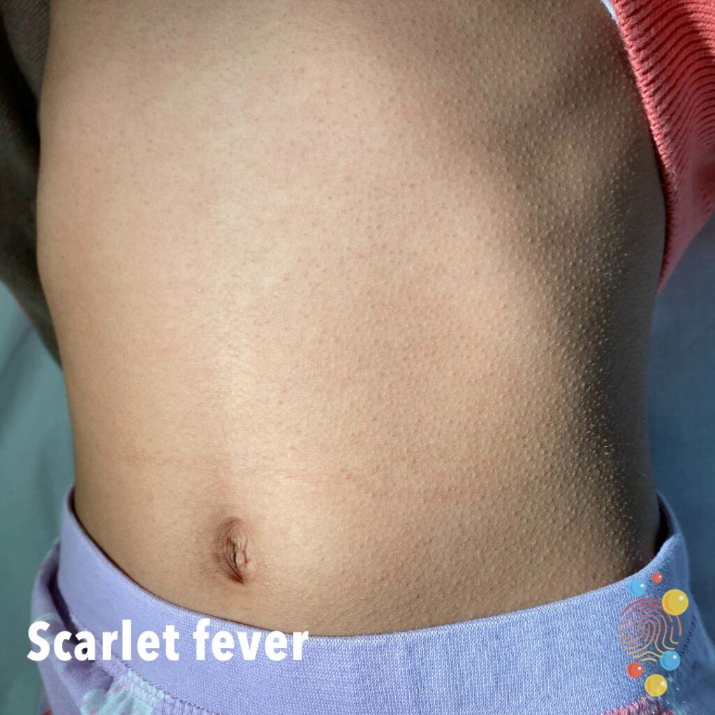
Scarlet Fever
Learn more about scarlet fever
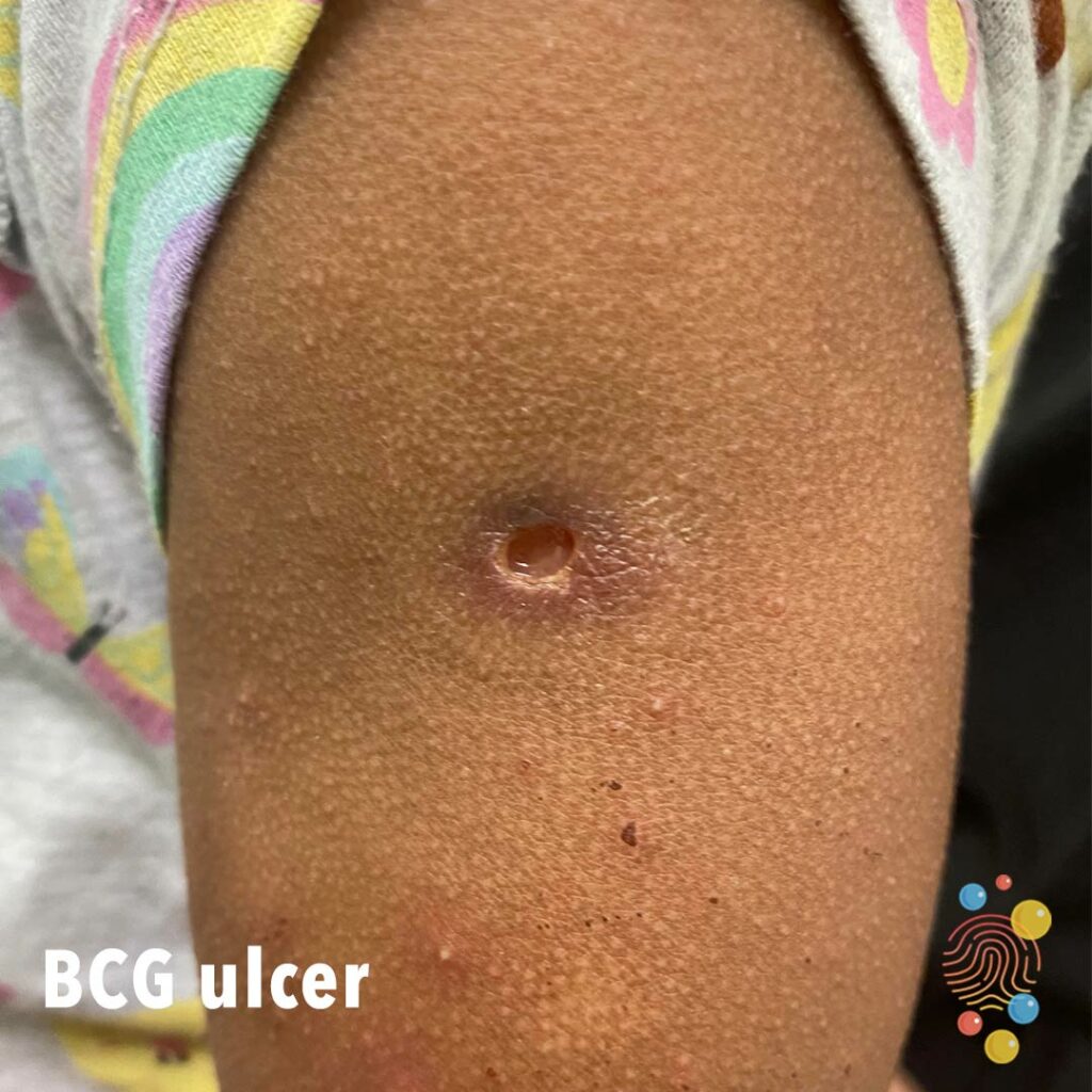
BCG Ulcer
Learn more about BCG

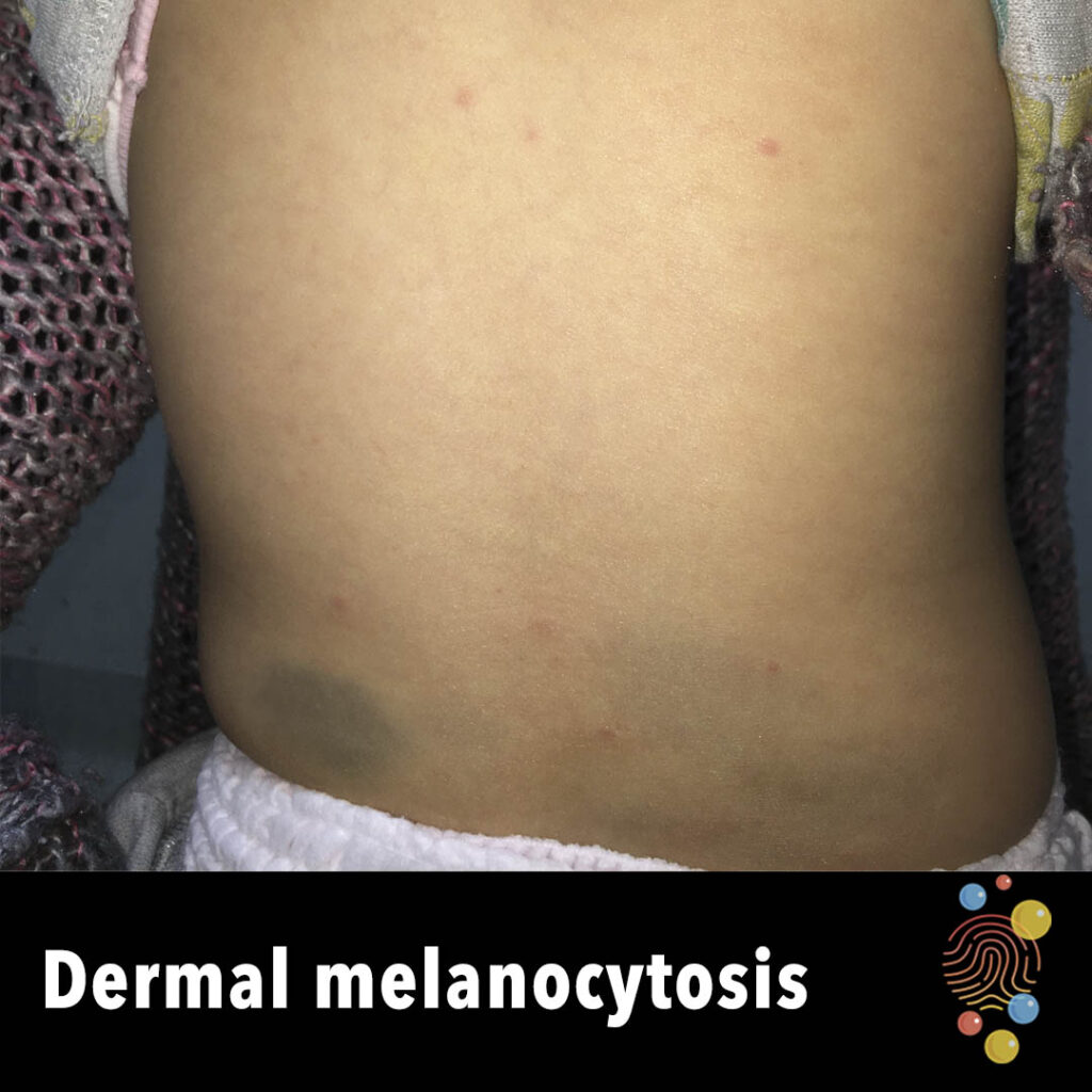
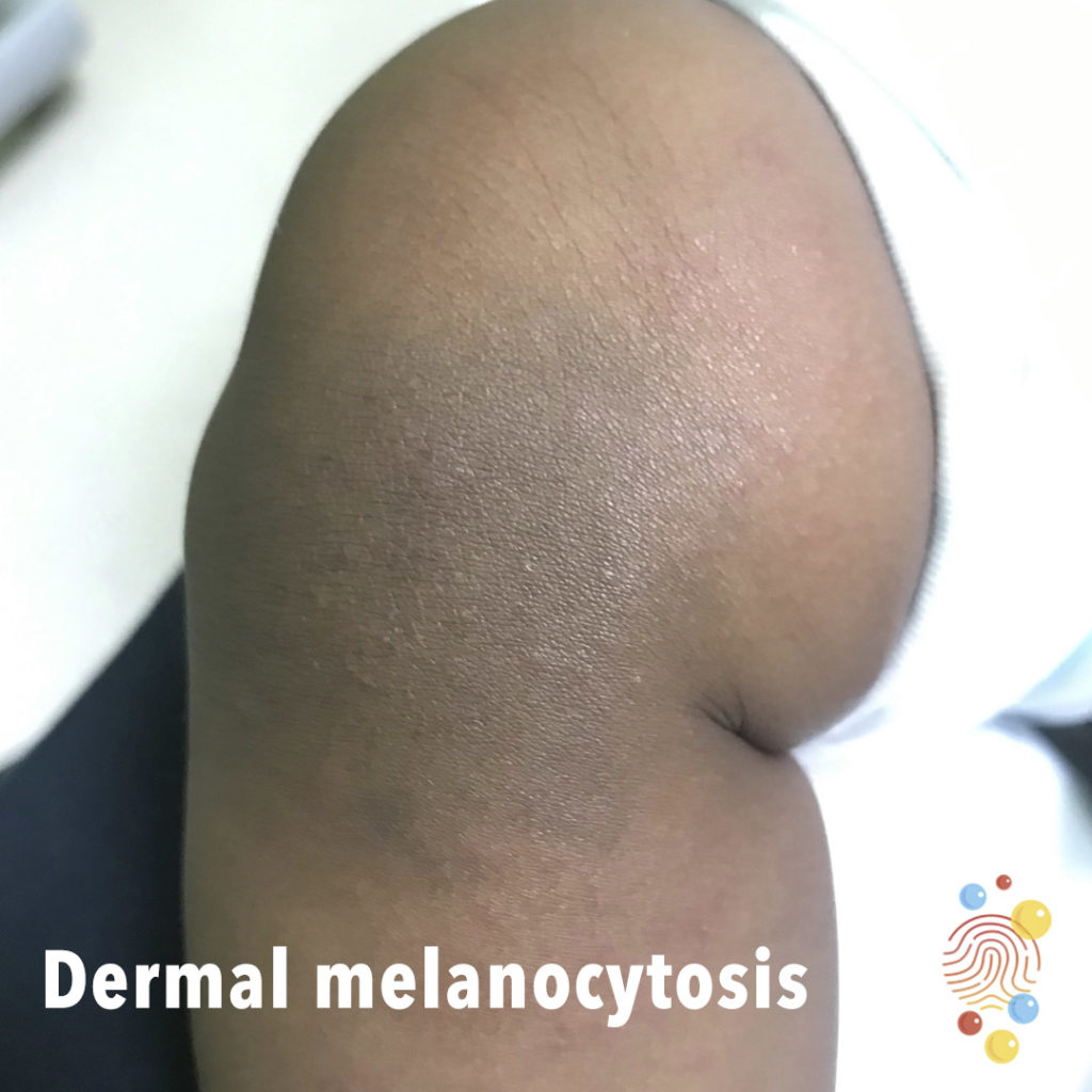
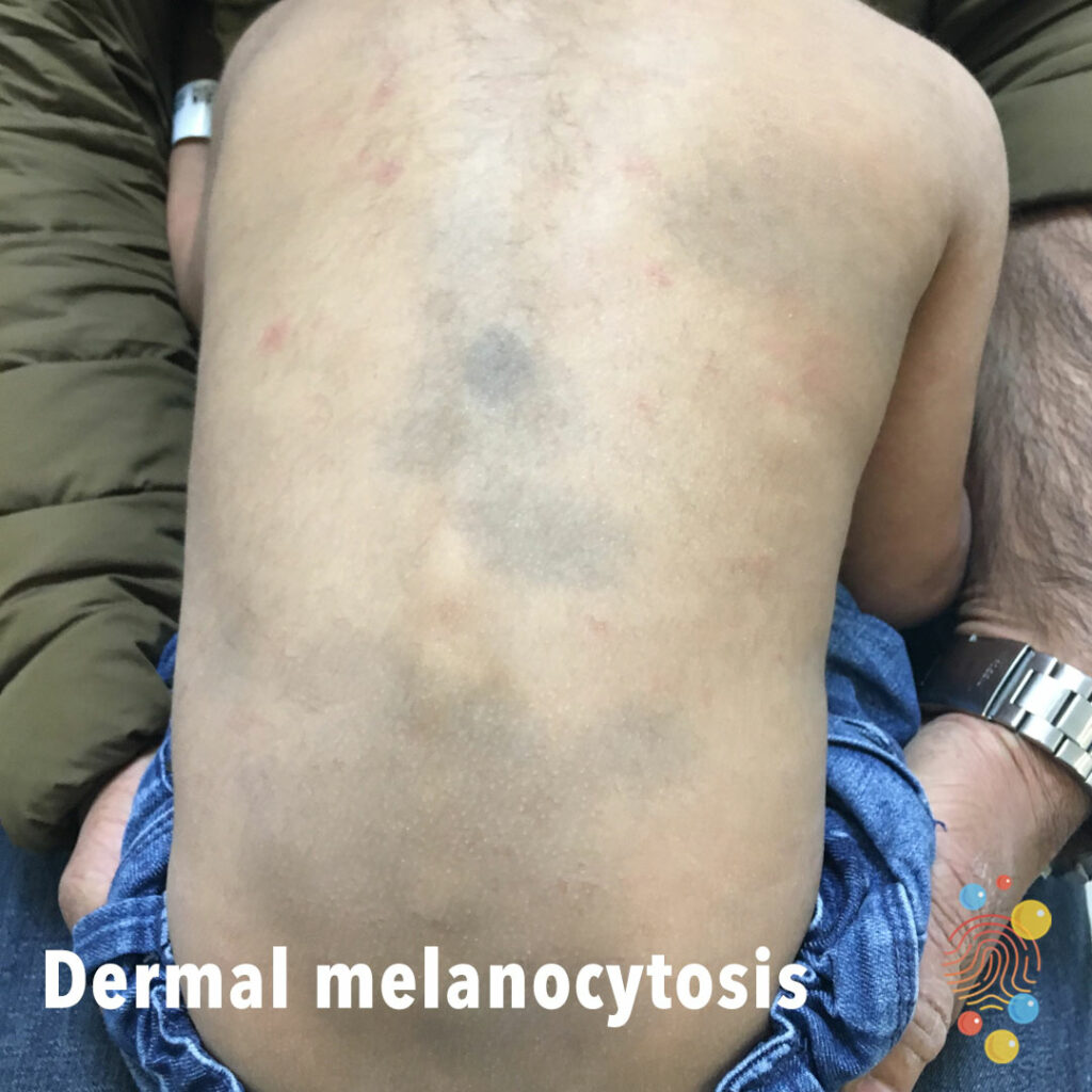
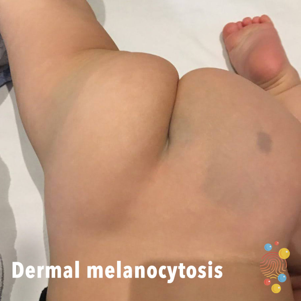
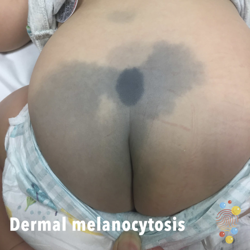
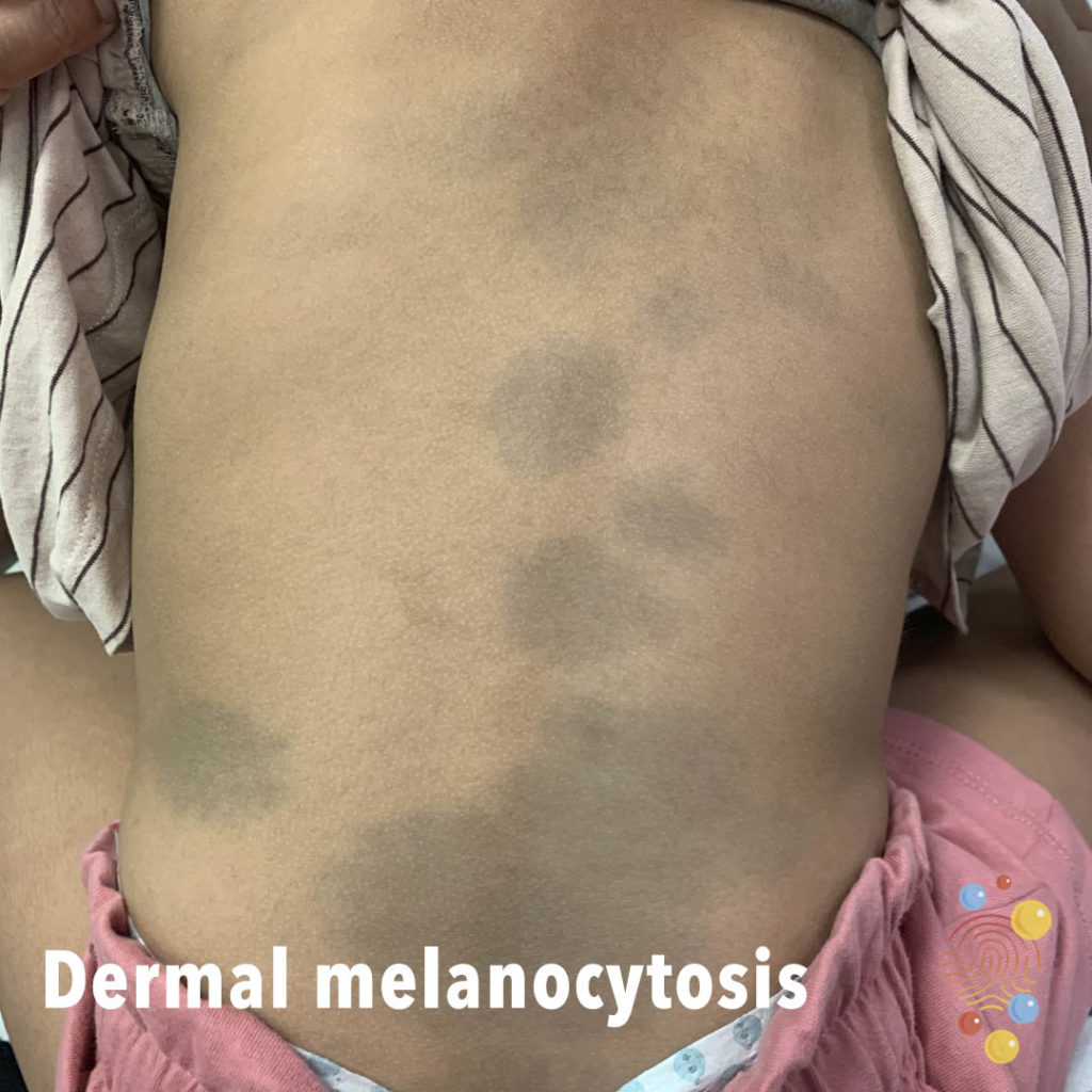
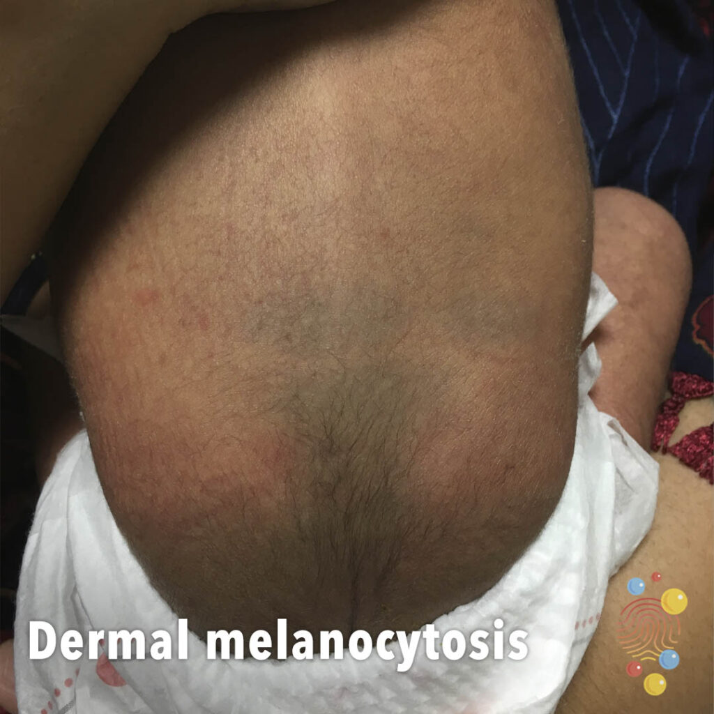
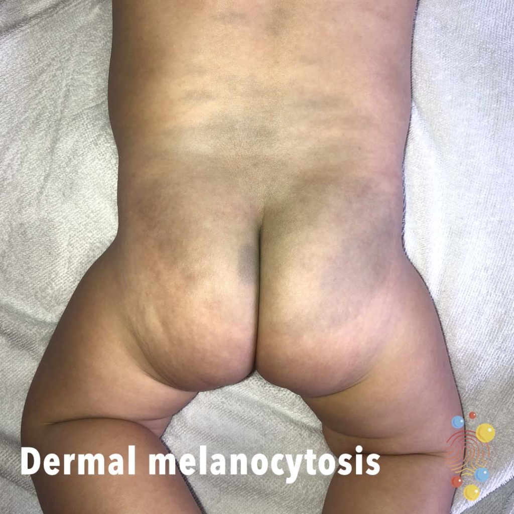
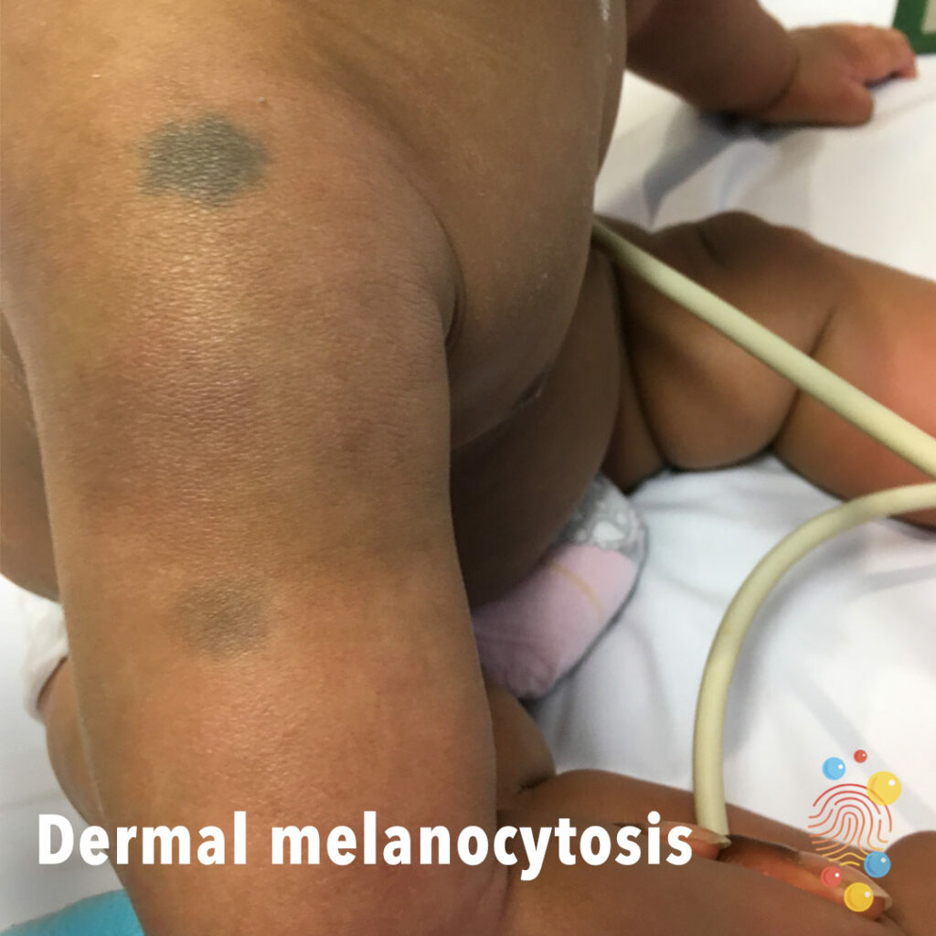
Dermal Melanocytosis
Learn more about dermal melanocytosis

Dental Abscess
Learn more about abscesses
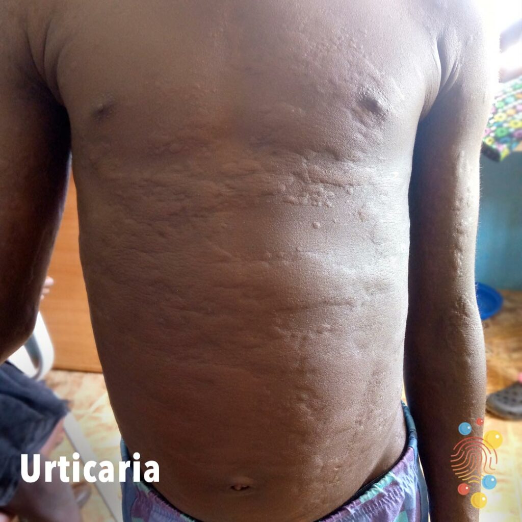
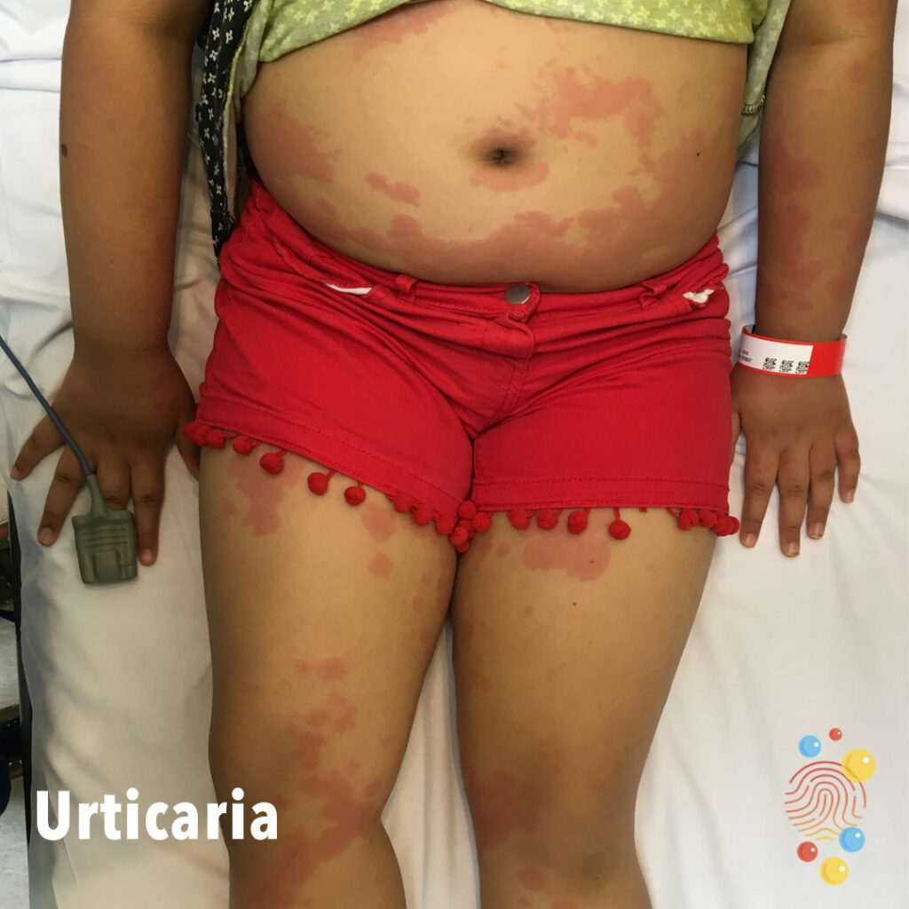
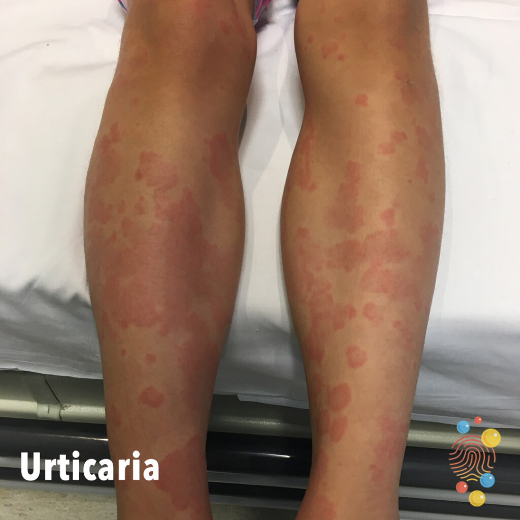
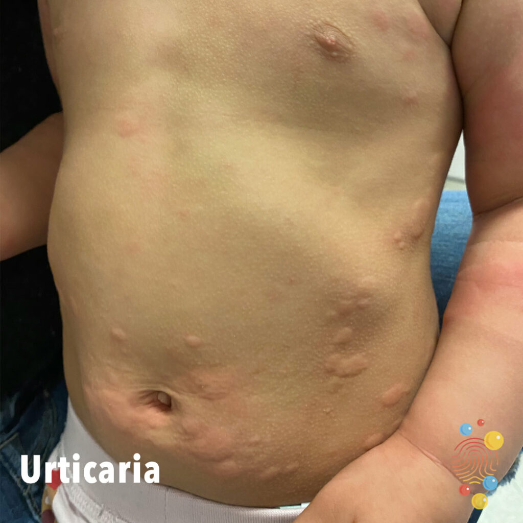
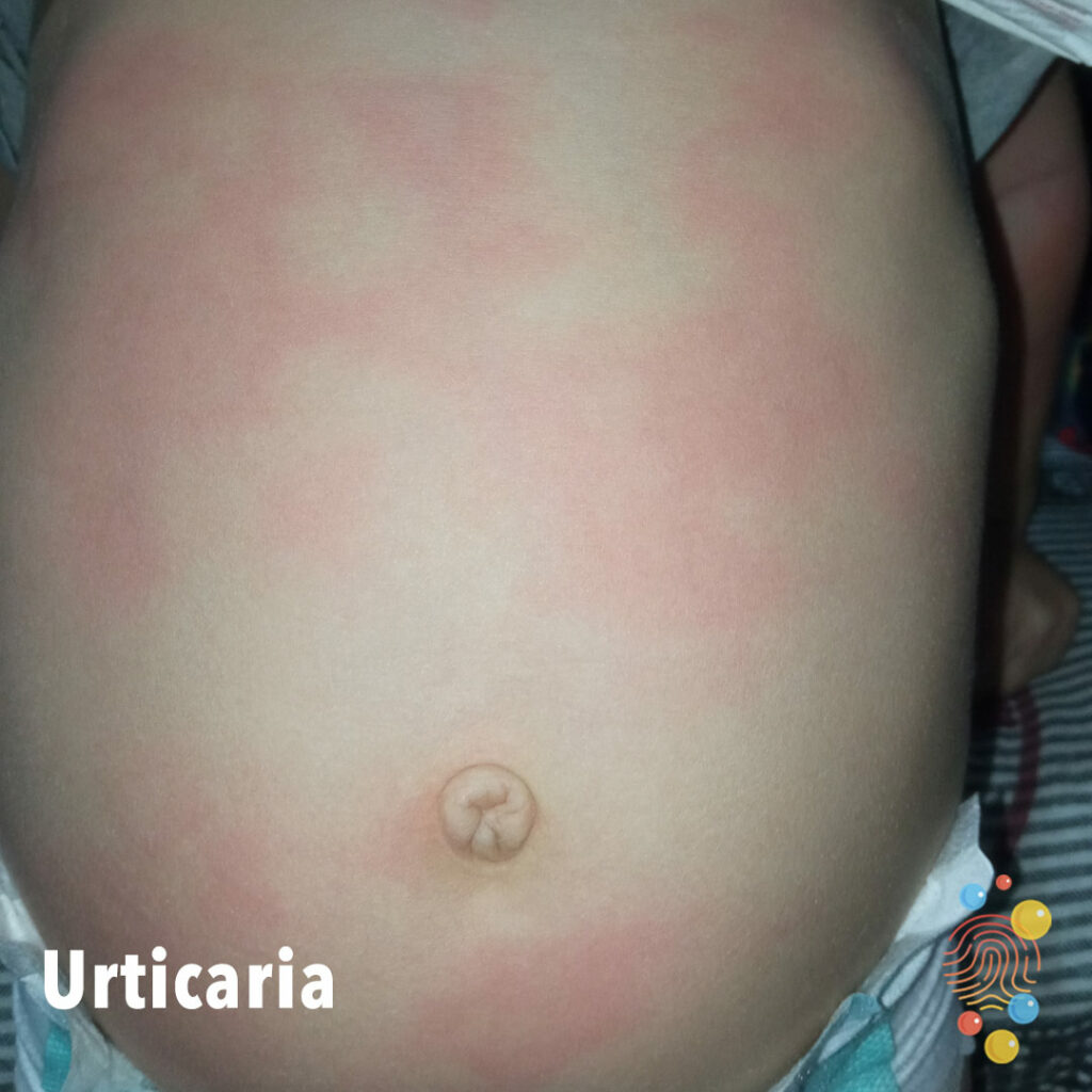
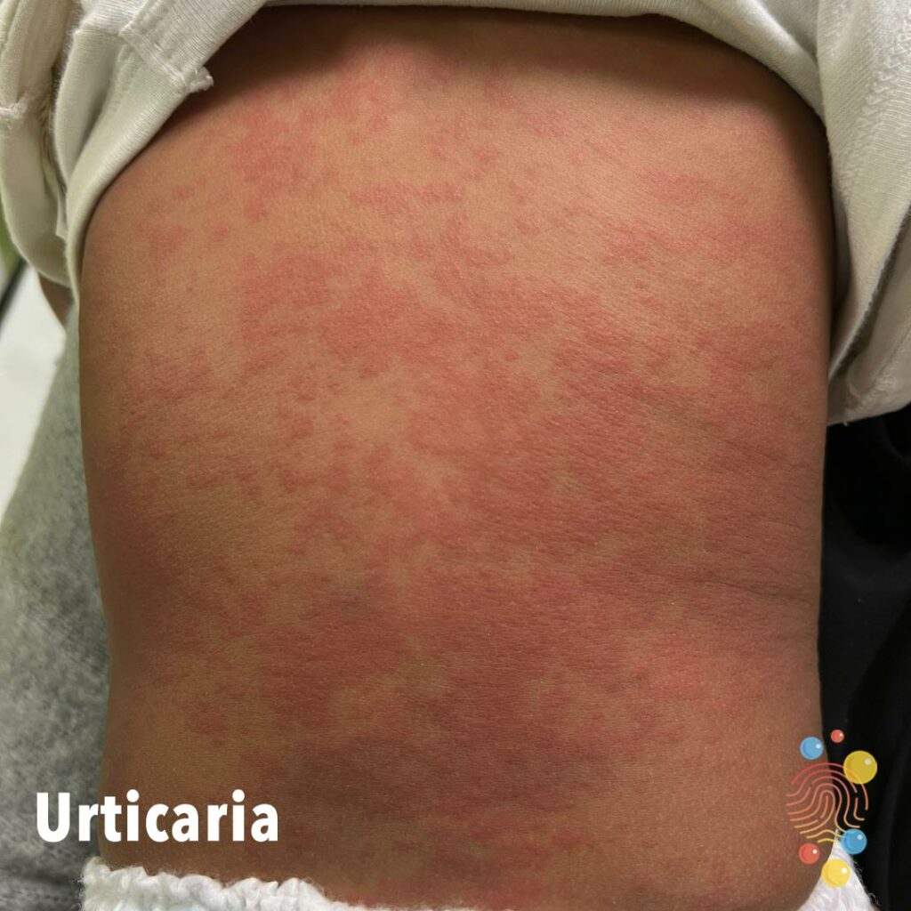
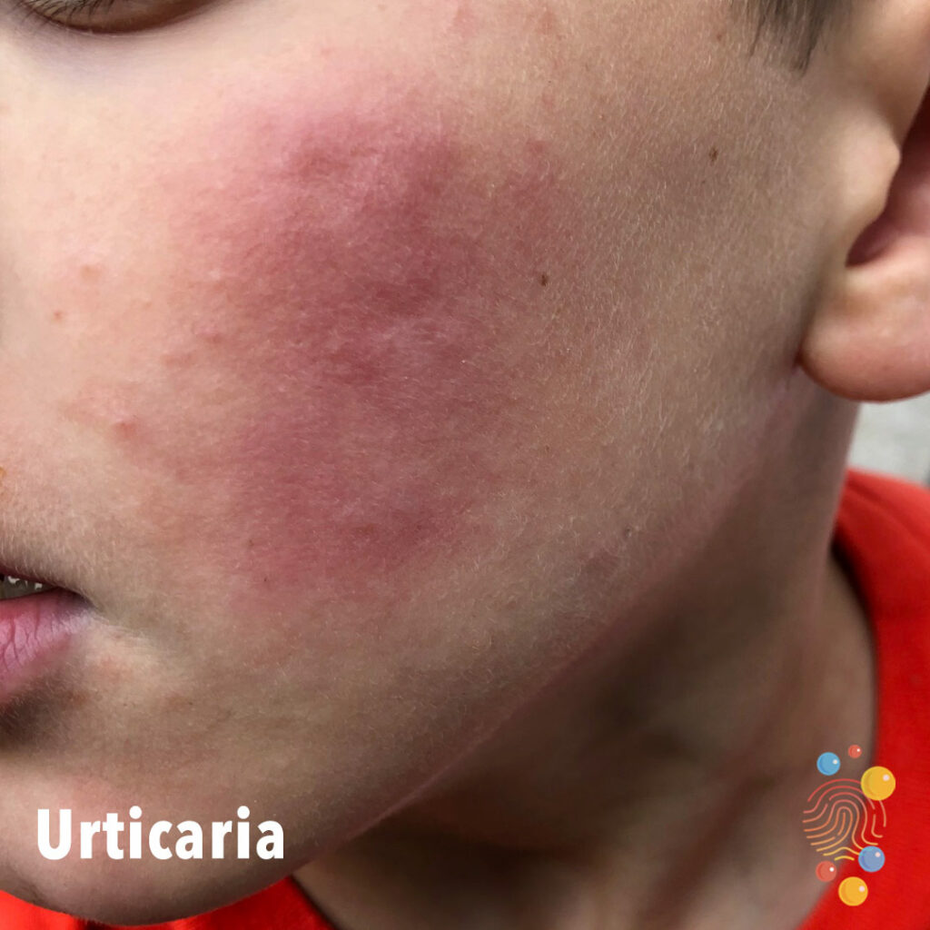
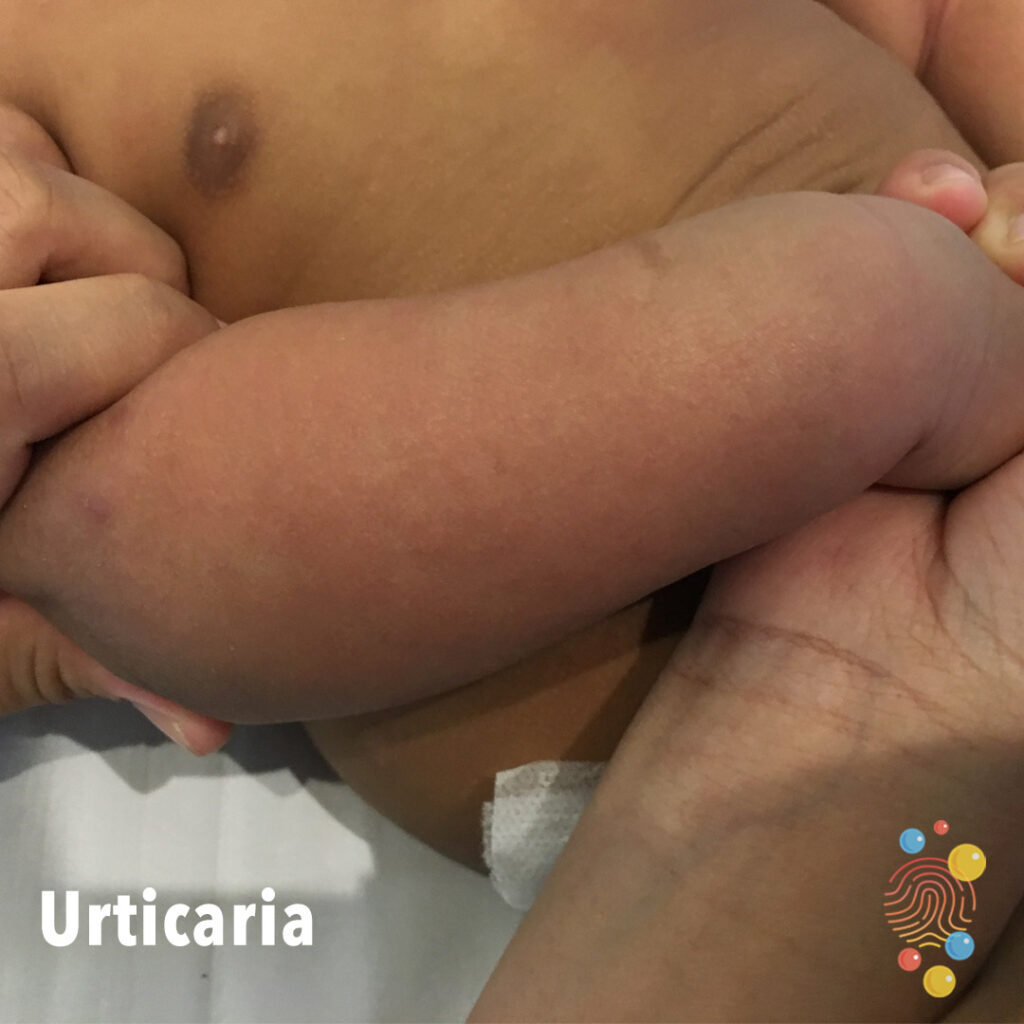
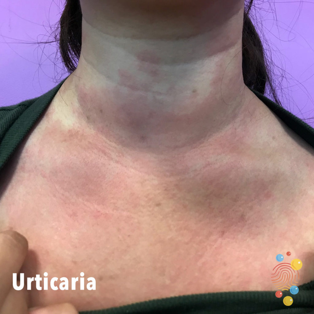
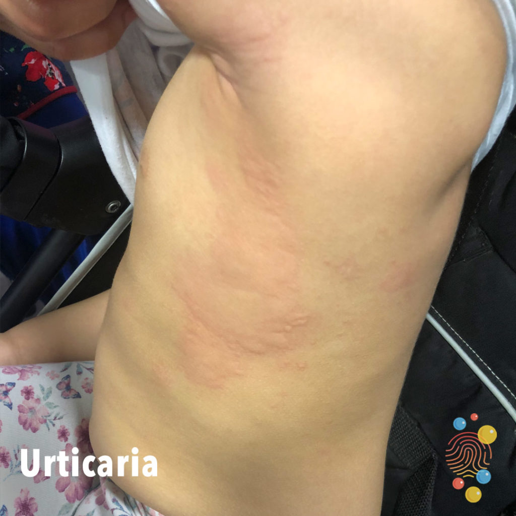
Urticaria
Learn more about urticaria
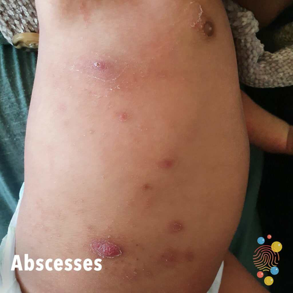
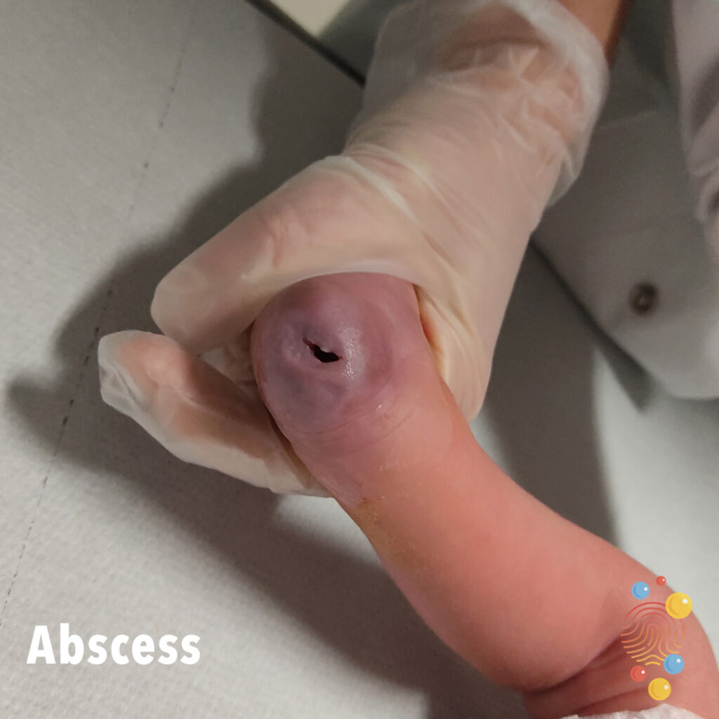
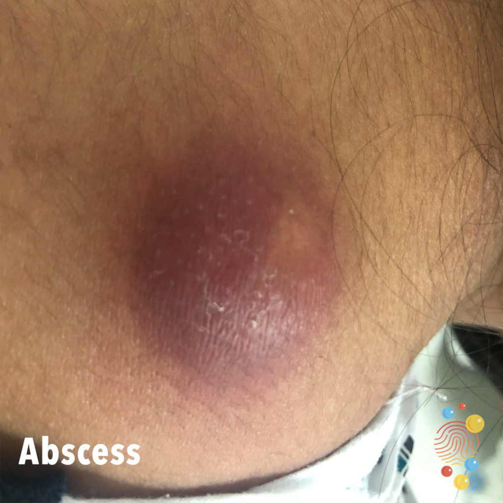
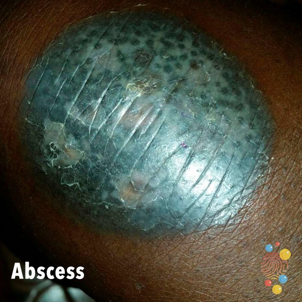
Abscesses
Learn more about abscesses
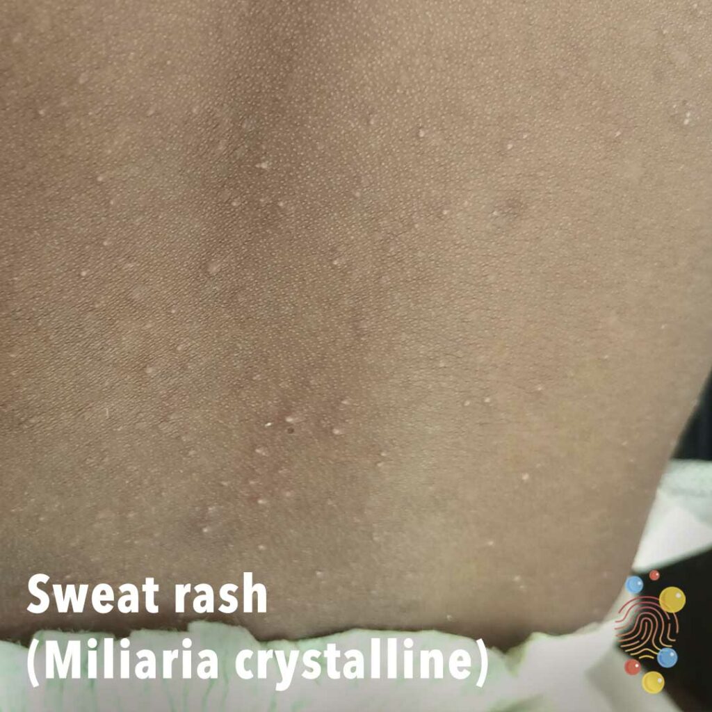
Sweat Rash (Miliaria Crystalline)
Learn more about miliaria

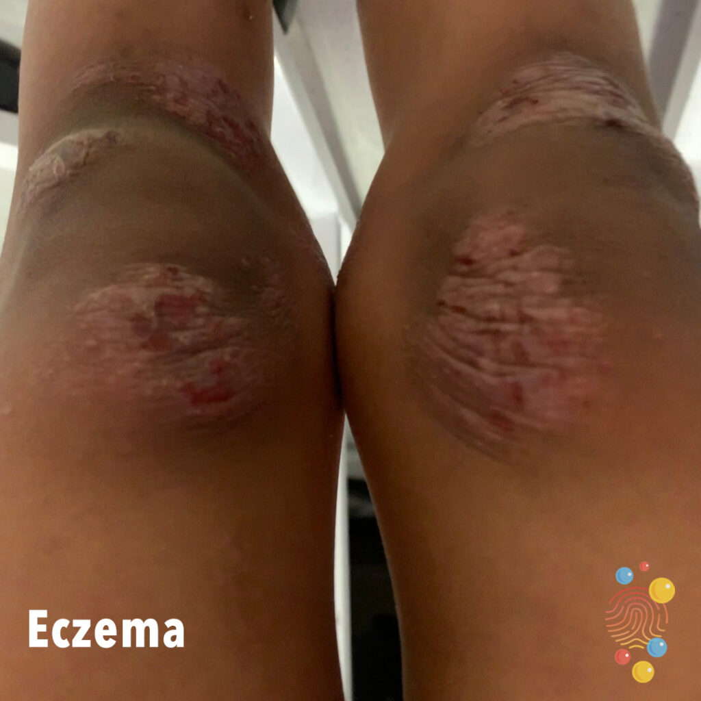
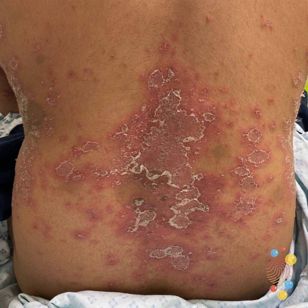
Eczema
Learn more about eczema
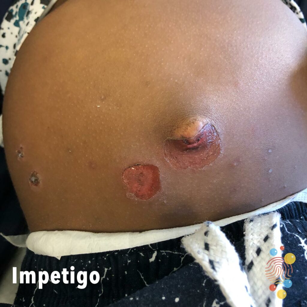
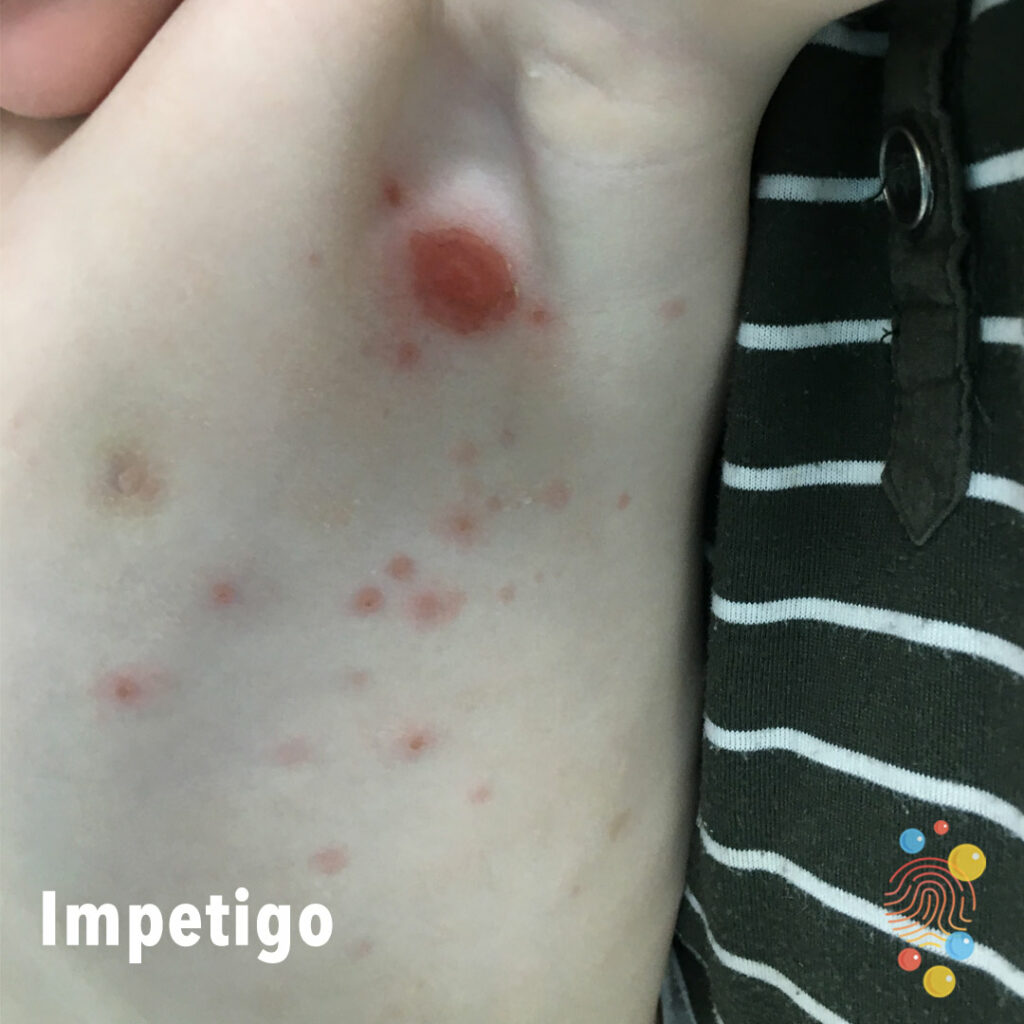
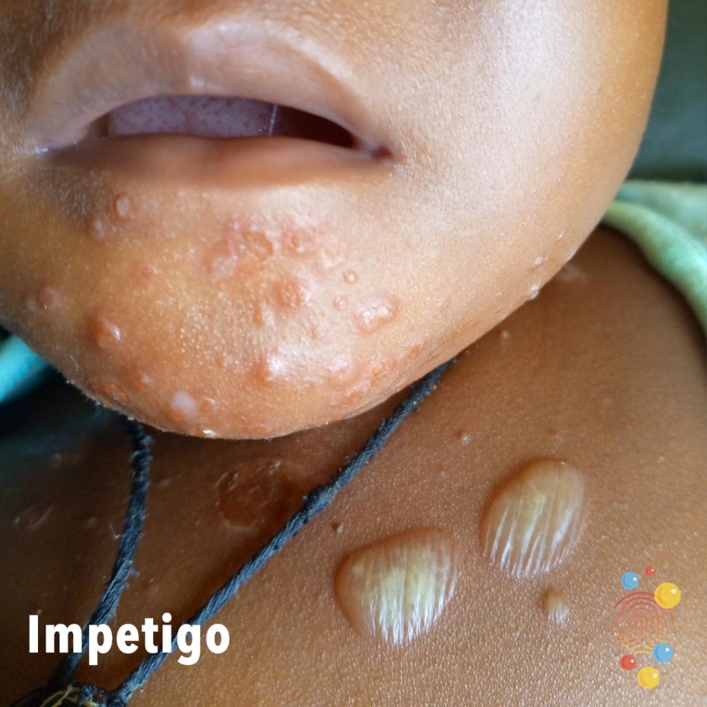
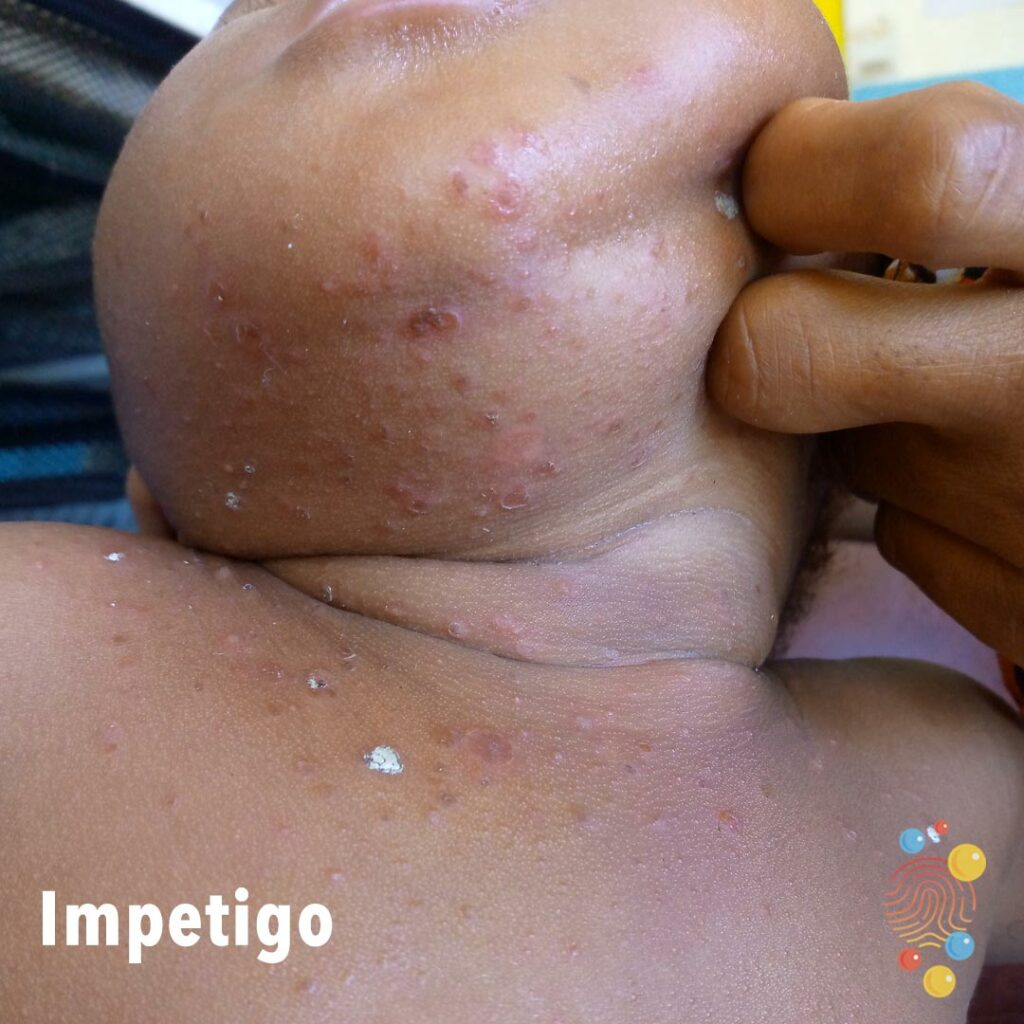
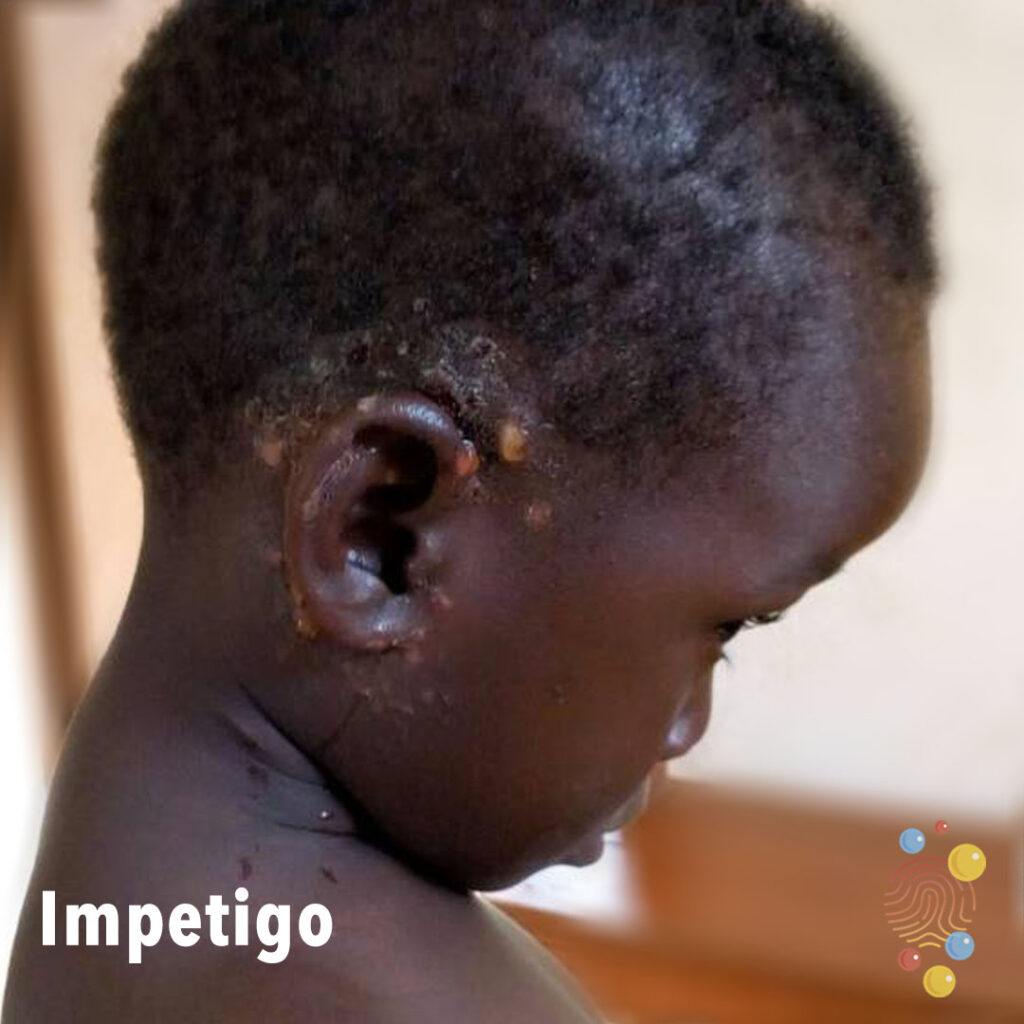
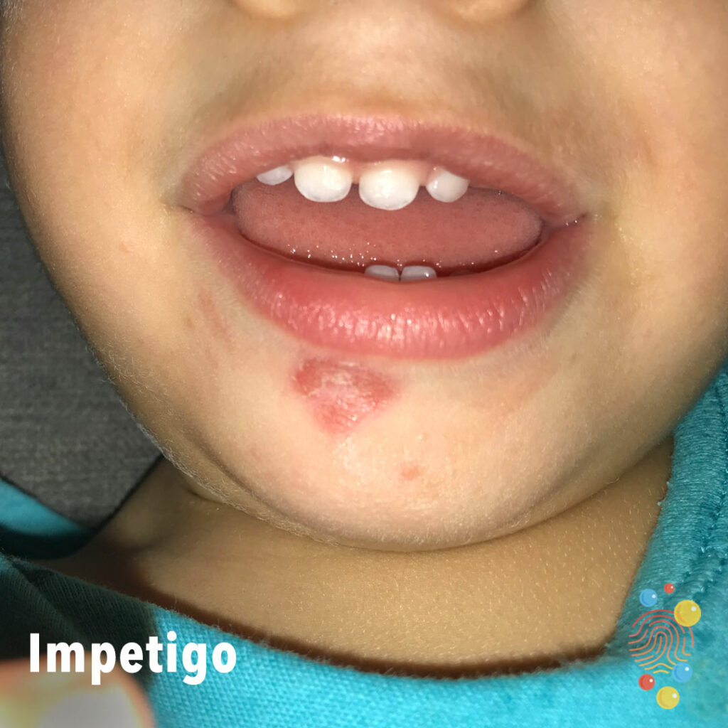
Impetigo
Learn more about impetigo
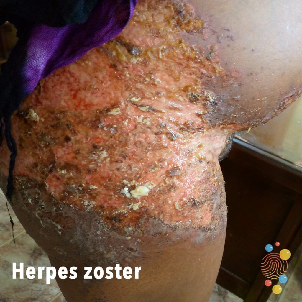
Herpes Zoster
Learn more about herpes zoster
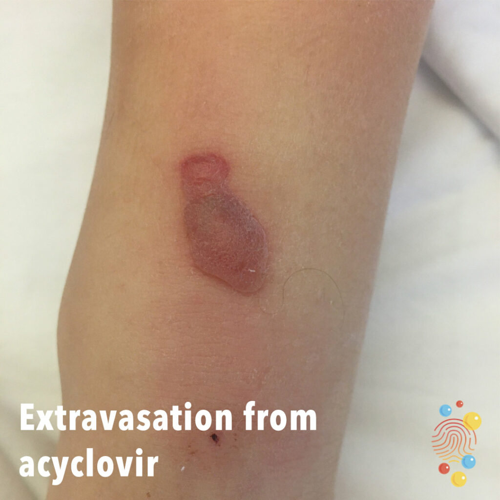
Extravasation From Acyclovir
Learn more about extravasation

Urticaria
Learn more about urticaria
Eczema
Learn more about eczema

Normal Umbilical Cord
Normal umbilical cord

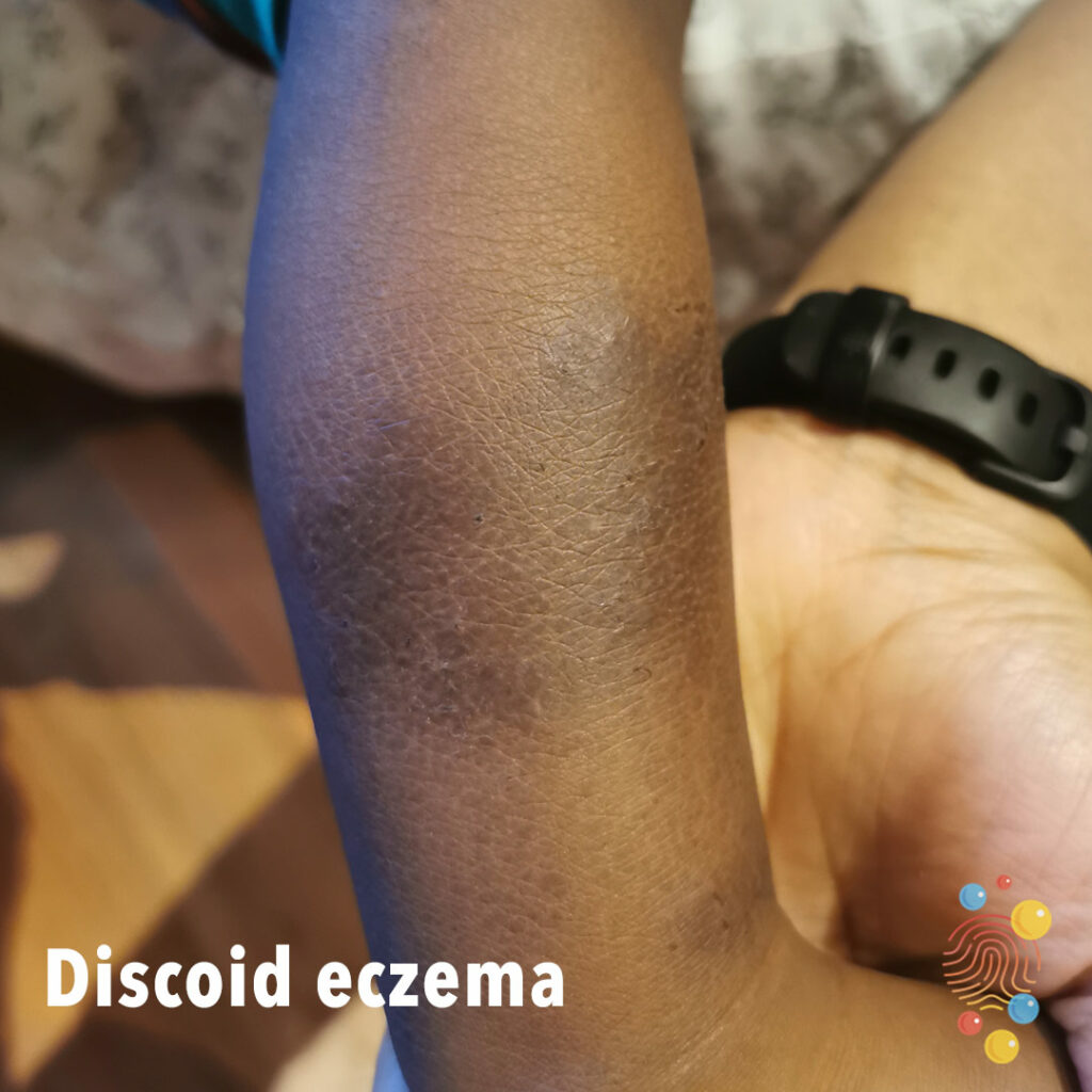
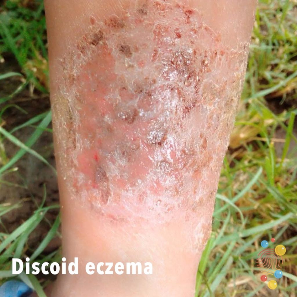
Discoid eczema
Learn more about eczema
Urticaria
Learn more about urticaria
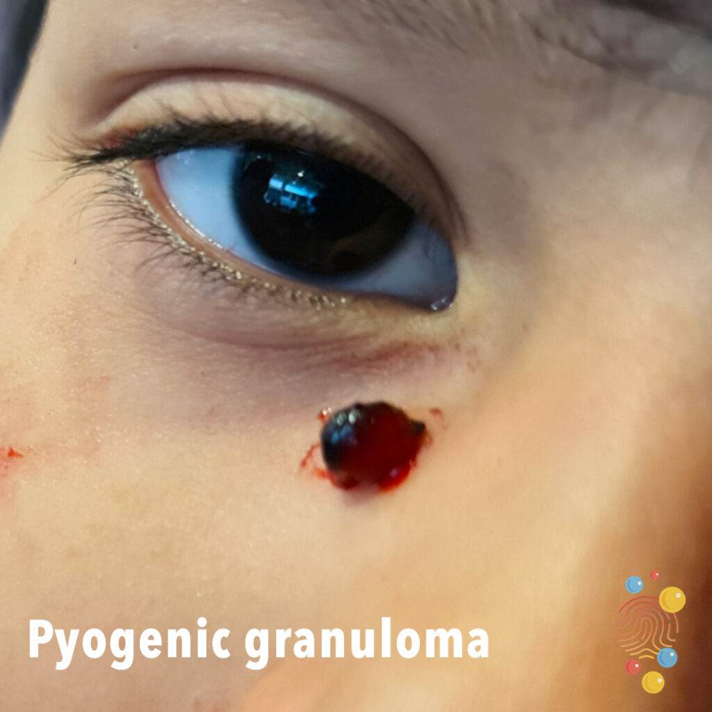
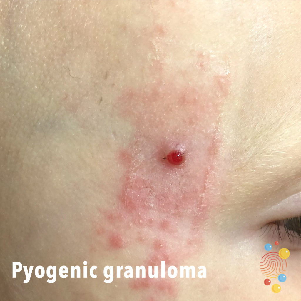
Pyogenic granuloma
Learn more about pyogenic granulomas

Torn upper lip frenulum
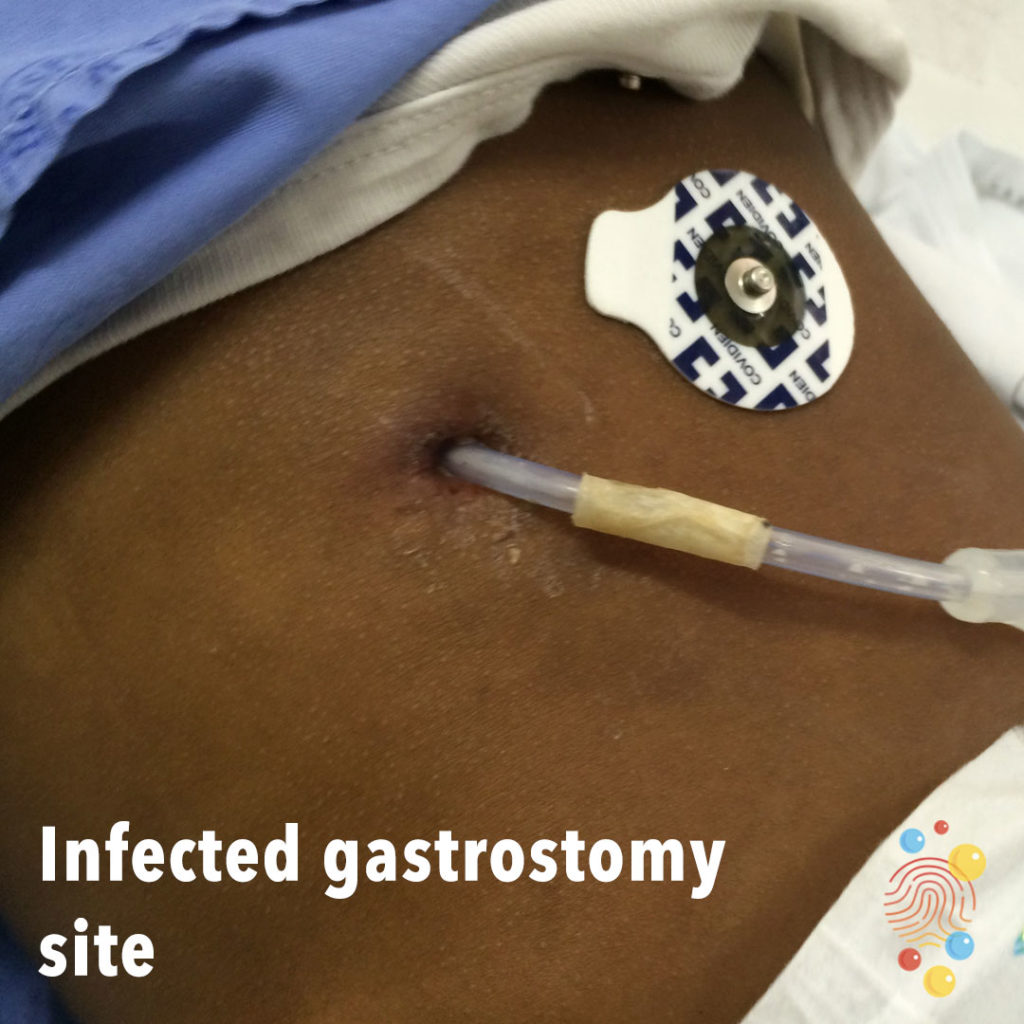
Infected Gastrostomy Site
Learn more about gastrostomies
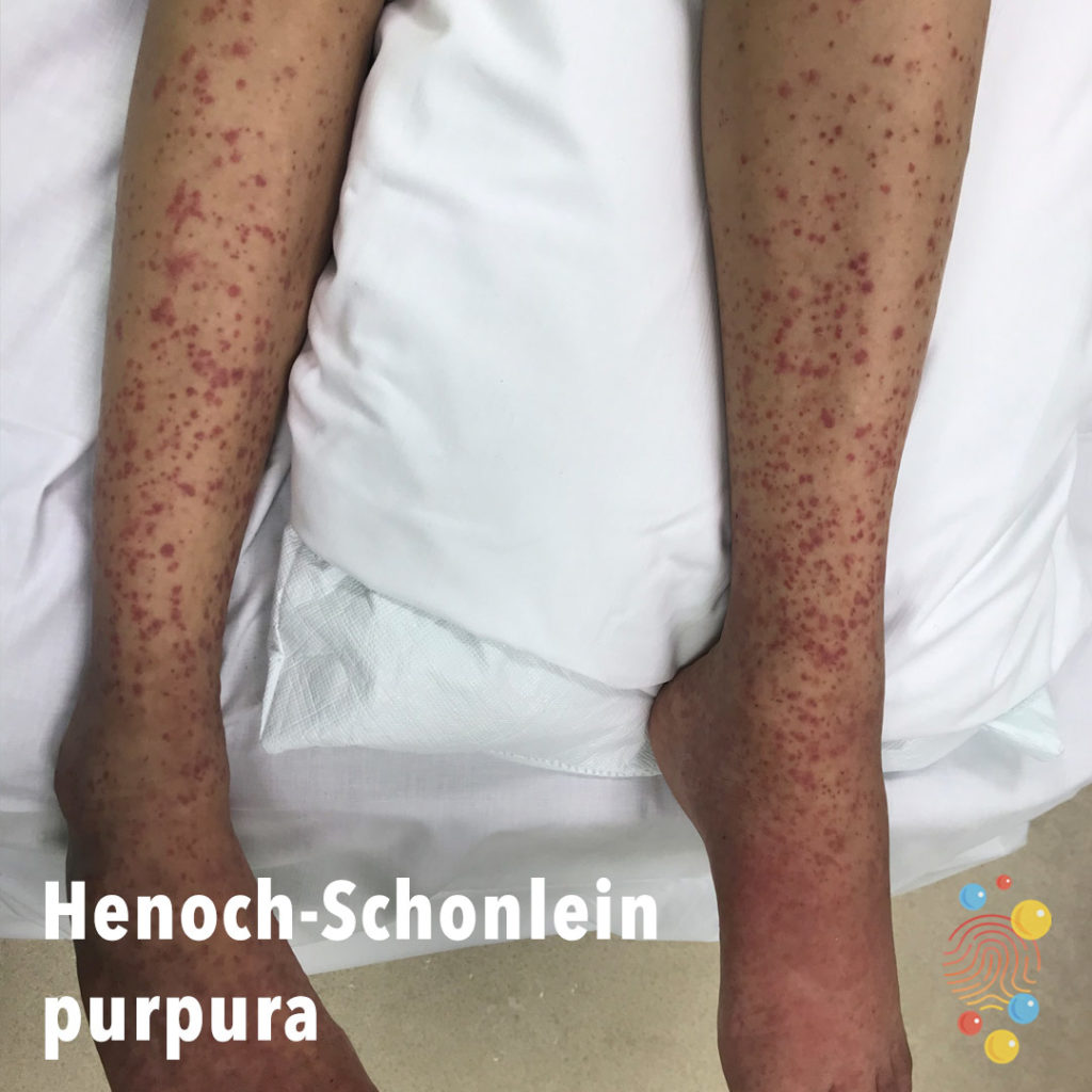
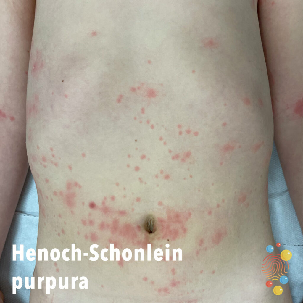
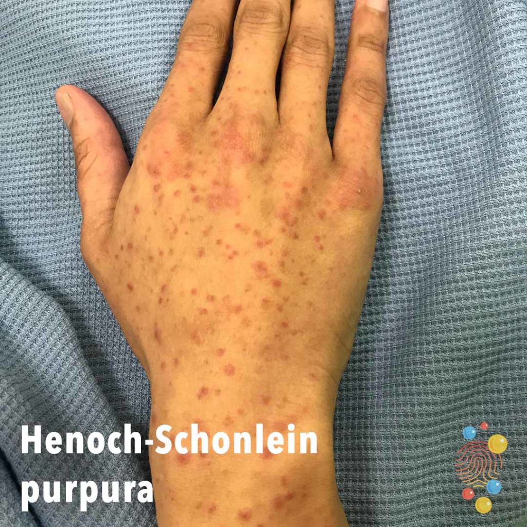
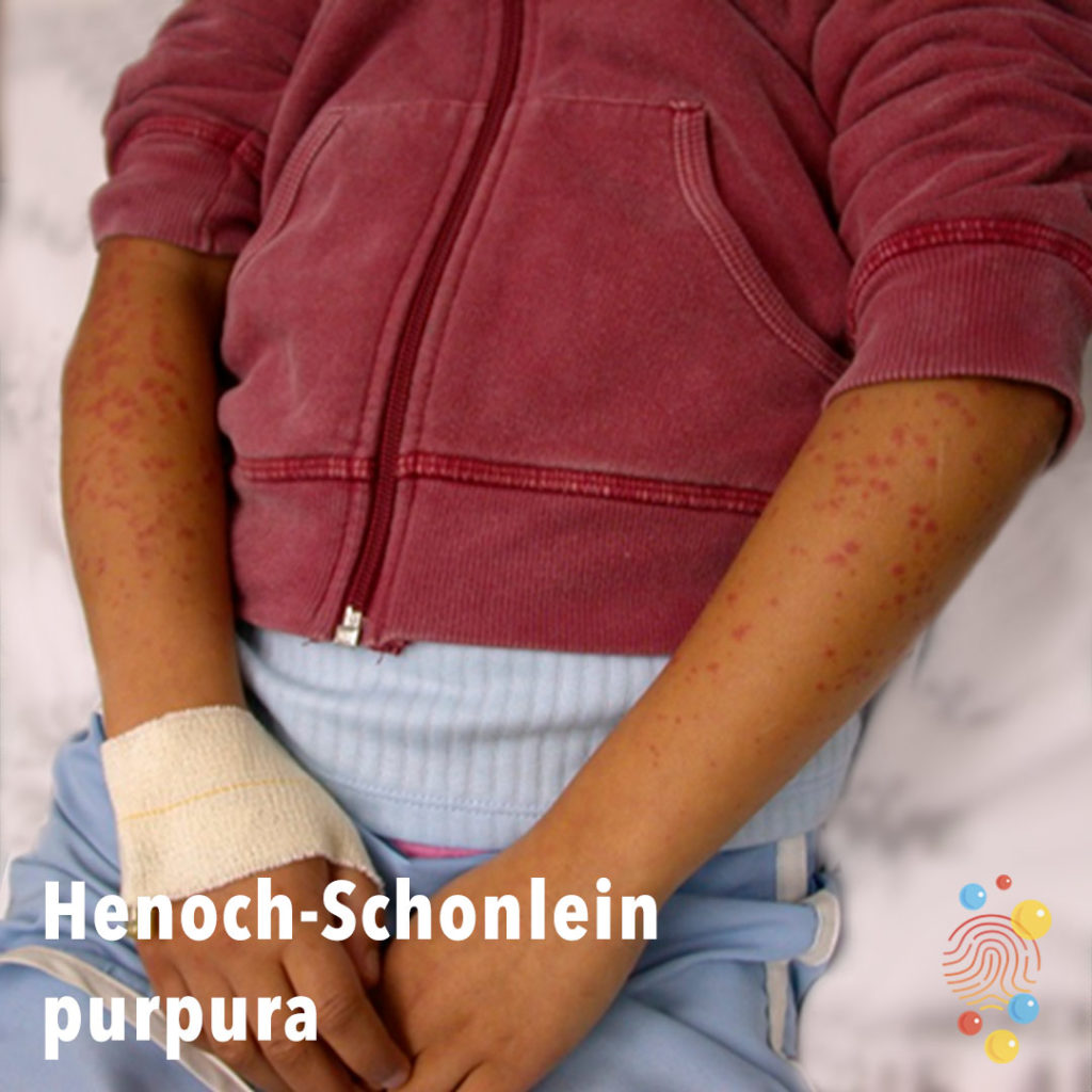
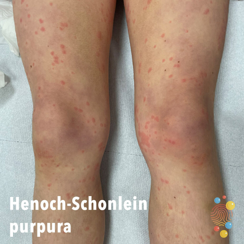
Henoch-Schonlein Purpura
Learn more about Henoch-Schonlein purpura

Eczema
Learn more about eczema

Omphalitis
Learn more about omphalitis
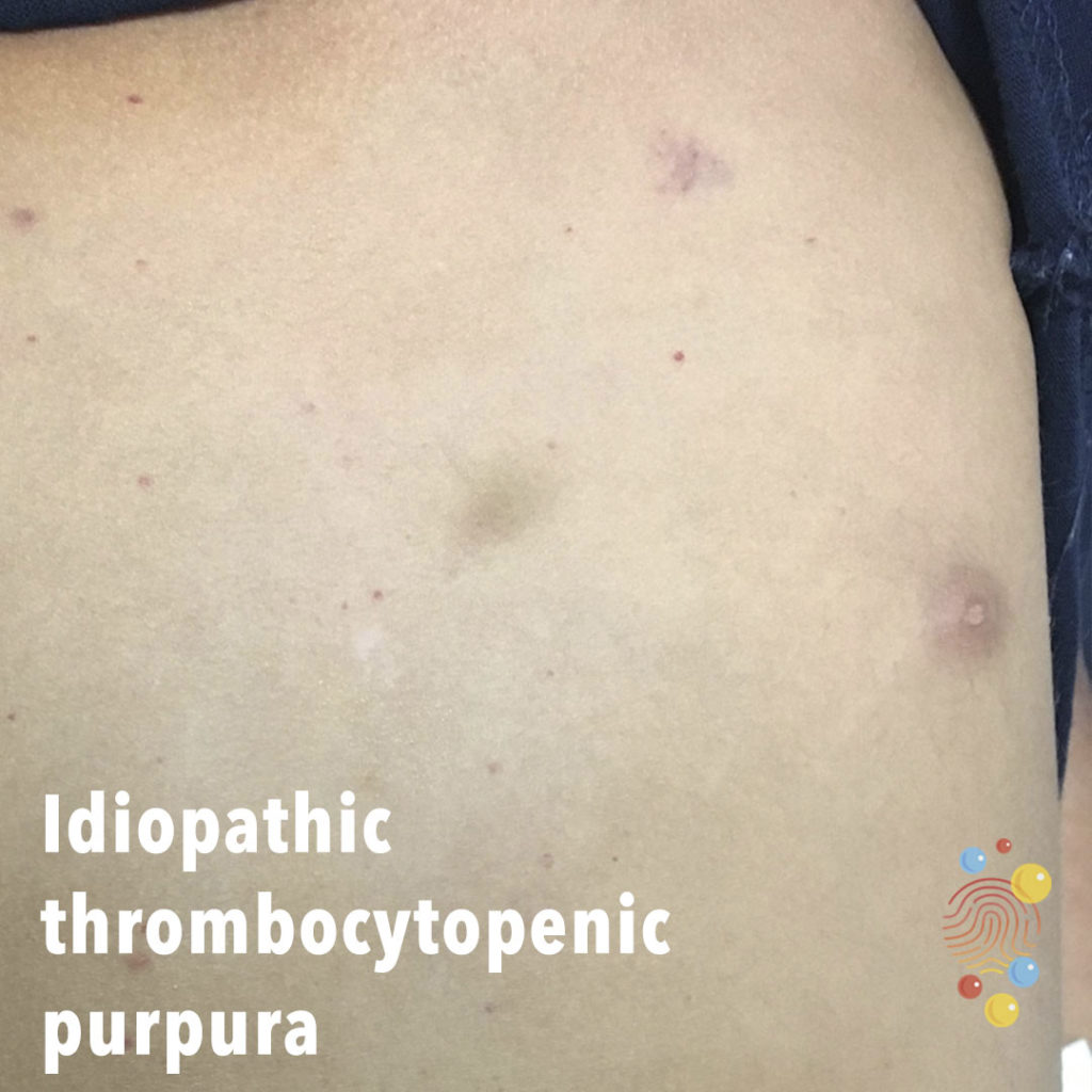
Idiopathic Thrombtocyopenic Purpura
Learn more about idiopathic thrombocytopenic purpura

Proximal Phalanx Fracture
left little finger proximal phalanx fracture

Accessory Digit
Learn more about accessory digits
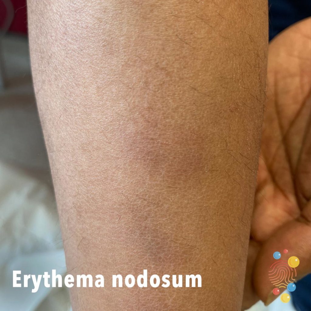
Erythema Nodosum
Learn more about erythema nodosum
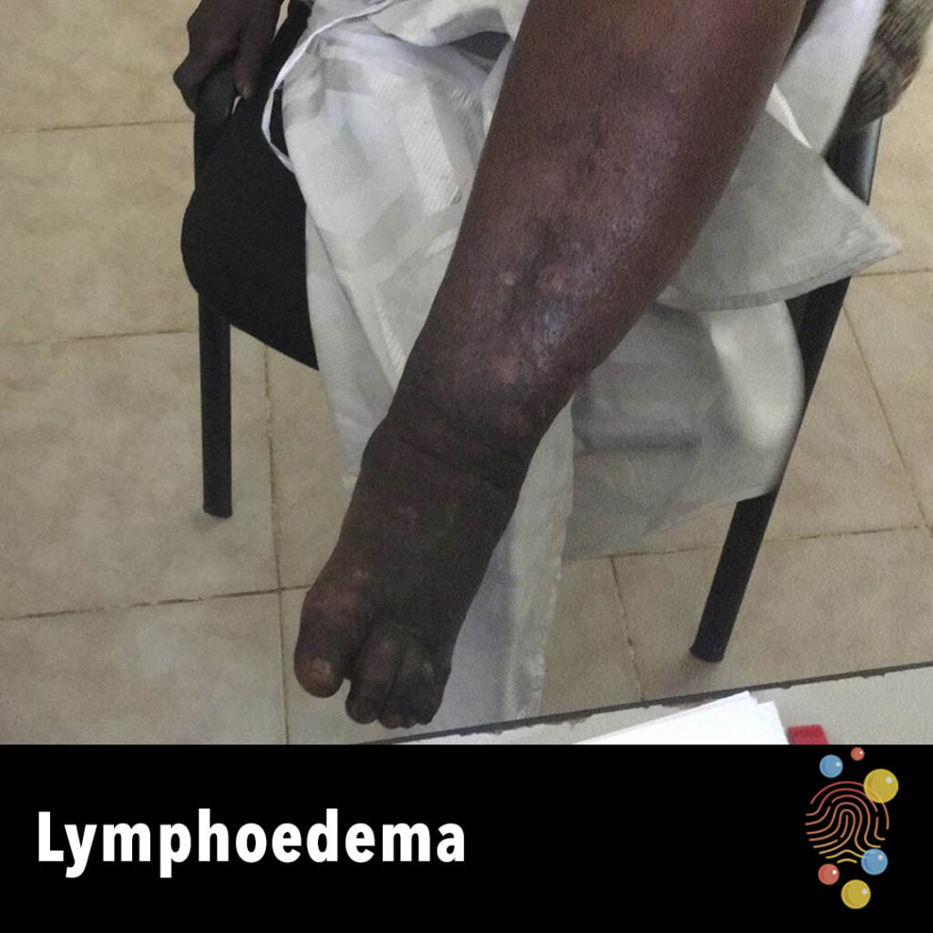
Lymphoedema
Learn more about lymphoedema

Superficial Infantile Haemangioma
Learn more about haemangiomas

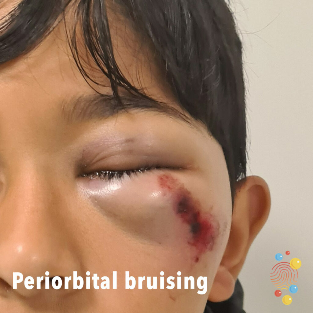
Periorbital bruising
A condition where blood pools in the tissues around the eyes, causing discoloration and bruising. It can appear as dark blue or purple bruises around the upper and lower eyelids

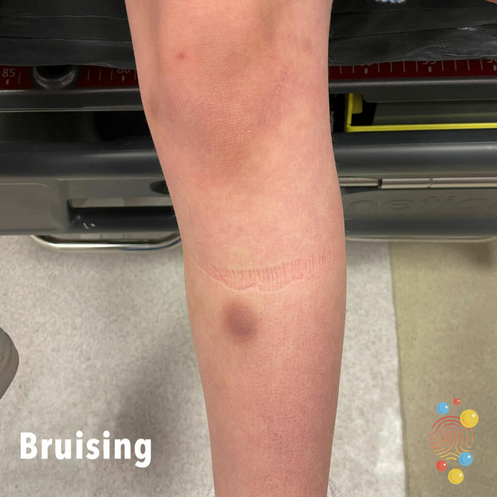
Bruise
Bruise to right knee from crawling
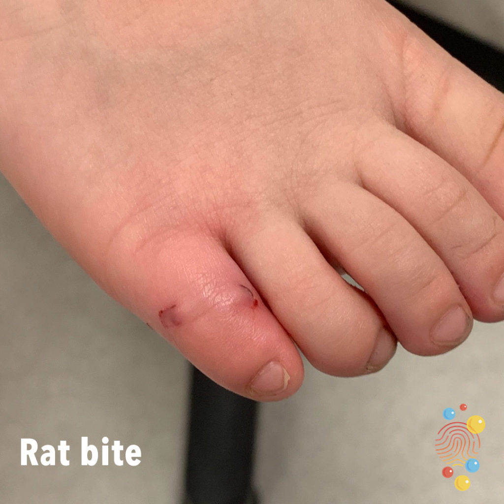
Rat Bite
Learn more about bites

Impetiginized Eczema
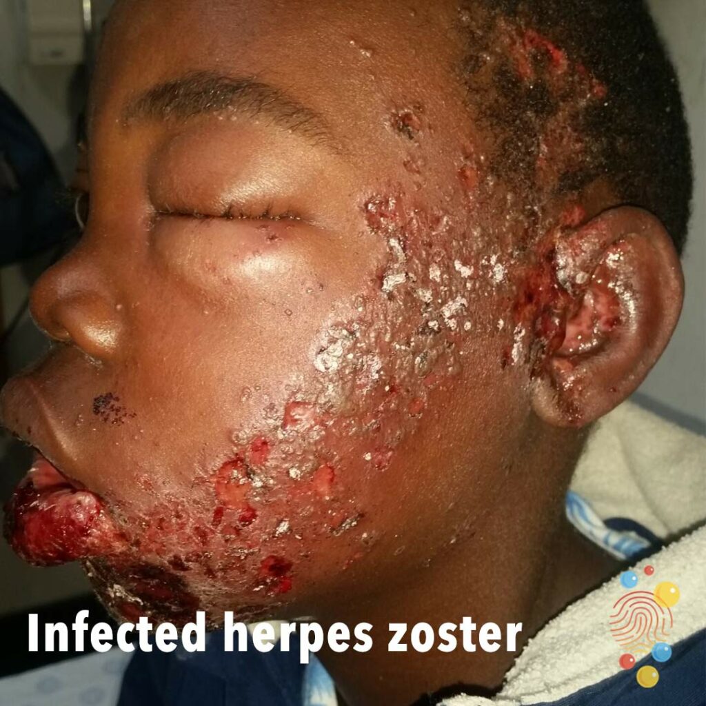
Infected herpes zoster
Learn more about herpes zoster
Abscess
Learn more about abscesses

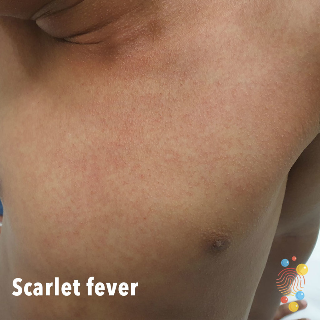
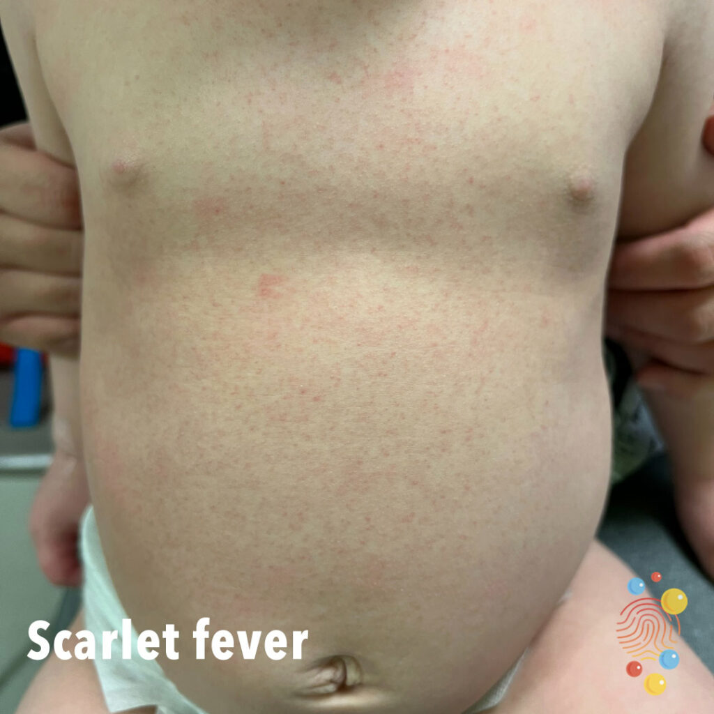
Scarlet Fever
Scarlet fever is a bacterial illness that develops in some people who have strep throat. Also known as scarlatina, scarlet fever features a bright red rash
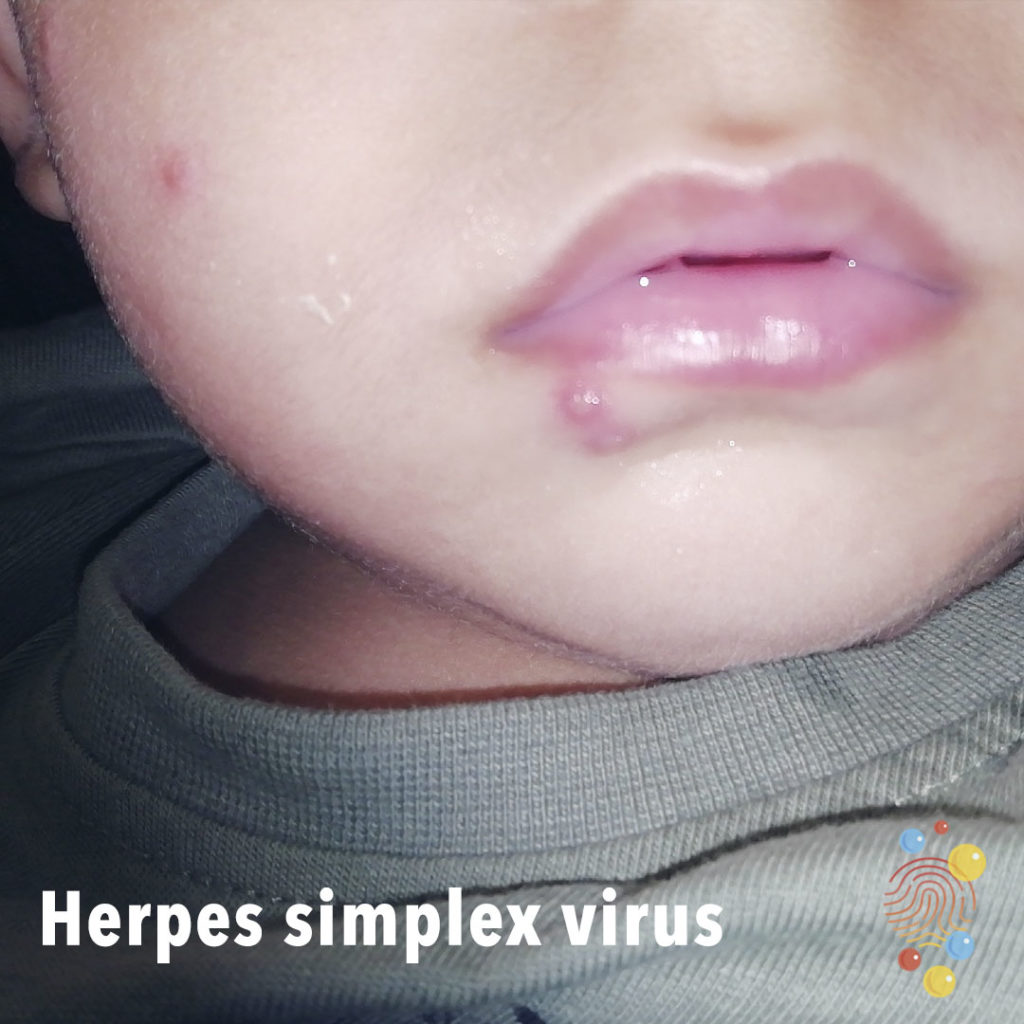
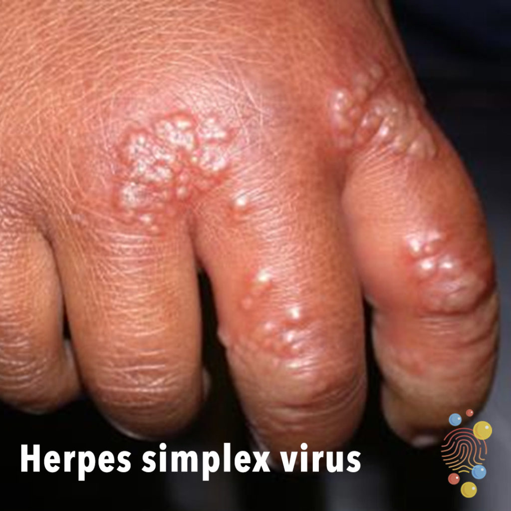
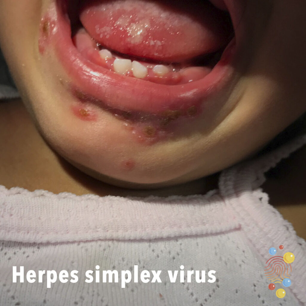
Herpes Simplex Virus
Learn more about herpes simplex virus
Dermal Melanocytosis
Learn more about dermal melanocytosis
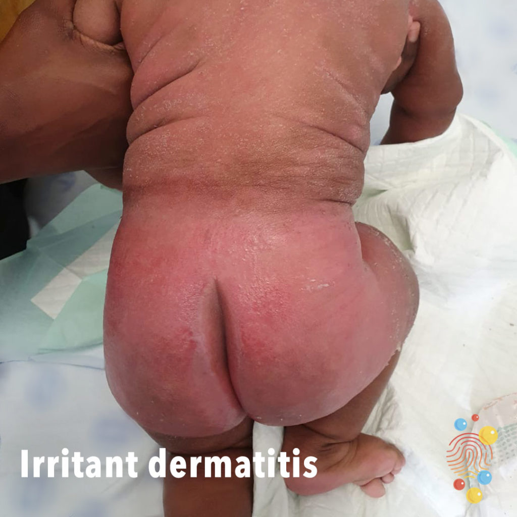
Irritant Dermatitis
Learn more about irritant dermatitis
Discoid eczema
Learn more about eczema

Conjunctivitis
Learn more about conjunctivitis
Impetigo
Learn more about bullous impetigo

Cellulitis
Learn more about cellulitis
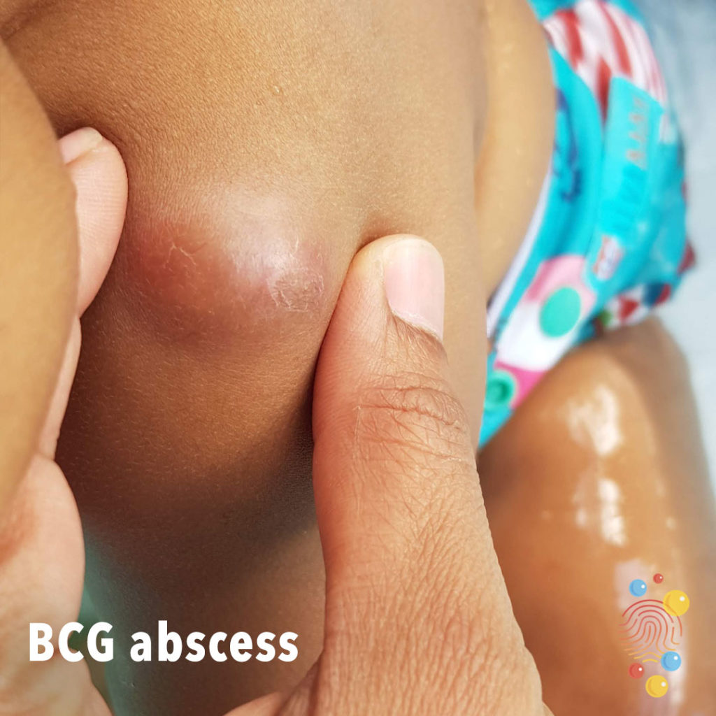
BCG Abscess
Learn more about BCGs
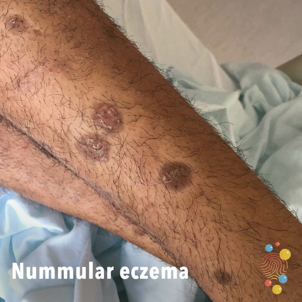
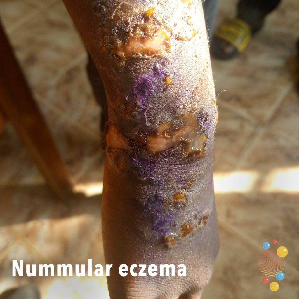
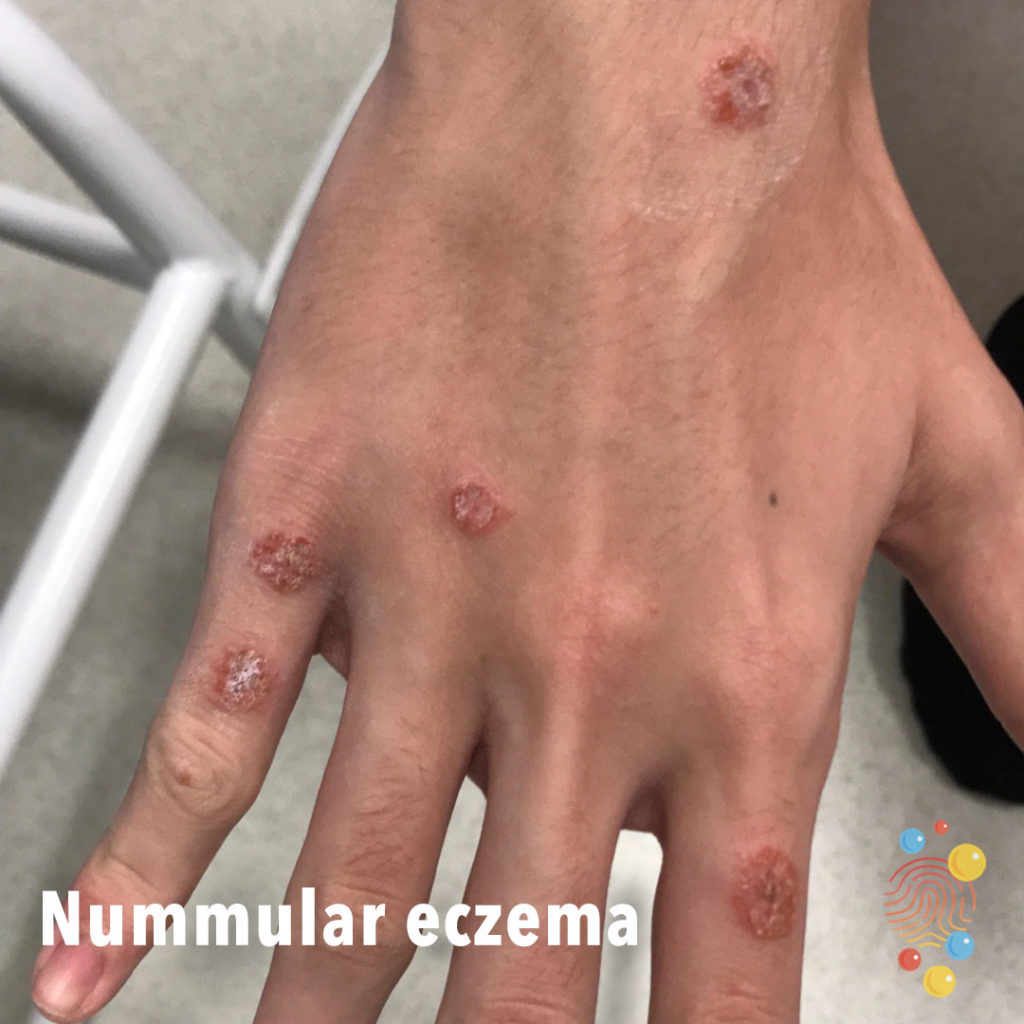
Nummular Eczema
Learn more about eczema
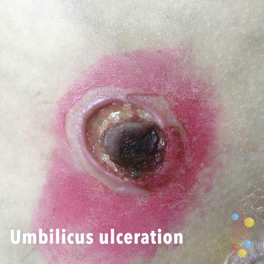
Umbilicus Ulceration
Learn more about ulcers
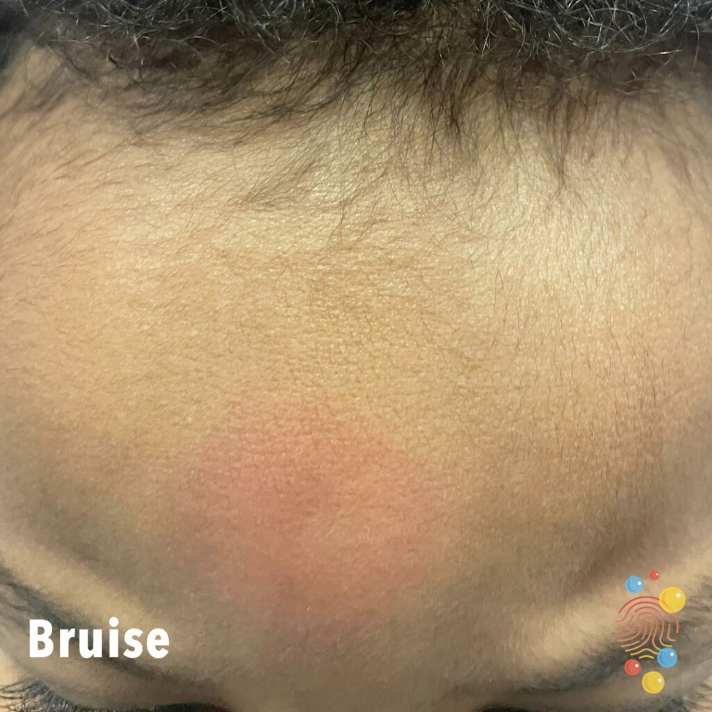
Bruise
Central forehead bruise.
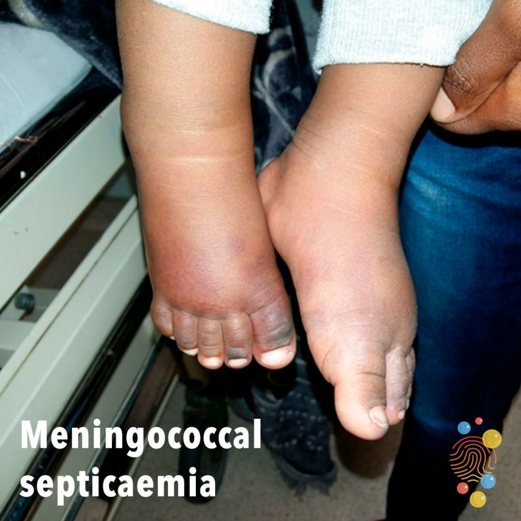
Meningococcal Septicaemia
Learn more about meningococcal septicaemia
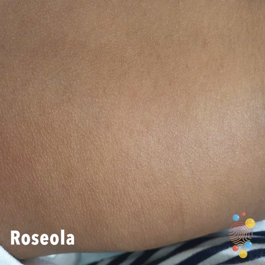
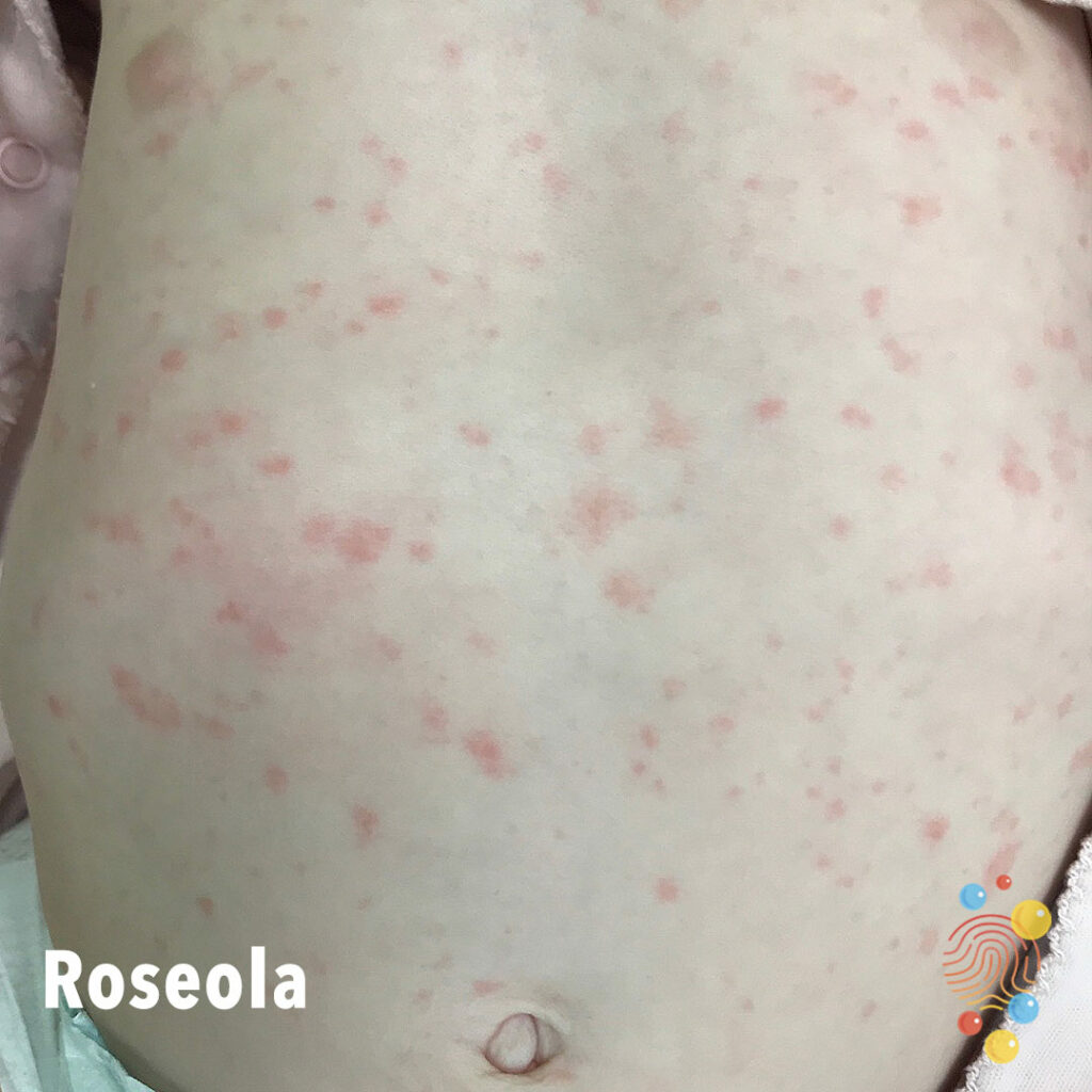
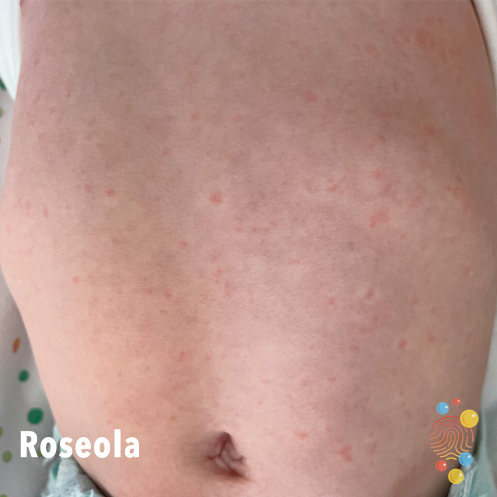
Roseola
Learn more about roseola

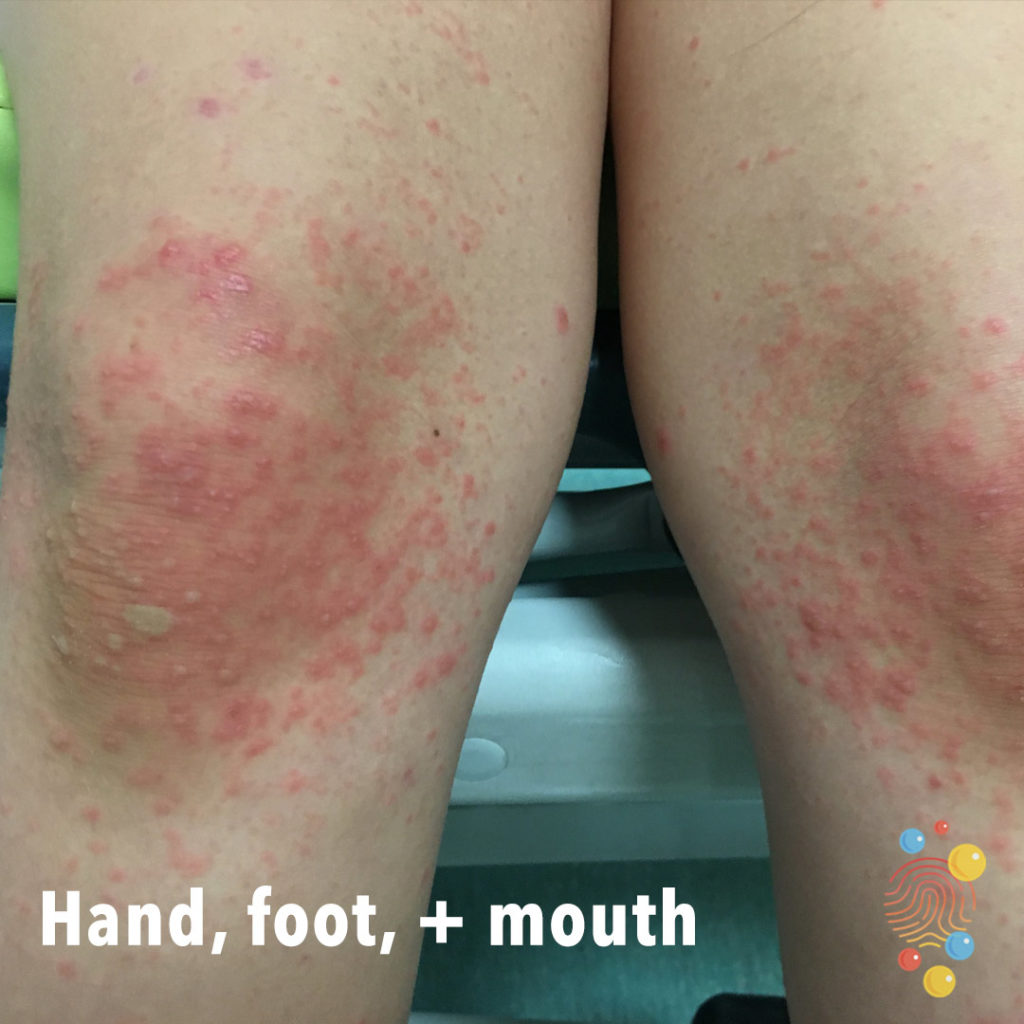
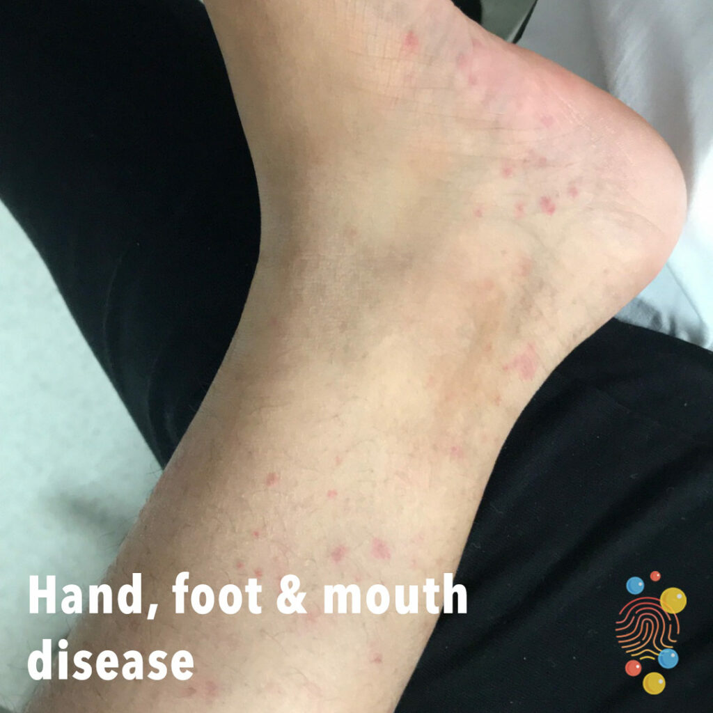
Hand, Foot, + Mouth
Learn more about hand, foot, + mouth disease
Scarlet Fever

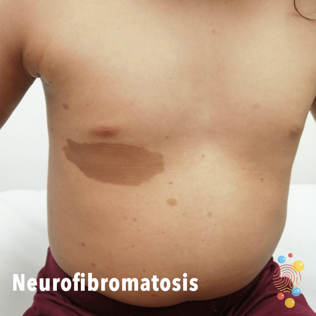
Neurofibromatosis
Multiple café-au-lait macules and axillary freckiling in a 4-year-old girl with NF1
Periorbital Bruising
a condition where blood pools in the tissues around the eyes, causing discoloration and bruising. It can appear as dark blue or purple bruises around the upper and lower eyelids
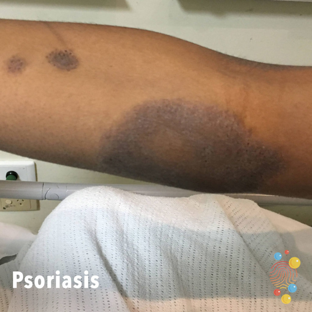
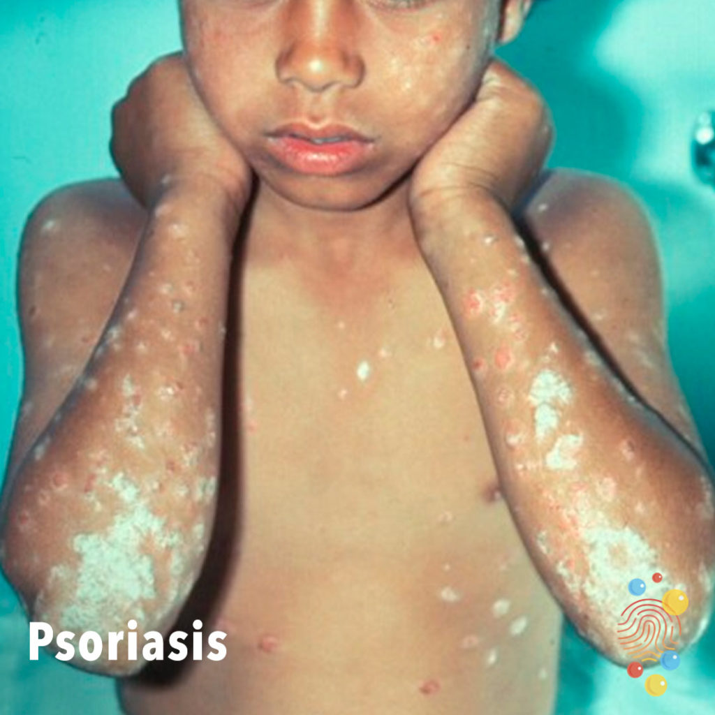
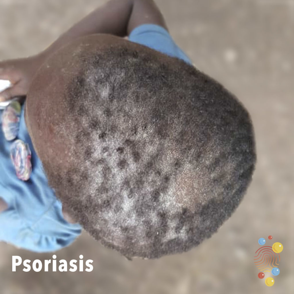
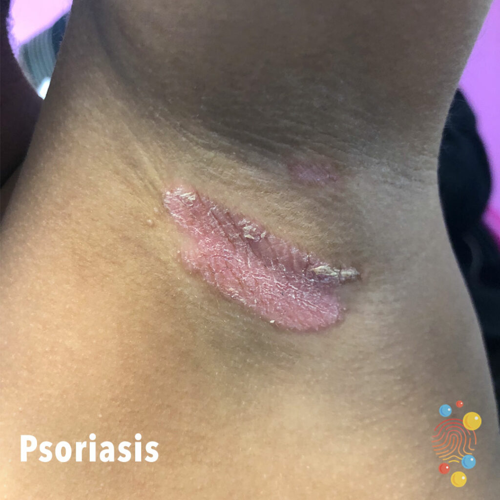
Psoriasis
Learn more about psoriasis
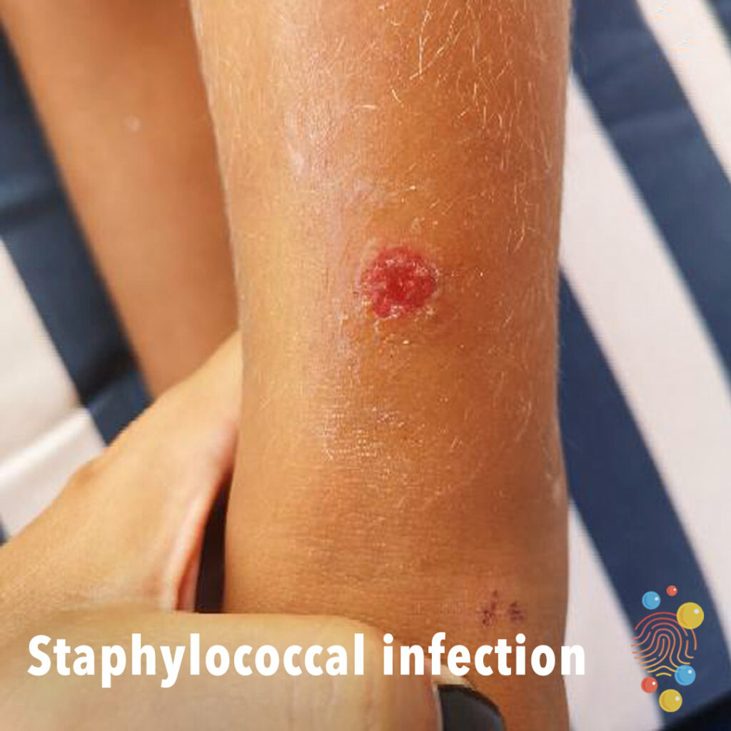
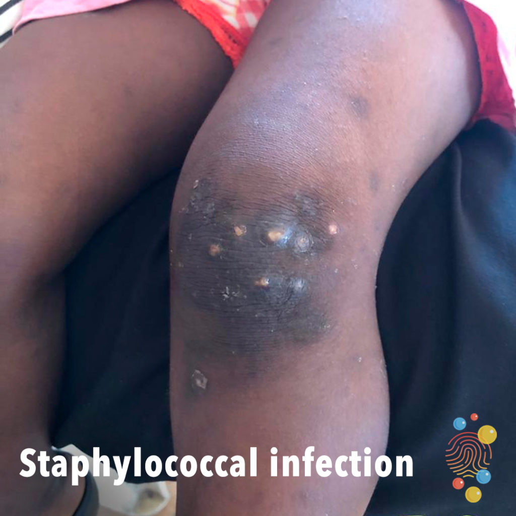
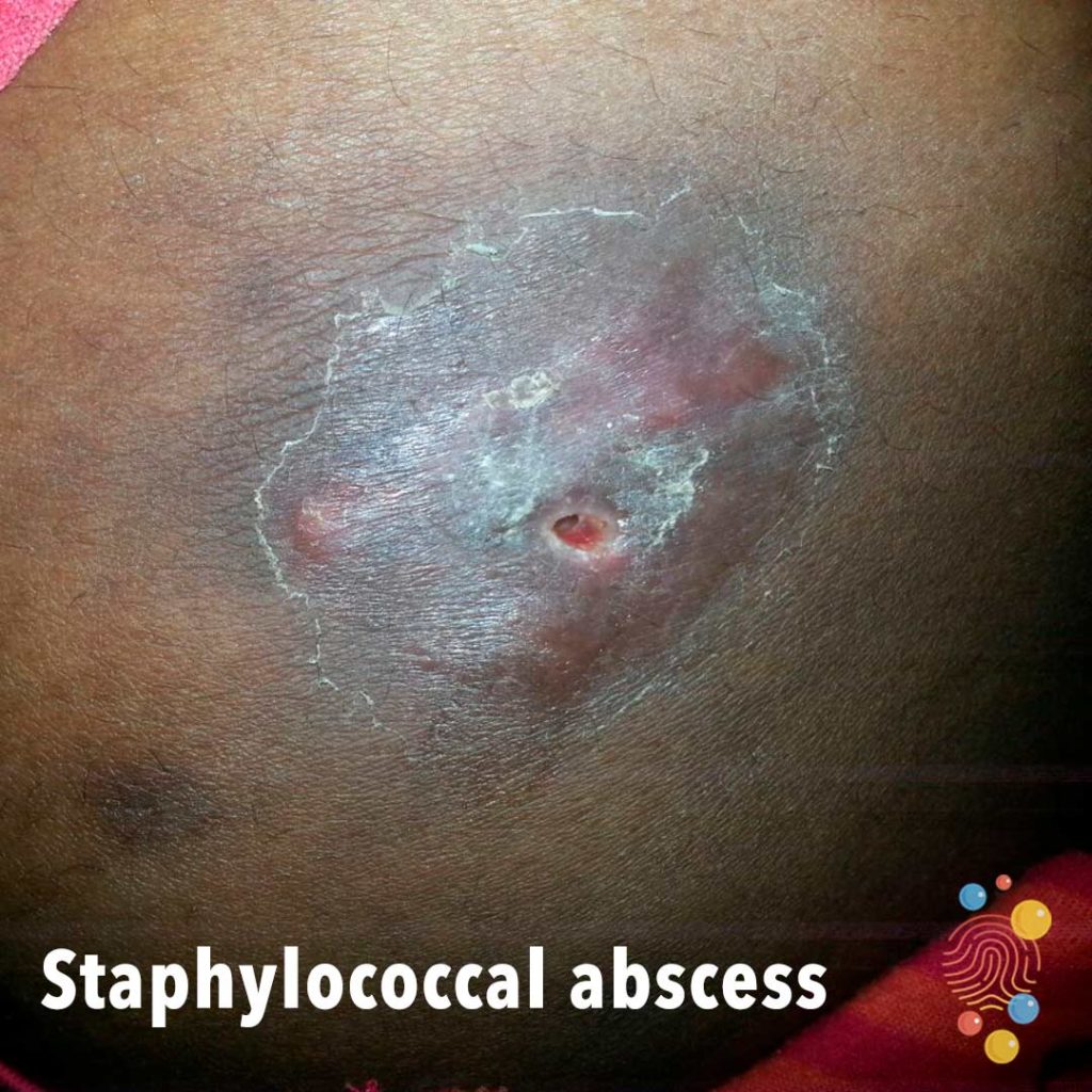
Staphylococcal Infection
Learn more about staphylococcal infection
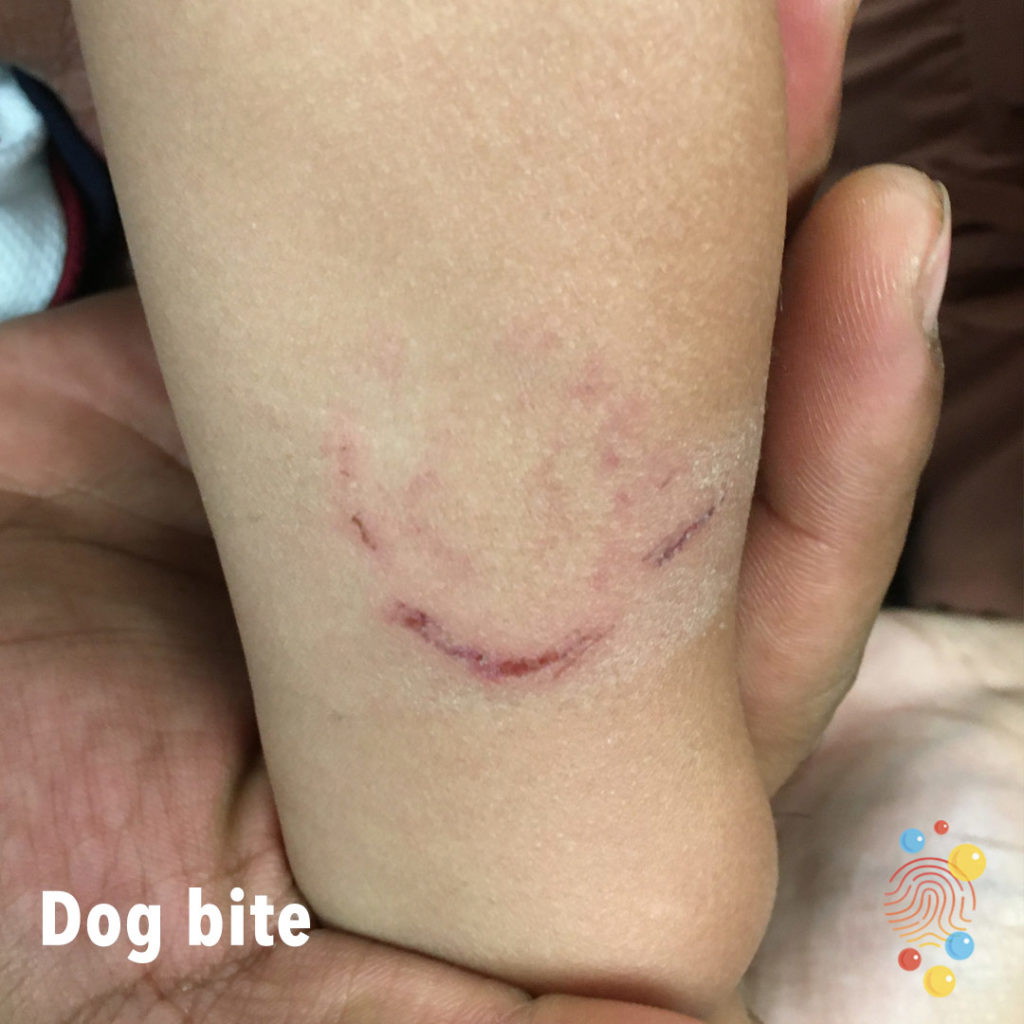
Dog Bite
Learn more about bites
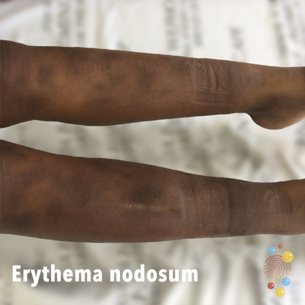
Erythema Nodosum
Learn more about erythema nodosum
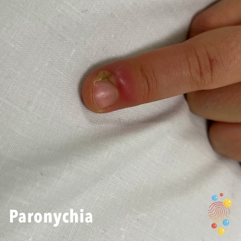
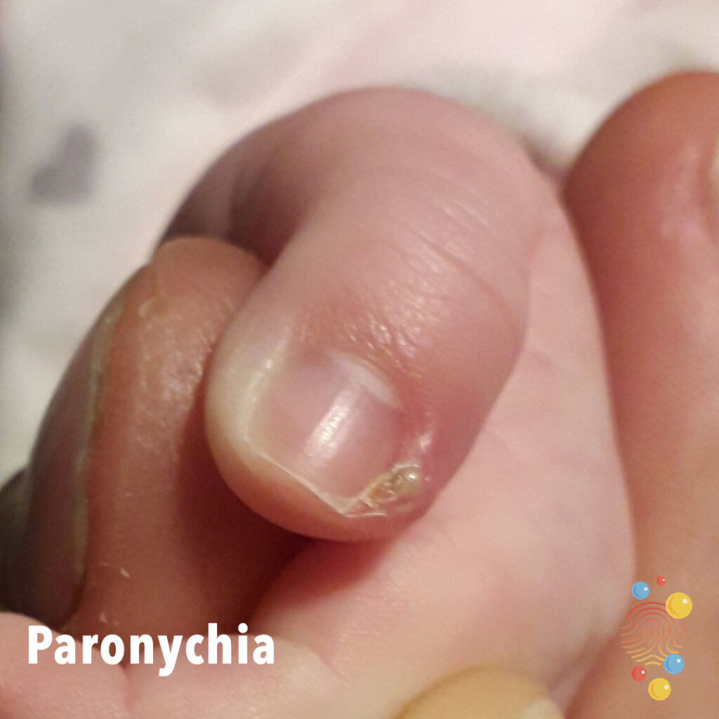
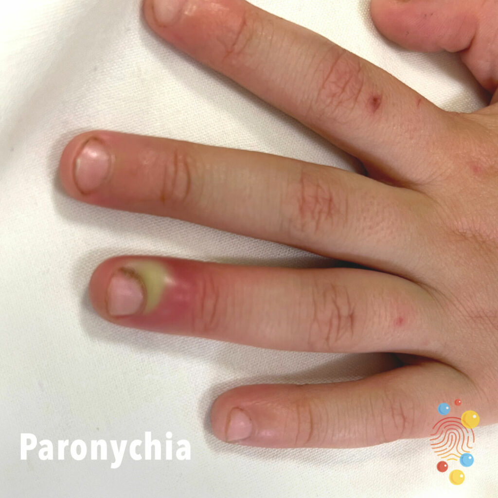
Paronychia
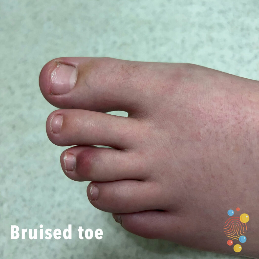
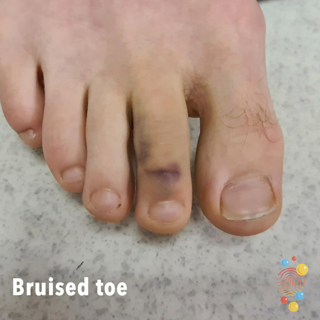
Bruised Toe
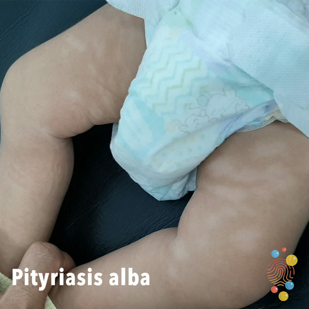
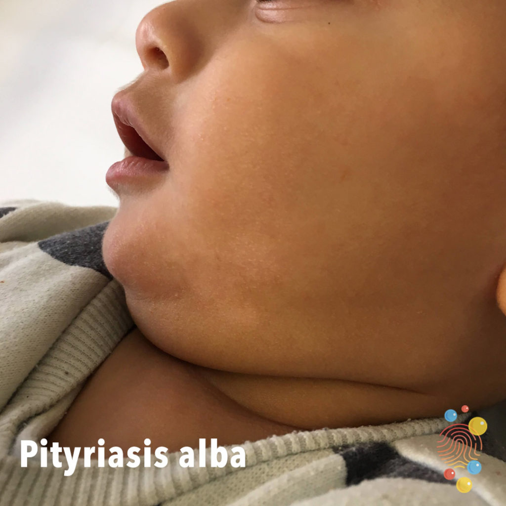
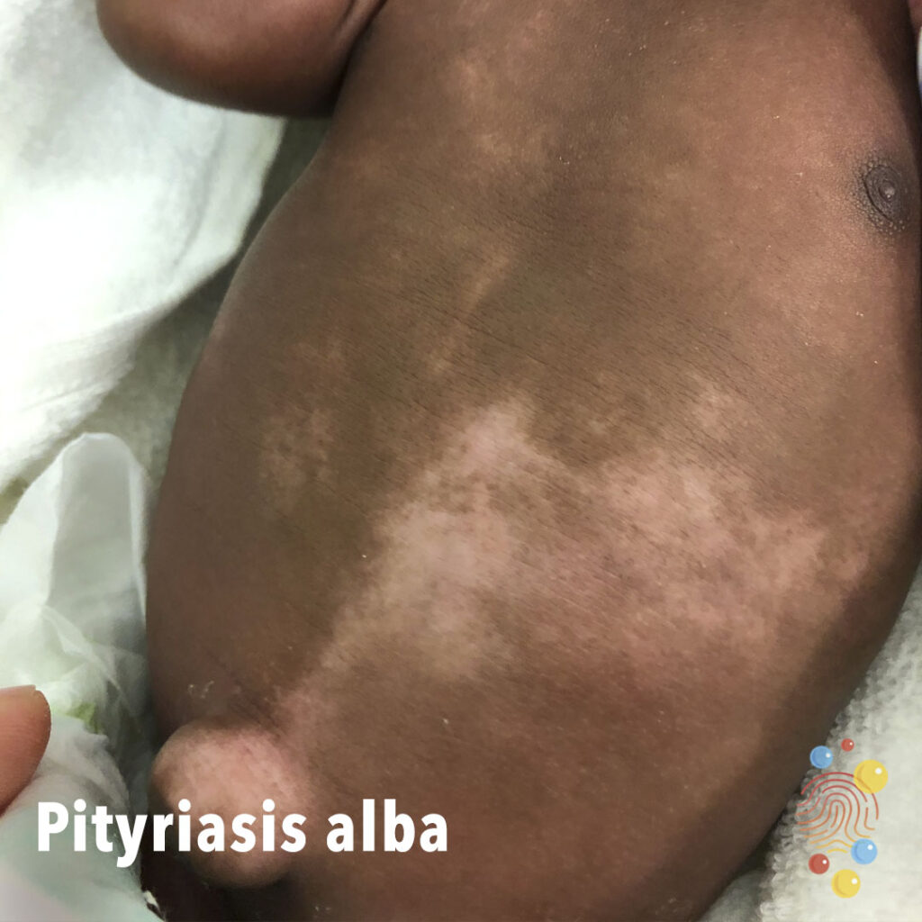
Pityriasis Alba
Learn more about pityriasis alba
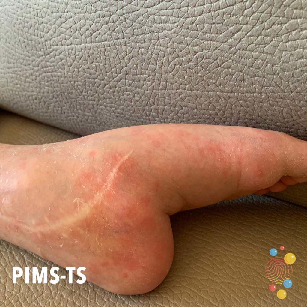
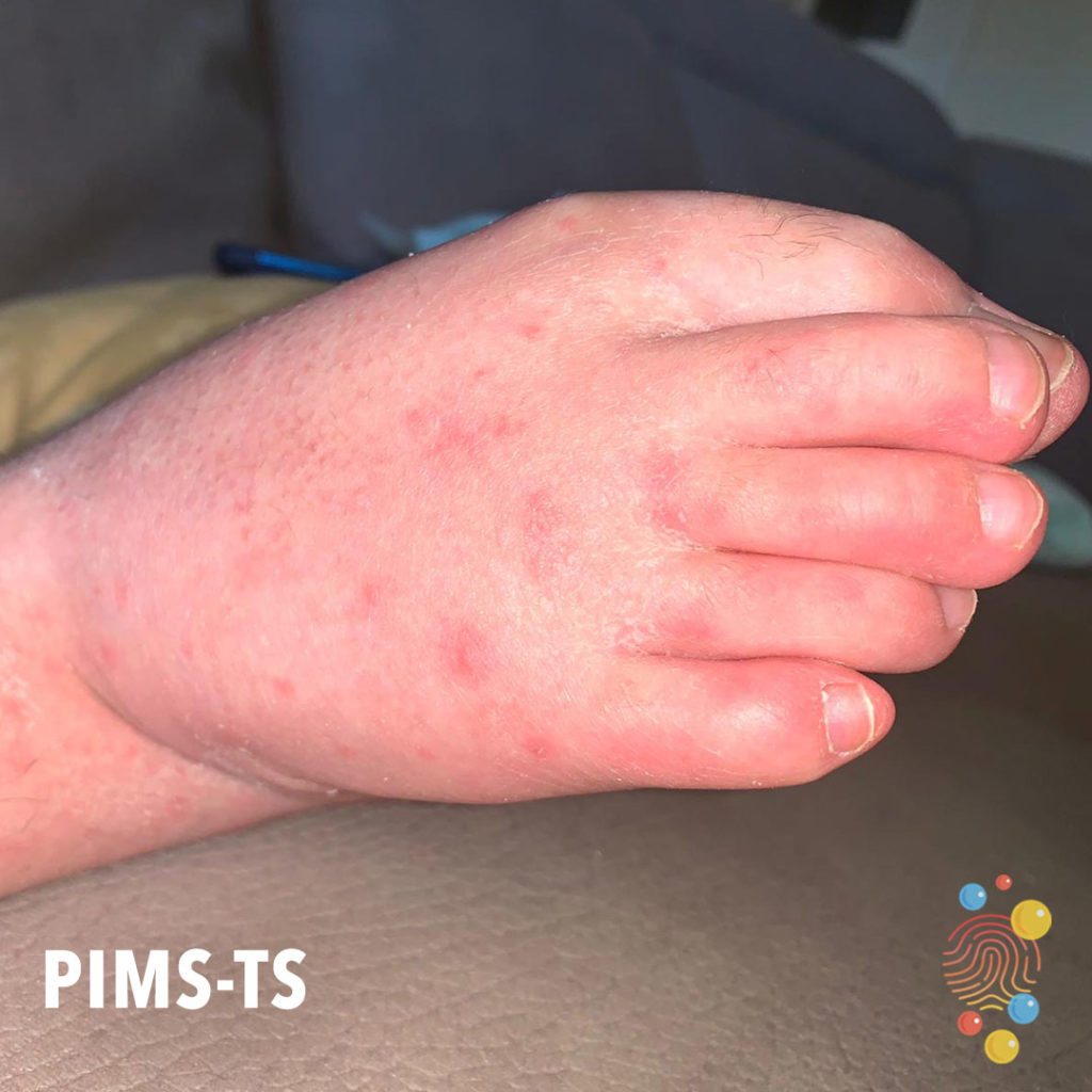
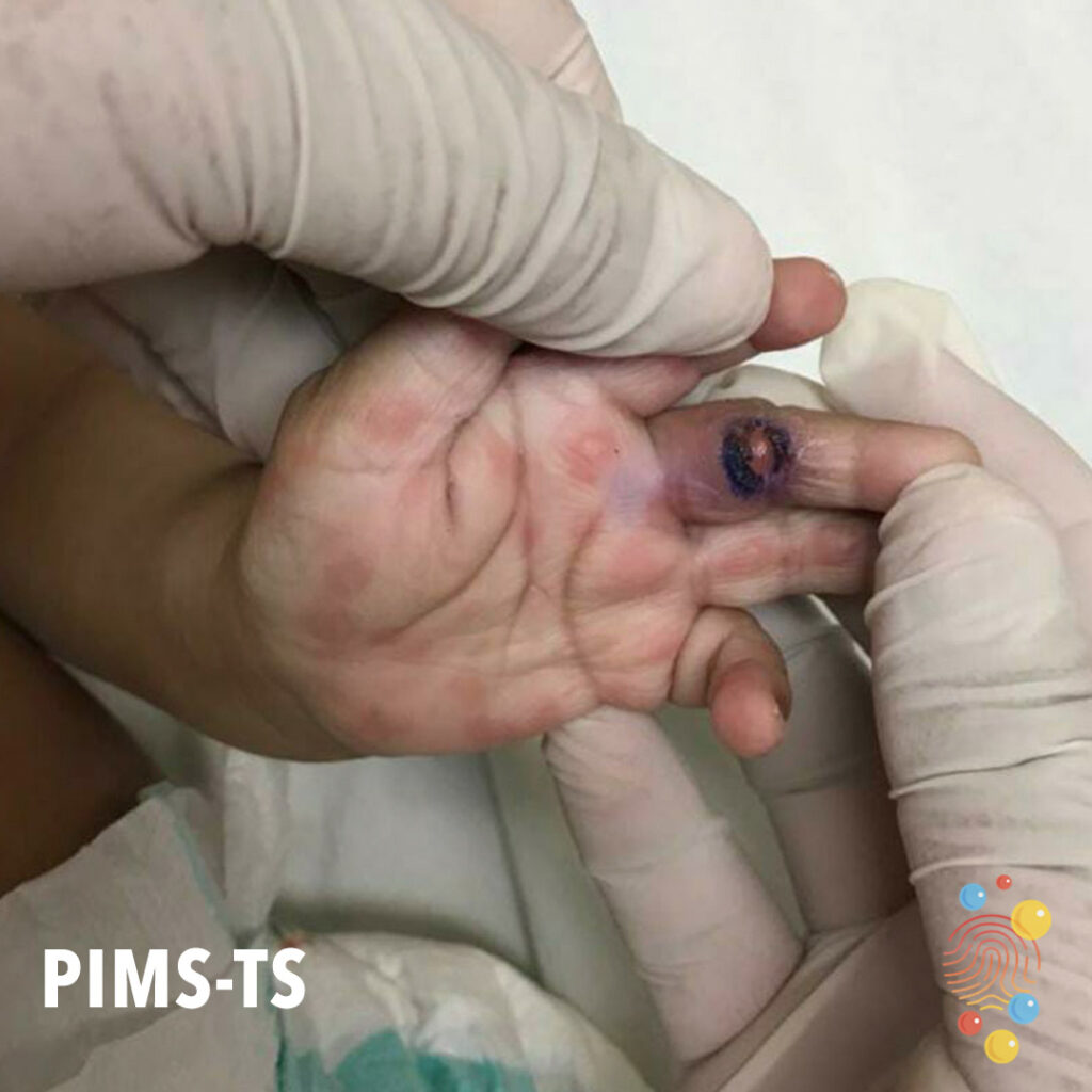
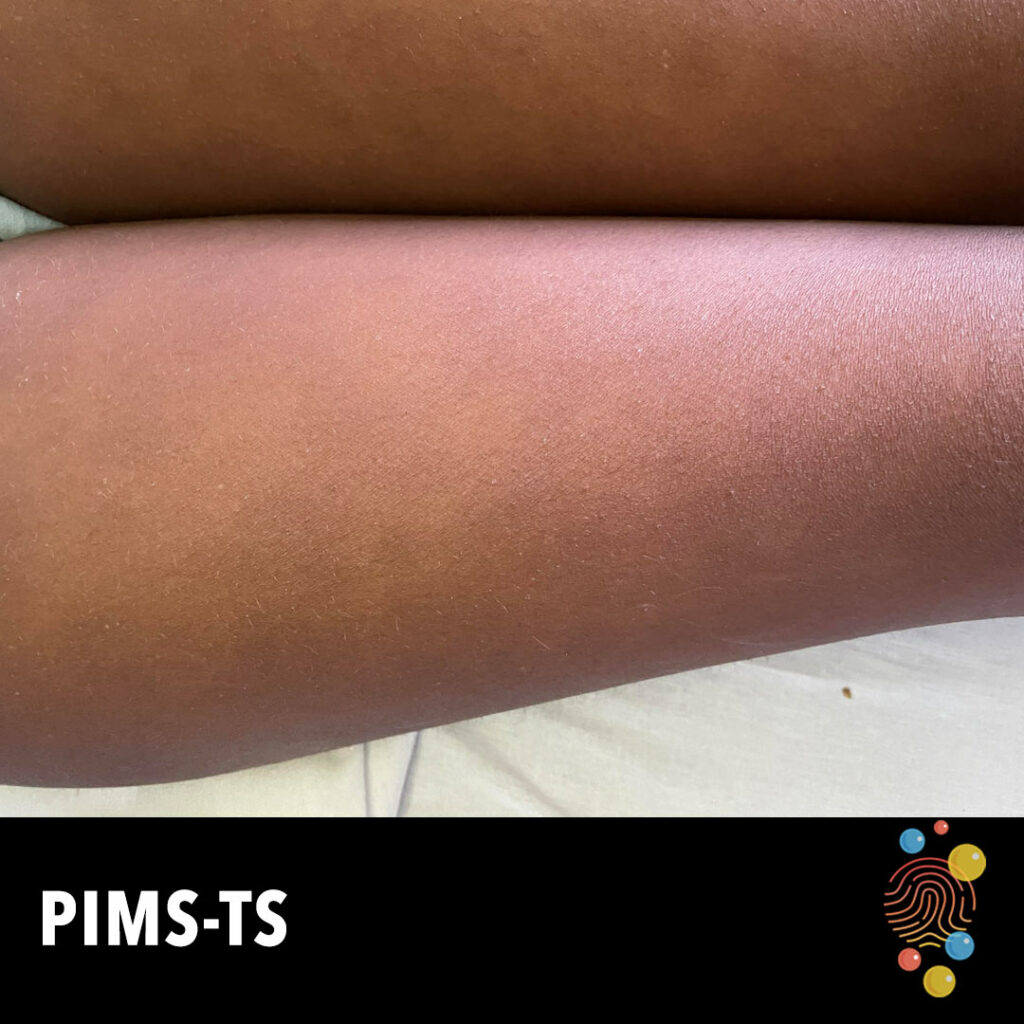
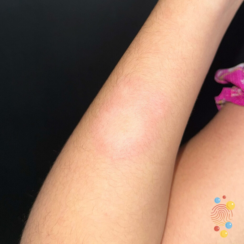
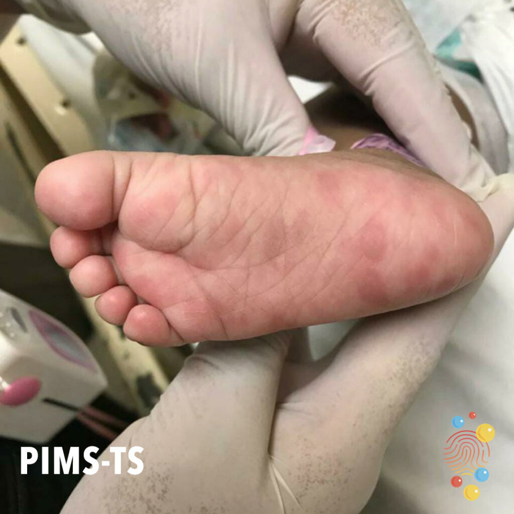
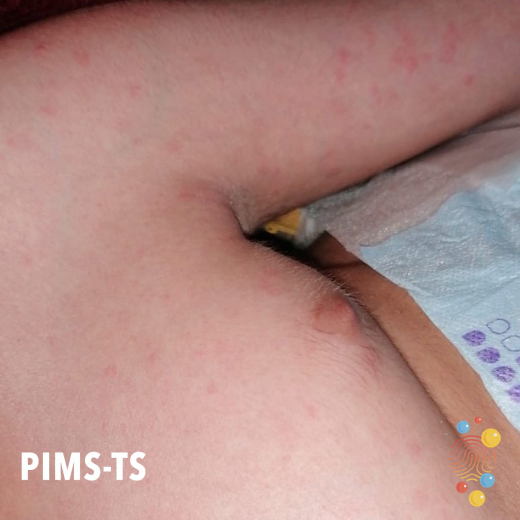
PIMS-TS
Scar overlying the medial malleolus of the left foot. Scattering of erythematous papules, xerosis of the skin (fine overlying scale)
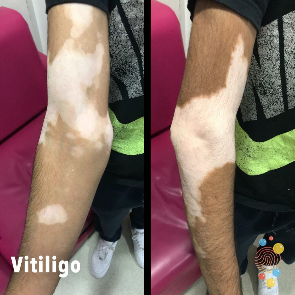
Vitiligo
Learn more about vitiligo

Miliaria Crystallina
Learn more about miliaria
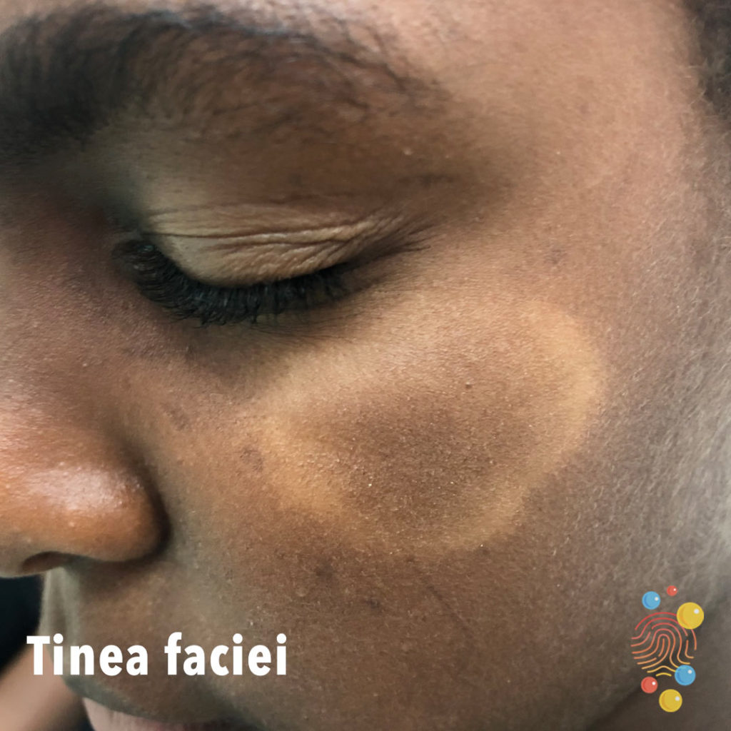
Tinea Faciei
Learn more about tinea faciei
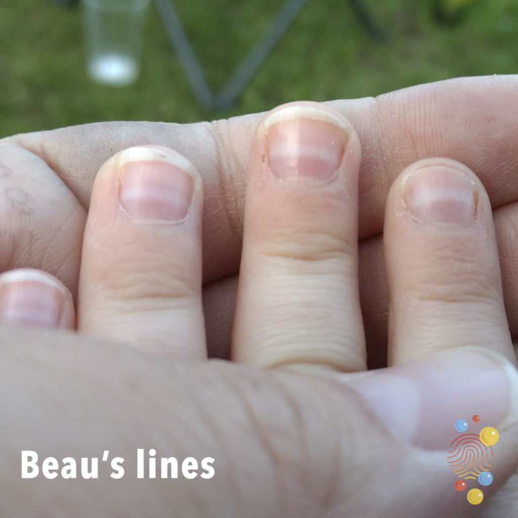
Beau’s Lines
Learn more about Beau’s lines
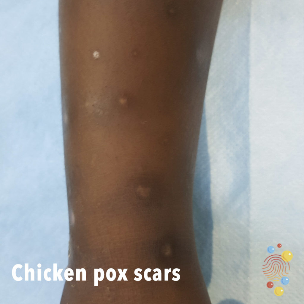
Chicken Pox Scars
Learn more about chicken pox

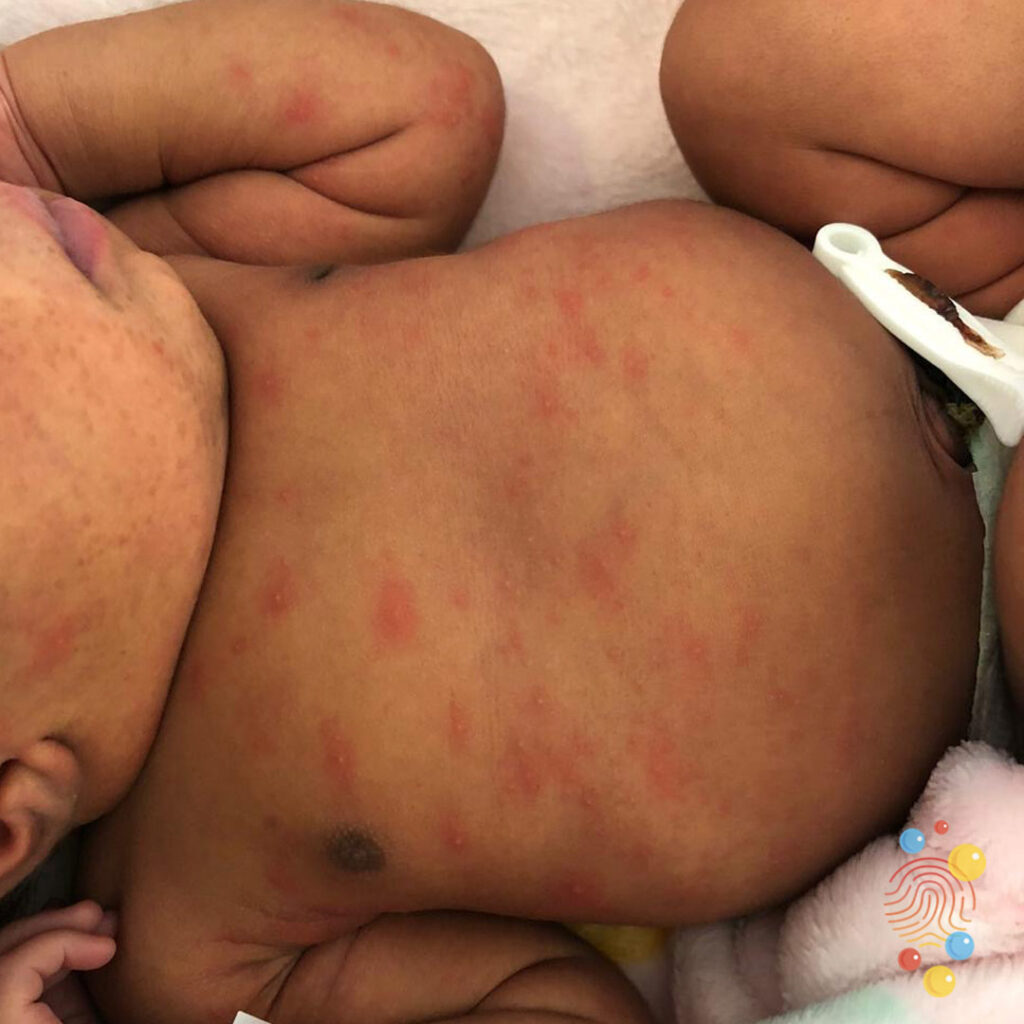
Gianotti Crosti

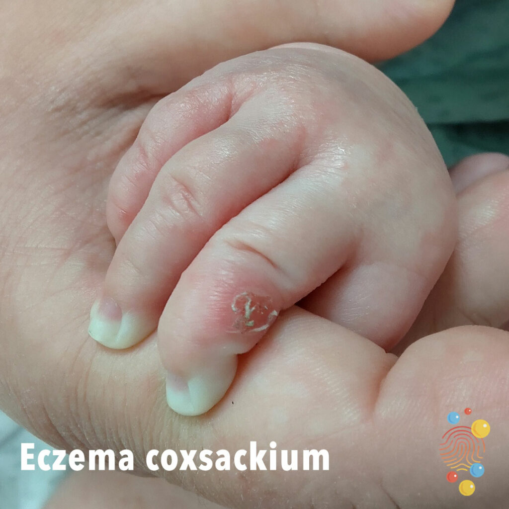
Eczema Coxsackium
Eruption of dark red macules, vesicles, and erosions distributed across areas previously affected by atopic dermatitis, with relative sparing of the trunk

Parvovirus
Bright red rash in symmetrical distribution
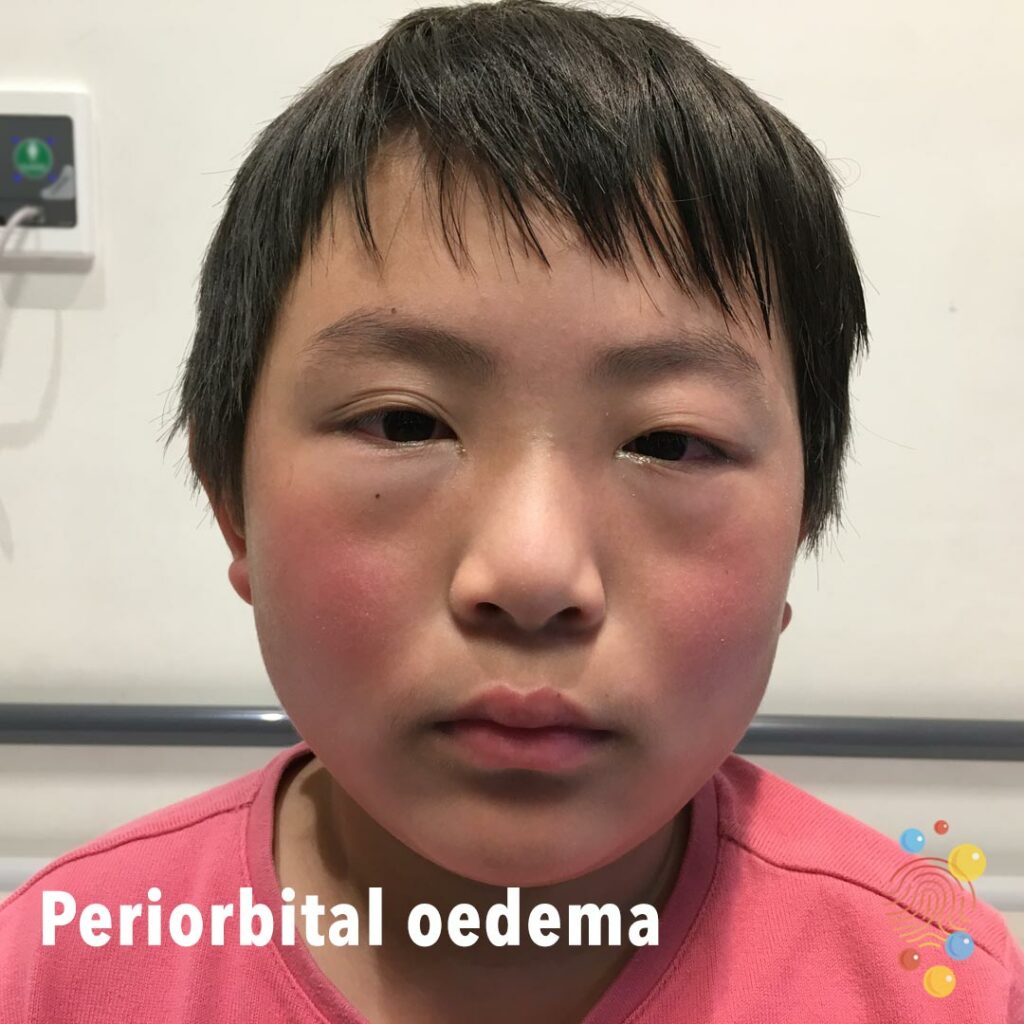
Periorbital Oedema
Learn more about periorbital oedema

Neurofibromatosis
Neurofibromatosis (NF) is a term that describes three genetic diseases caused by mutations in genes that lead to increased risk of developing tumors. Different types of neurofibromatosis lead to growth of different tumors (neurofibromas and schwannomas) in various parts of the body.
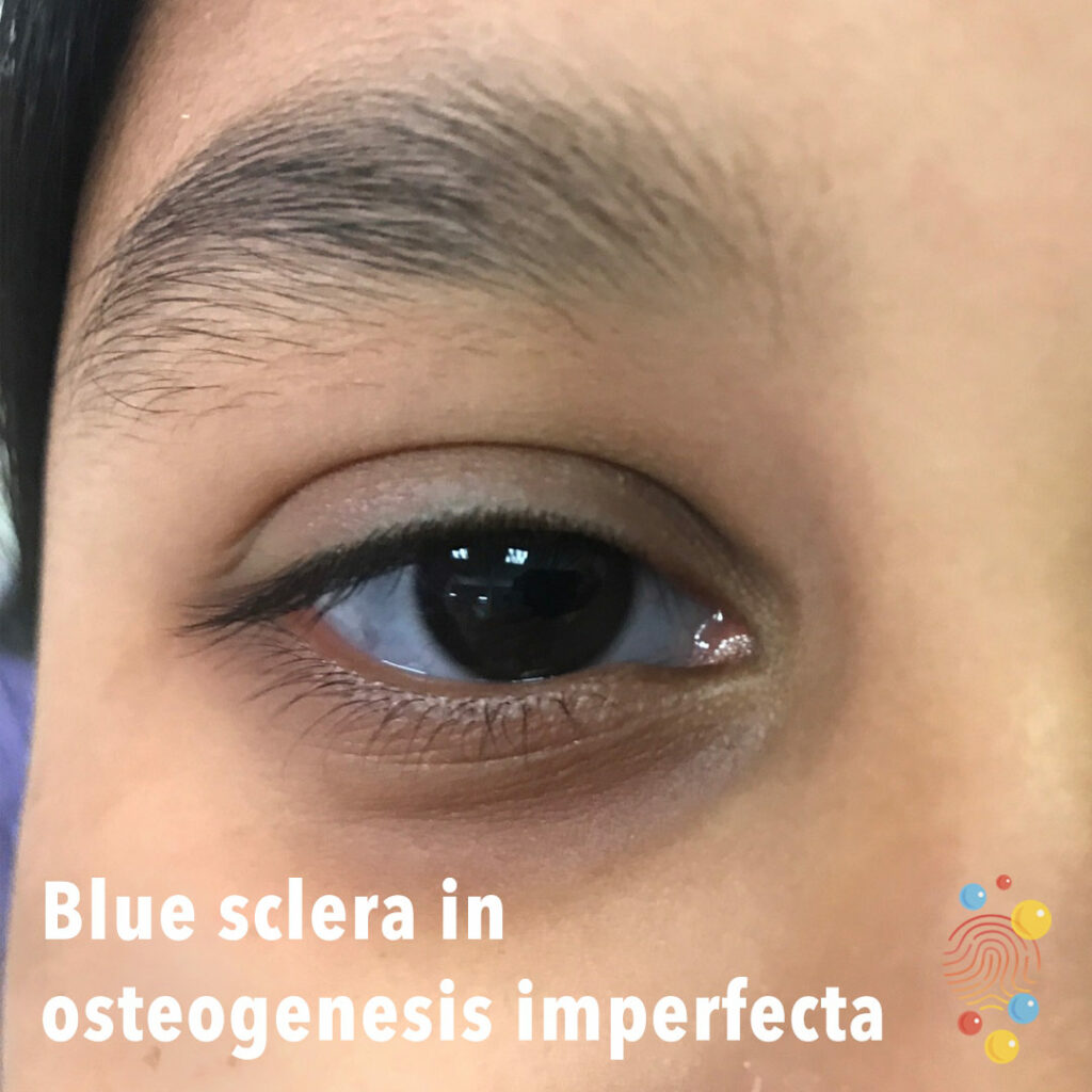
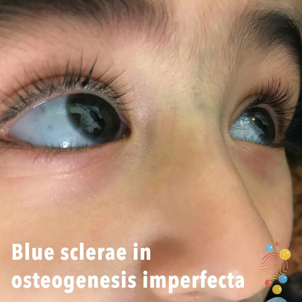
Blue sclera in osteogenesis imperfecta
Learn more about blue sclerae
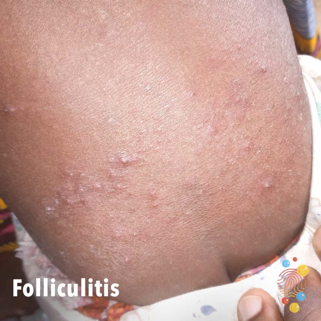
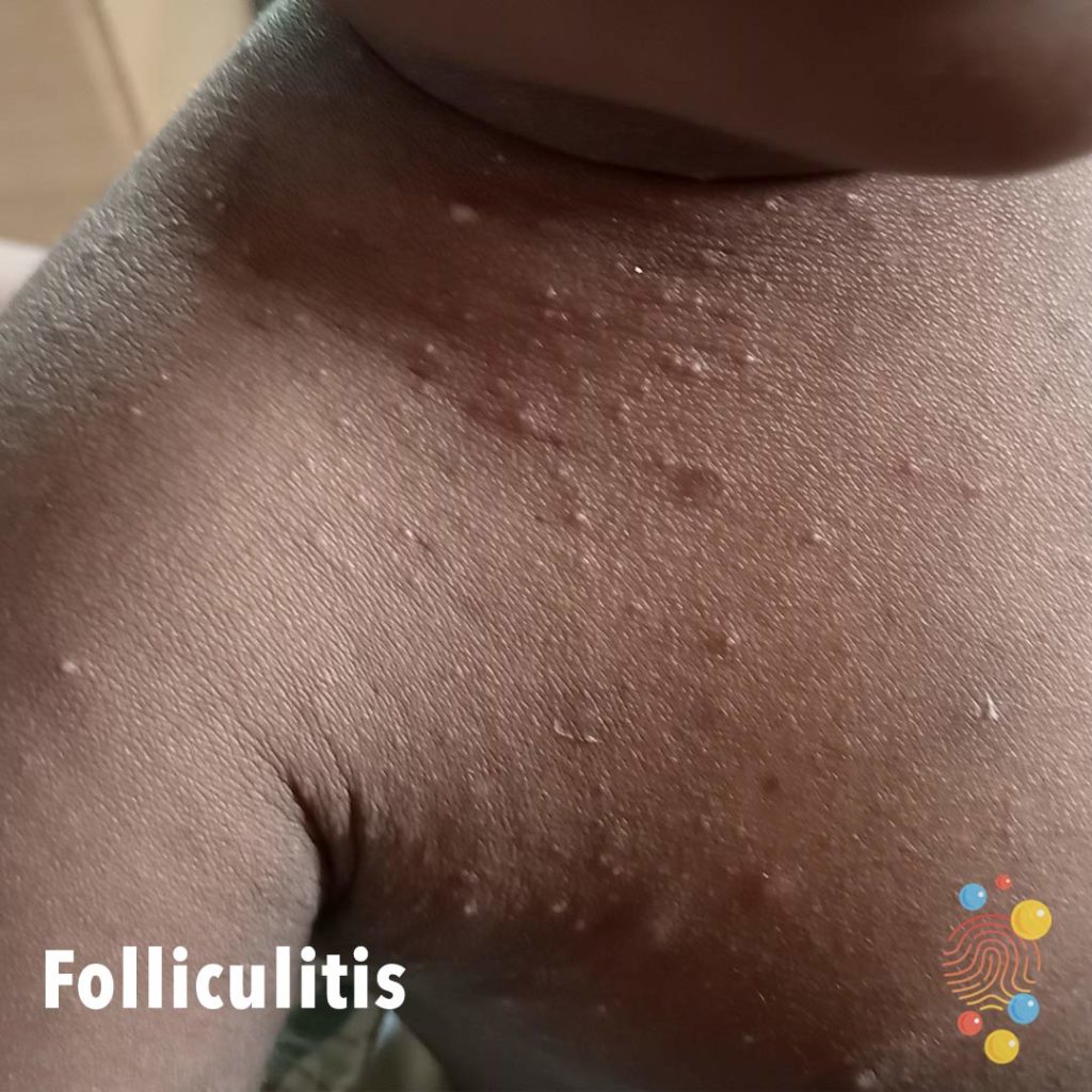
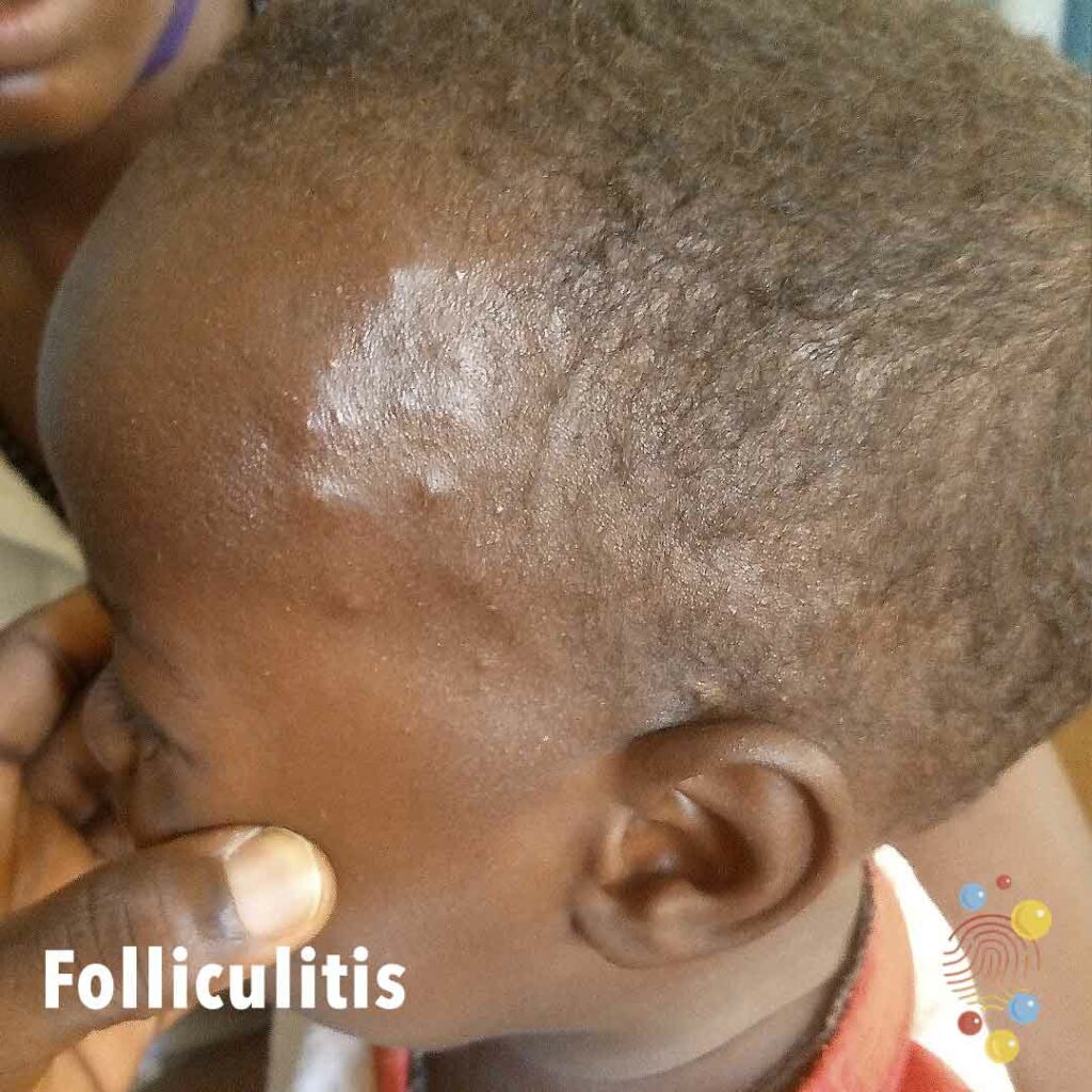
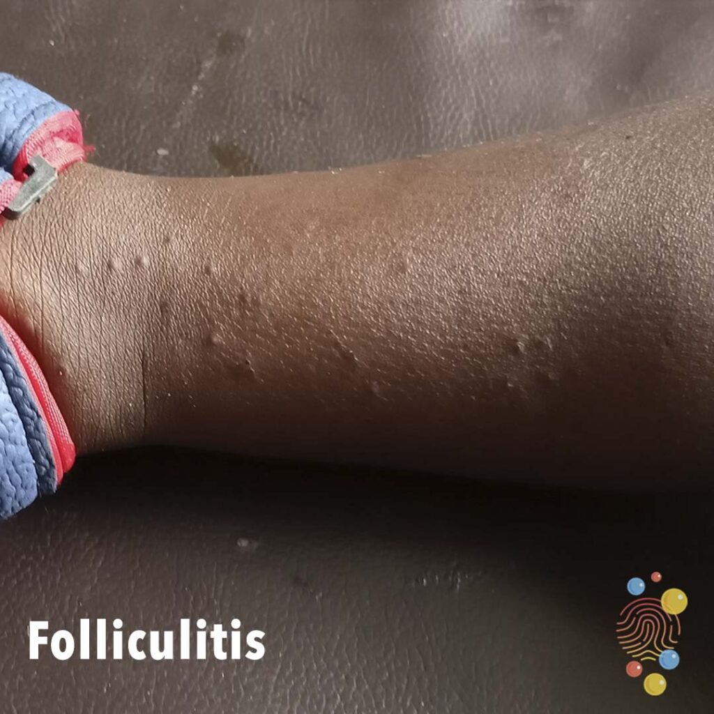
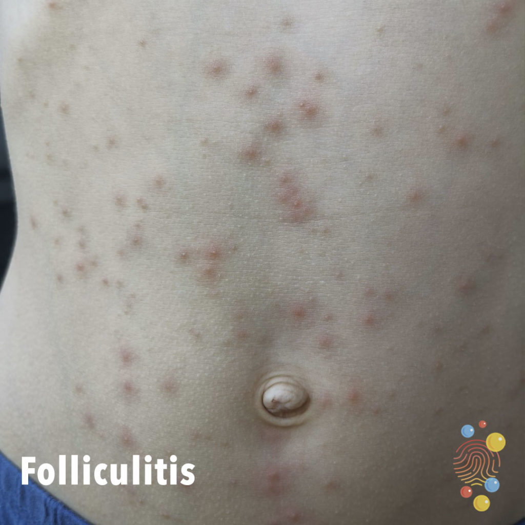
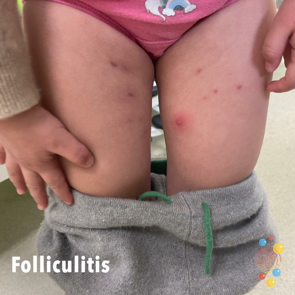
Folliculitis
Learn more about folliculitis
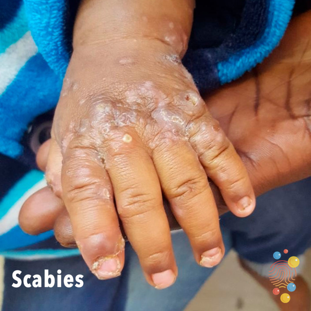
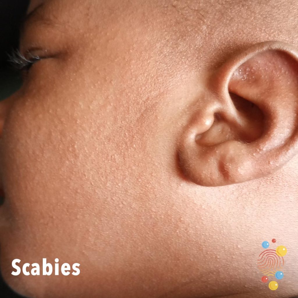
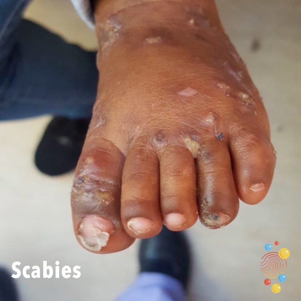
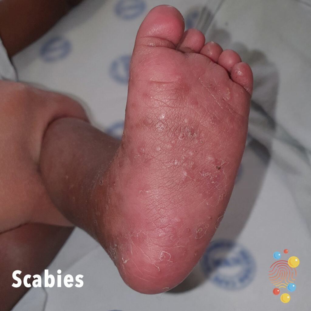
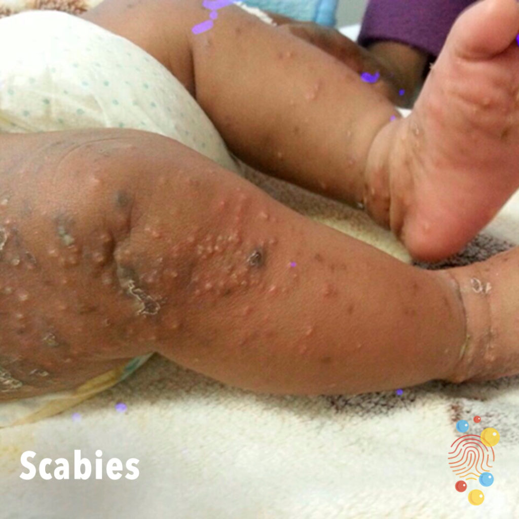
Scabies
Learn more about scabies
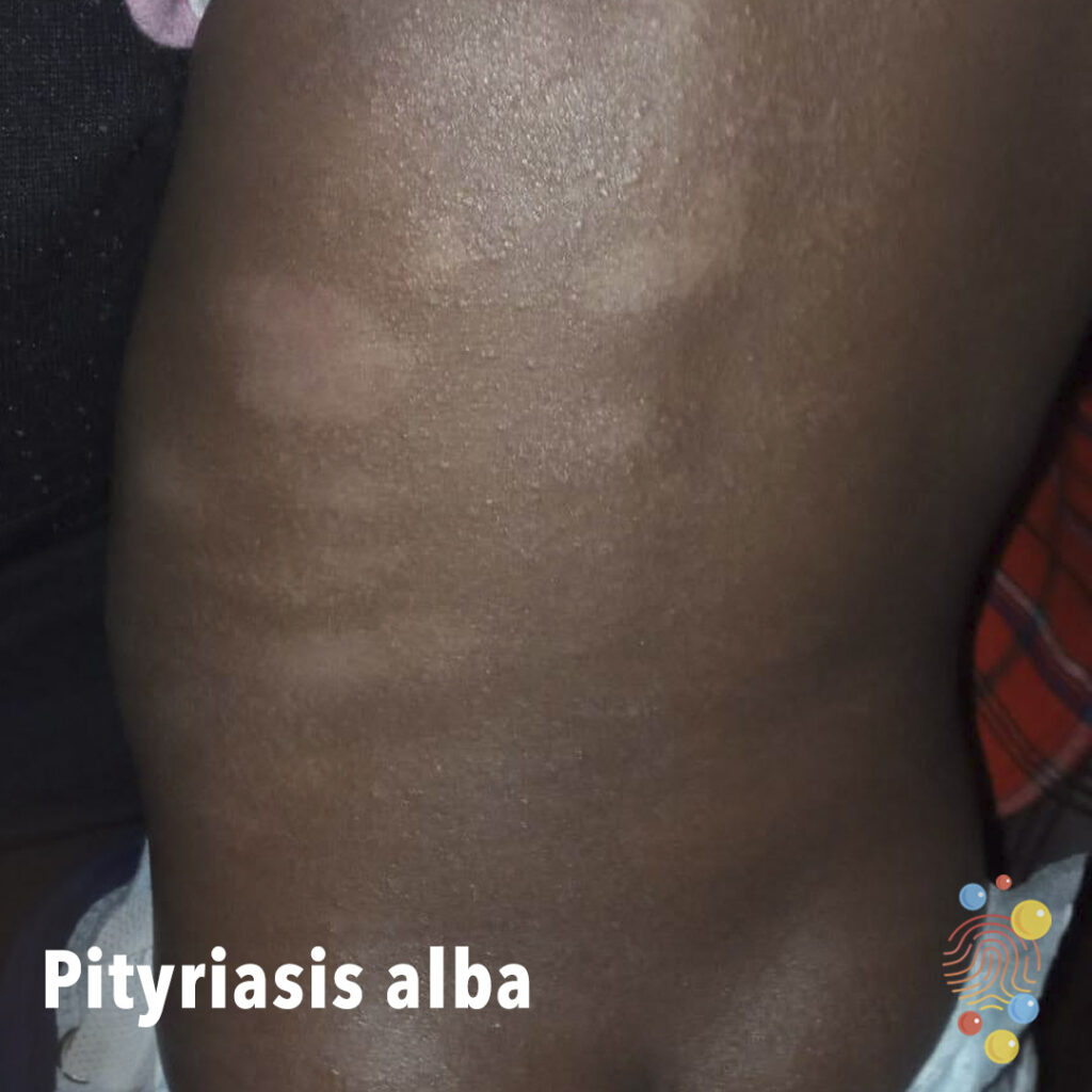
Pityriasis Alba
Learn more about pityriasis alba
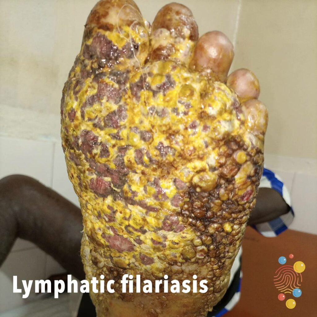
Lymphatic Filariasis
Learn more about lymphatic filariasis
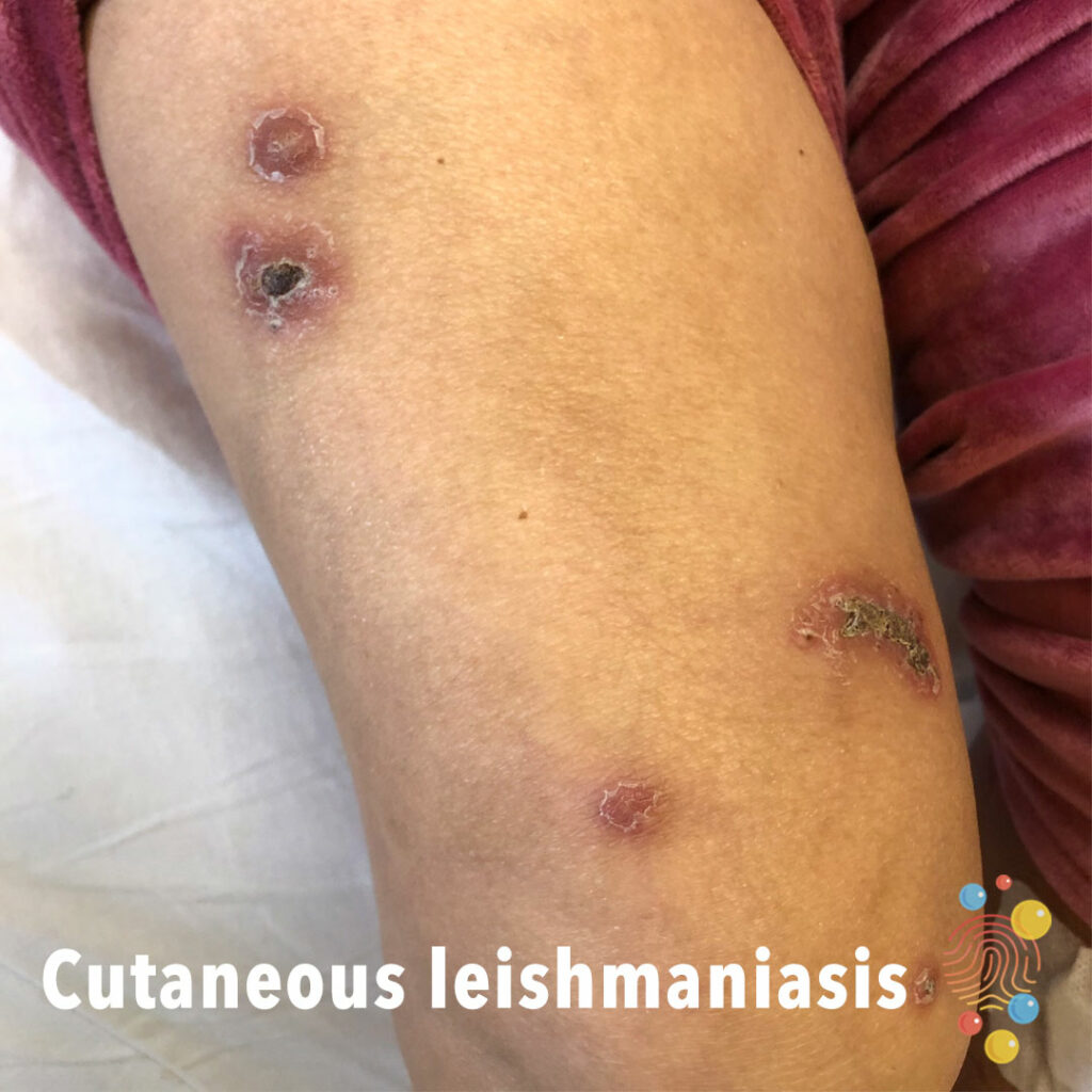
Cutaneous Leishmaniasis
Learn more about leishmaniasis
Herpes Simplex Virus
Learn more about herpes simplex virus
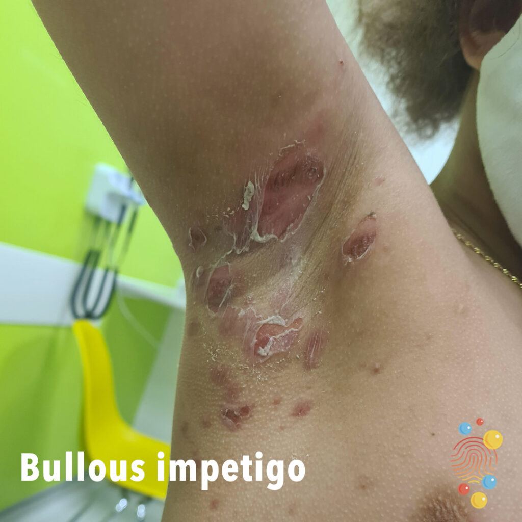
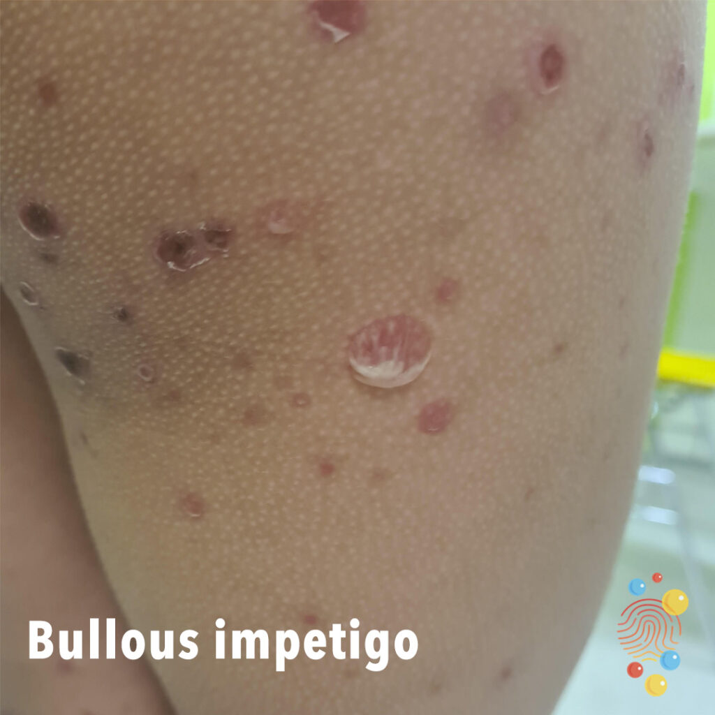
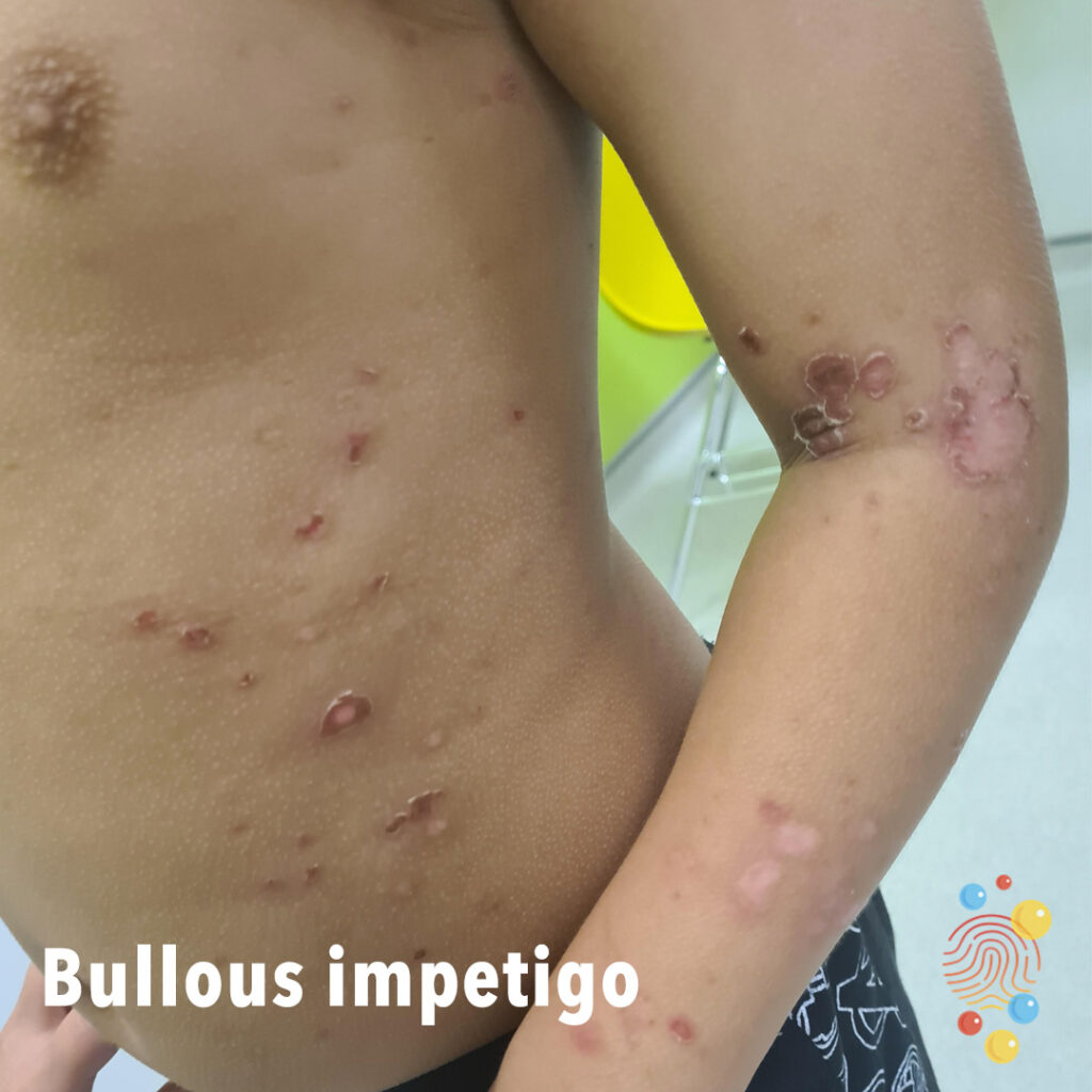
Bullous Impetigo
Bullous impetigo is a bacterial skin infection that causes large, fluid-filled blisters to appear on the body
Scabies
Learn more about scabies

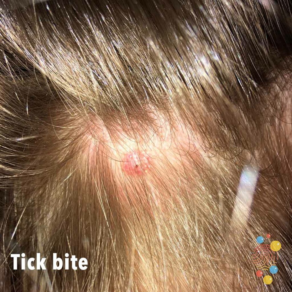
Tick Bite
Learn more about tick bites

Hand Foot And Mouth Disease
Learn more about hand, foot and mouth

Urticaria
Learn more about urticaria
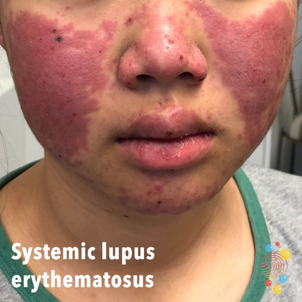
Systemic Lupus Erythematosus
Learn more about systemic lupus erythematosus
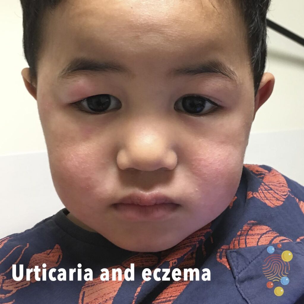
Urticaria And Eczema
Learn more about eczema
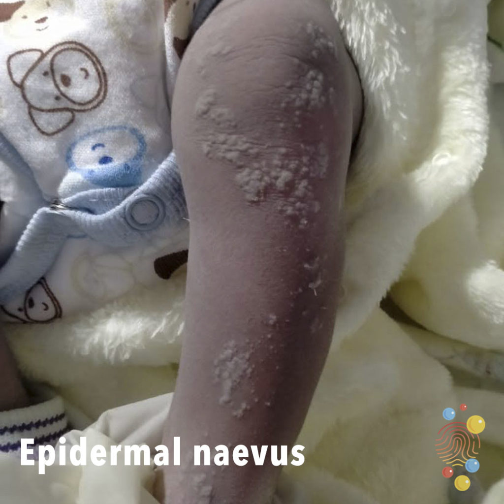
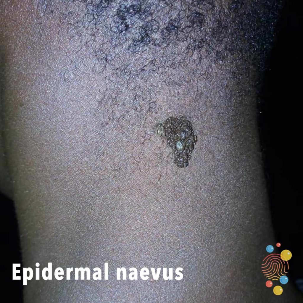
Epidermal Naevus
Learn more about epidermal naevus
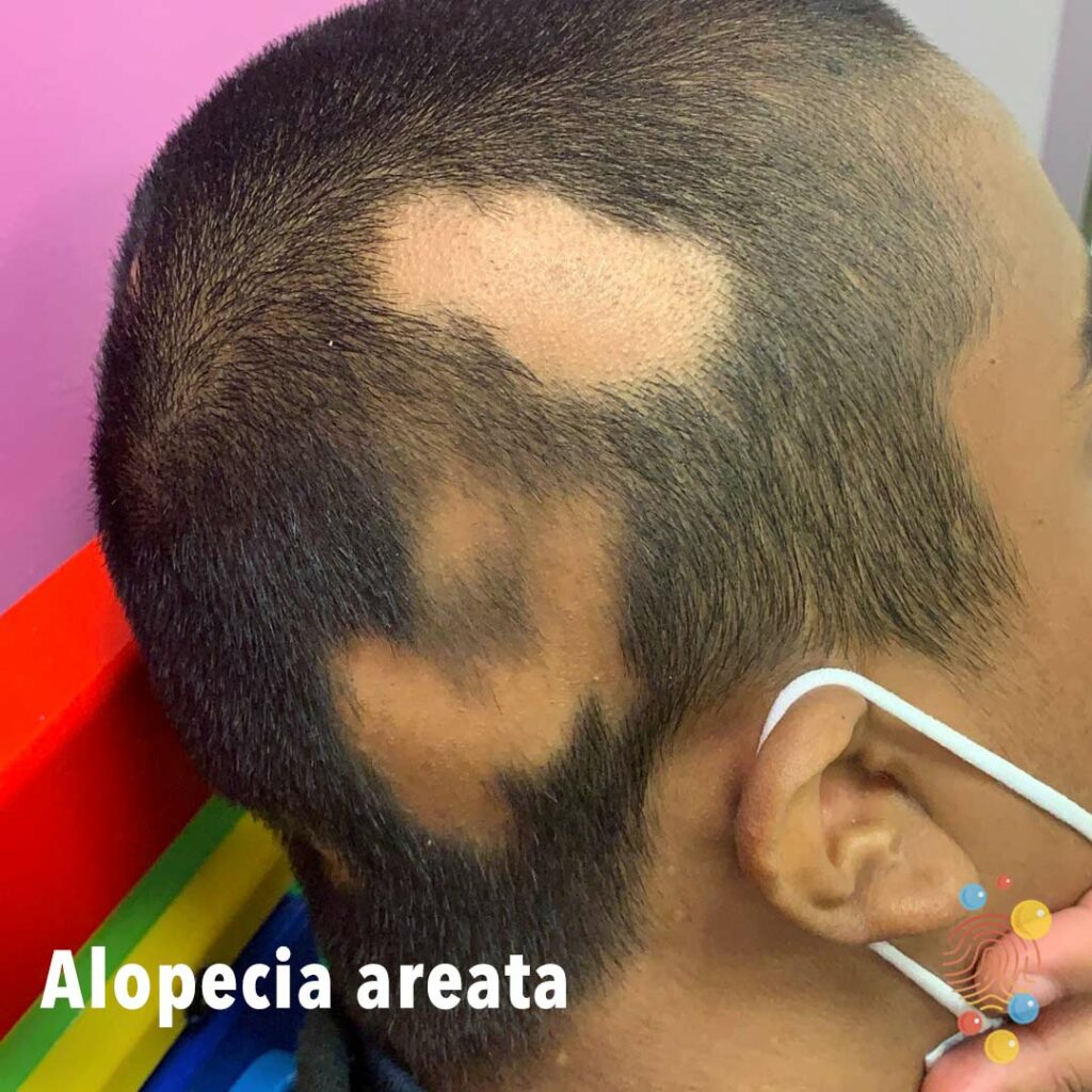
Alopecia
Learn more about alopecia areata

Mastoiditis
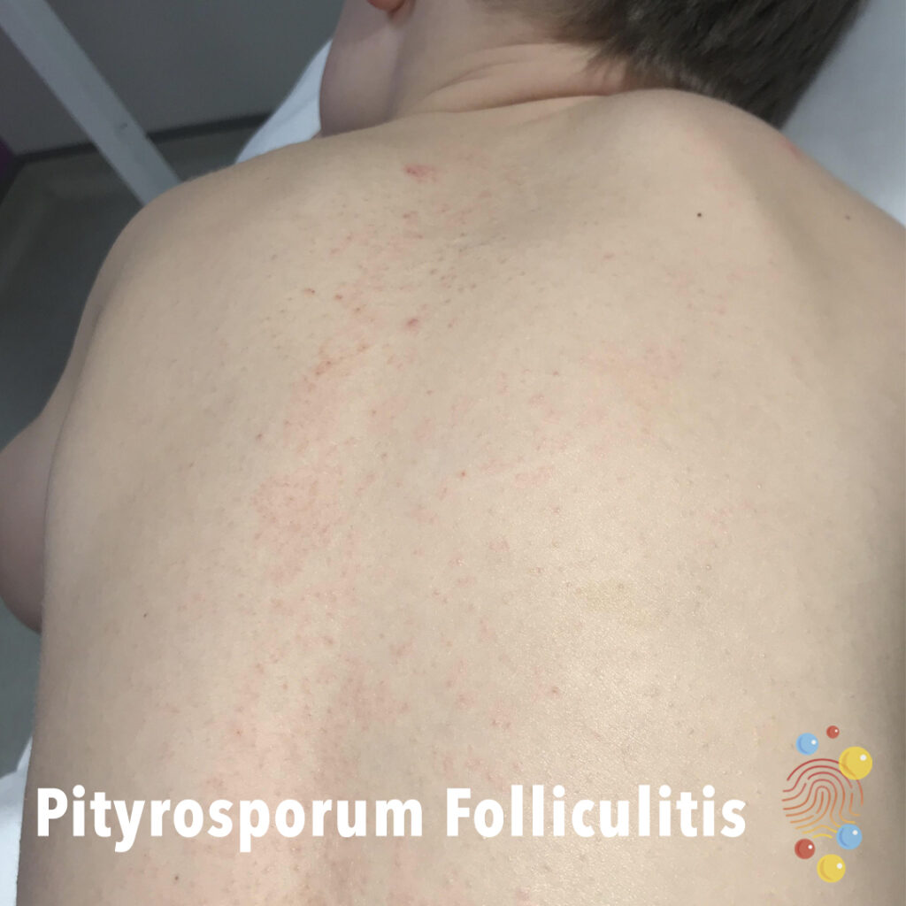
Pityrosporum Folliculitis
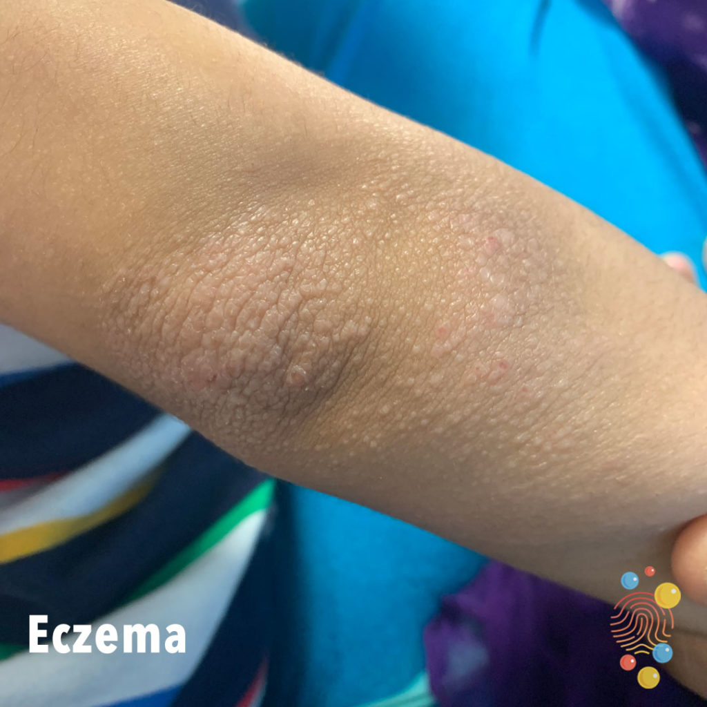
Eczema
Learn more about eczema
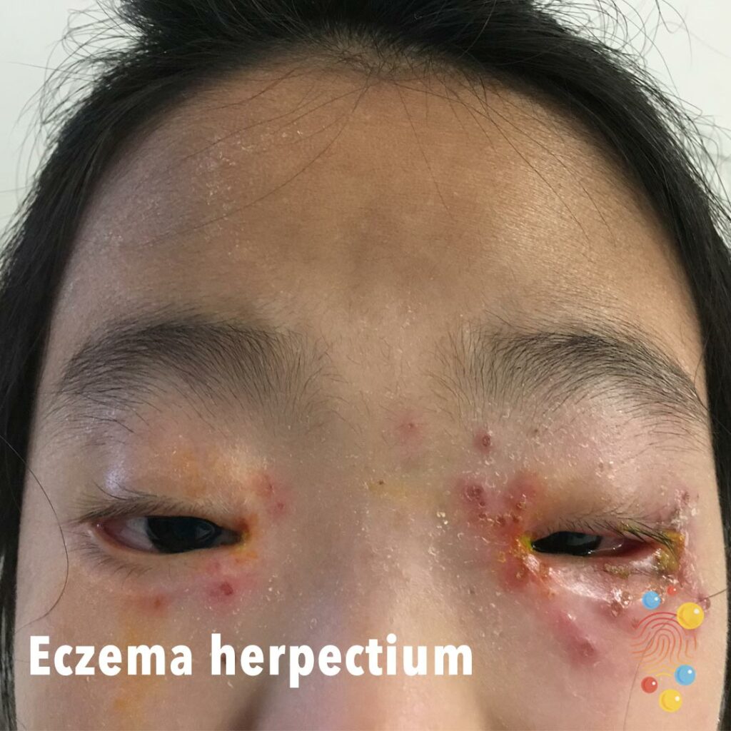
Eczema Herpectium
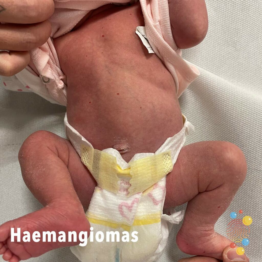
Haemangiomas
A haemangioma is a non-cancerous tumor that appears as a collection of abnormal blood vessels under or on the skin. They are also known as “strawberry marks” because of their red, purple, or blue color.

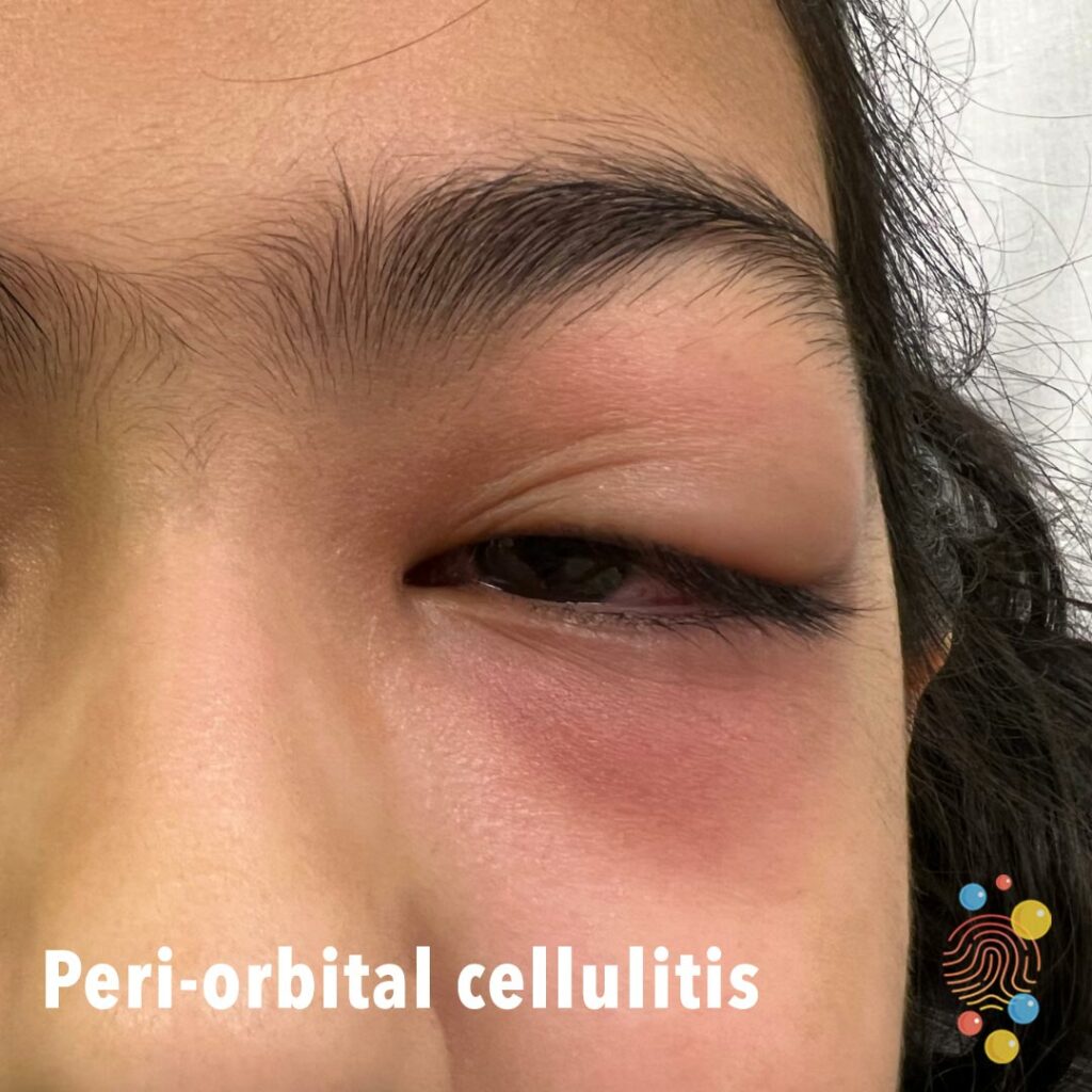
Periorbital cellulitis
Learn more about periorbital cellulitis
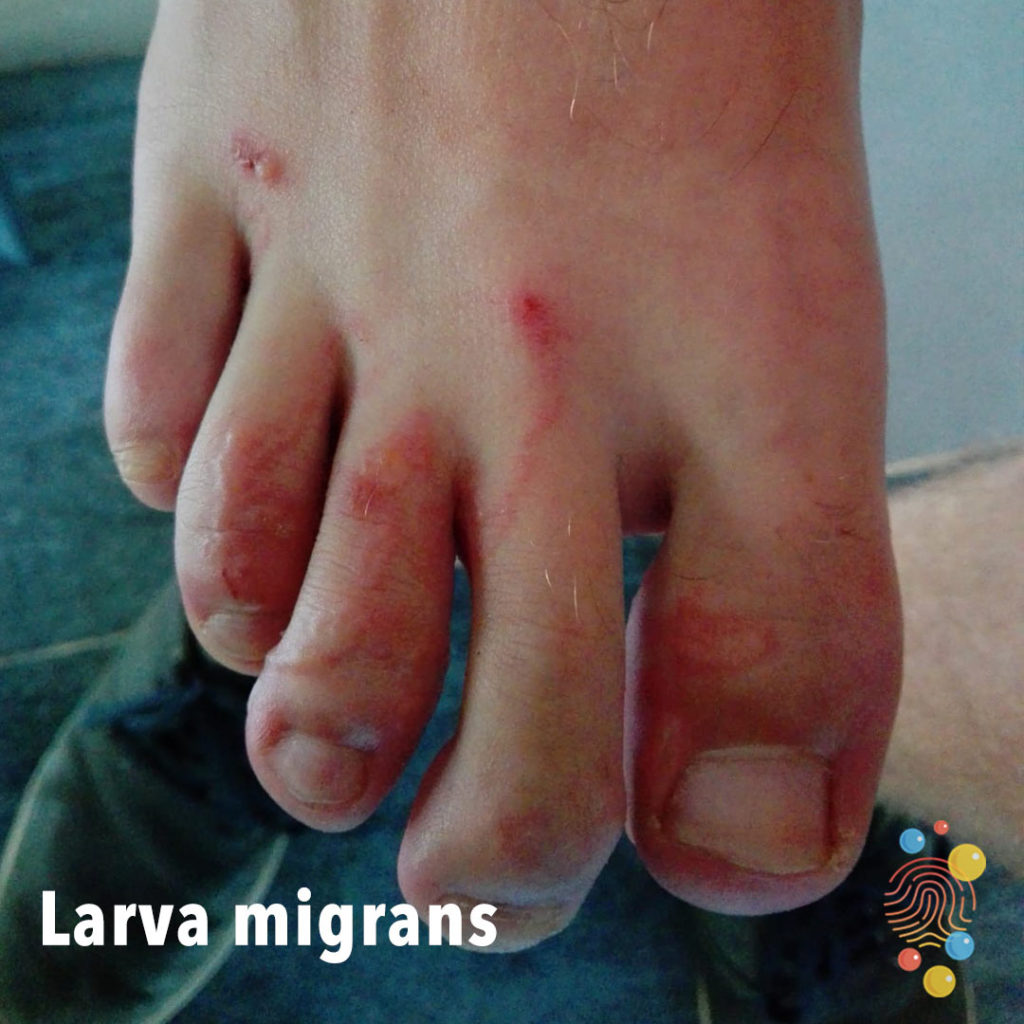
Larva Migrans
Learn more about larva migrans
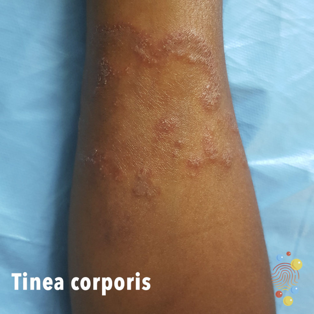
Tinea Corporis
Learn more about tinea corporis
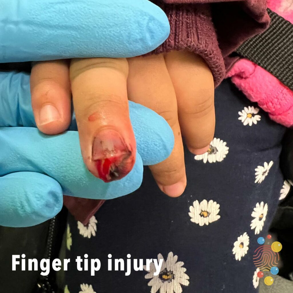
Finger Tip Injury
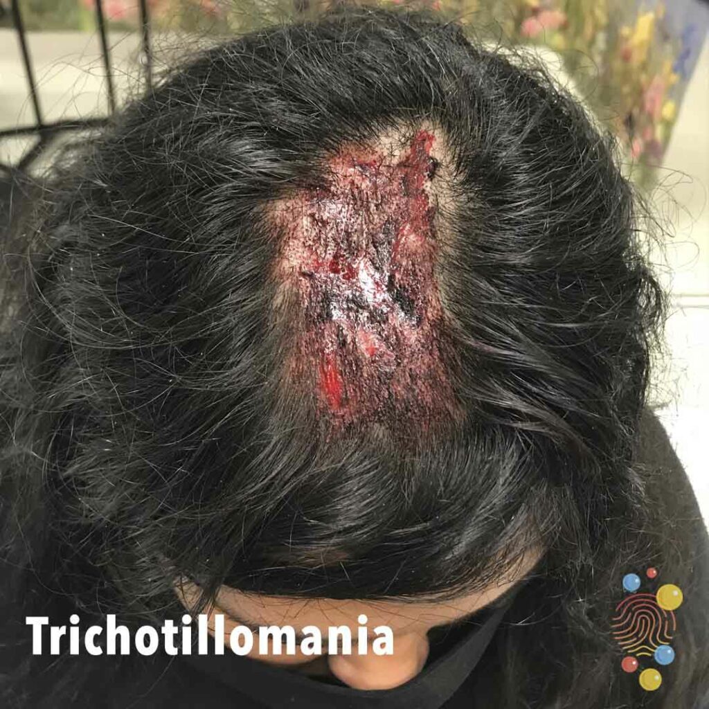
Trichotillomania
Learn more about trichotillomania

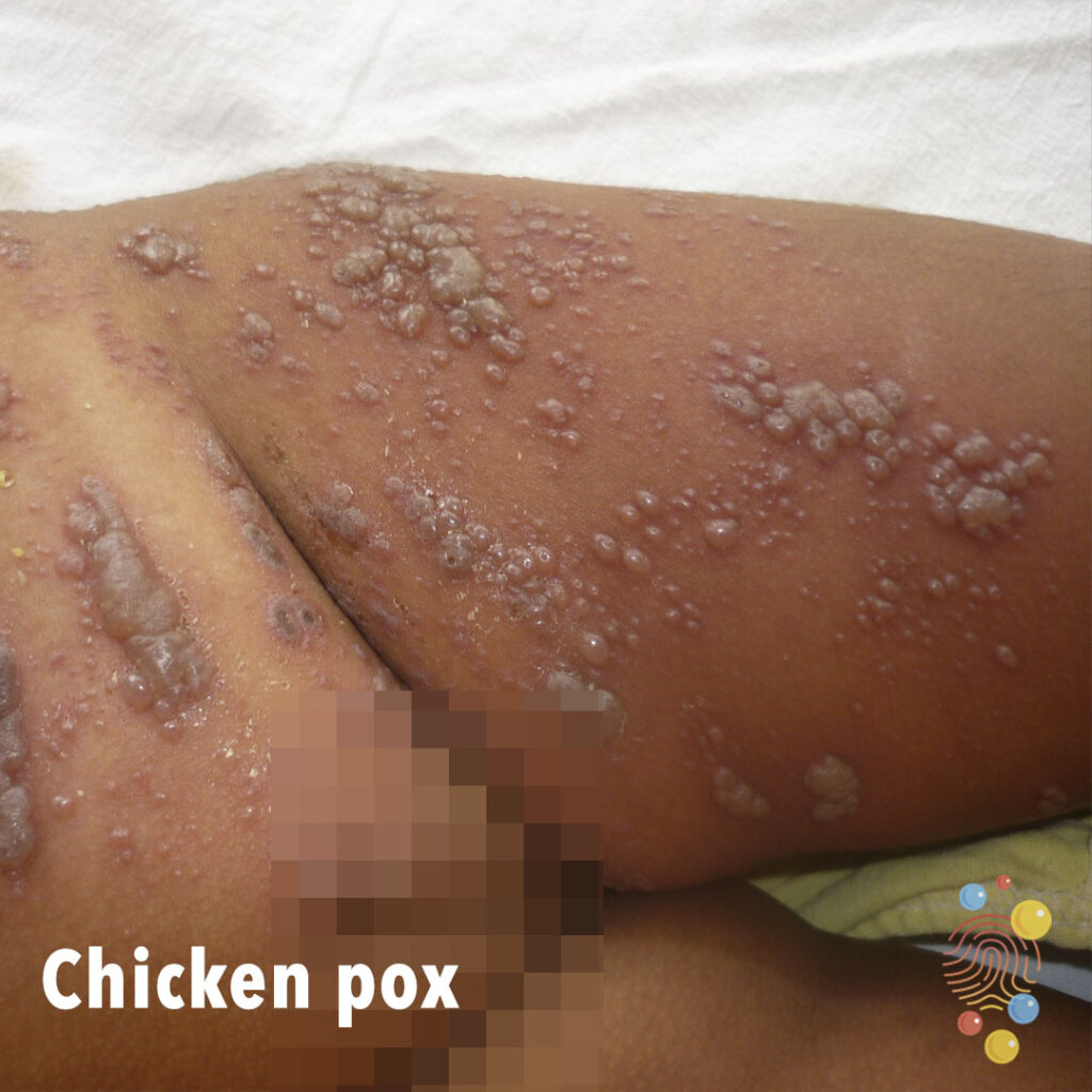
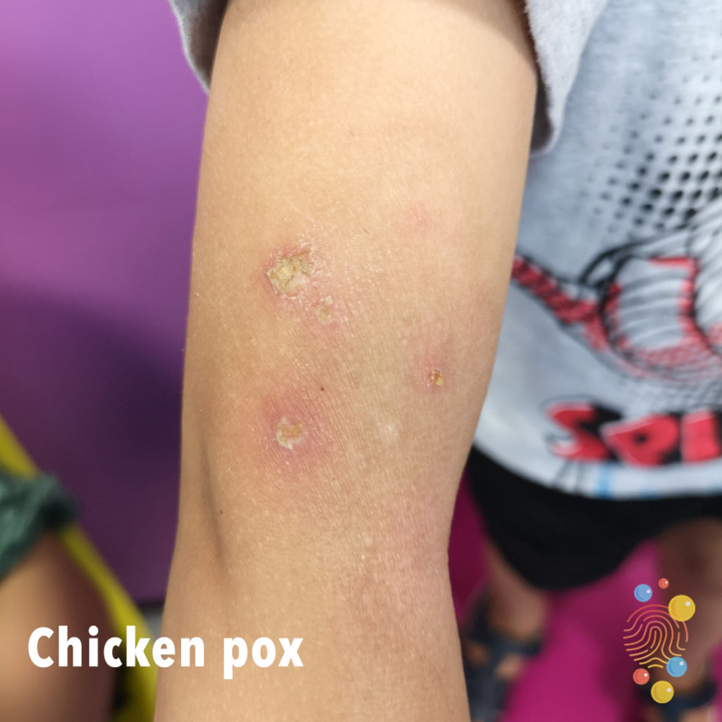
Chicken Pox
Learn more about chicken pox

Impetiginized Eczema
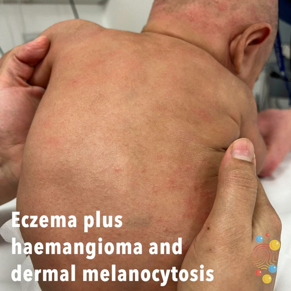
Eczema plus haemangioma and dermal melanocytosis
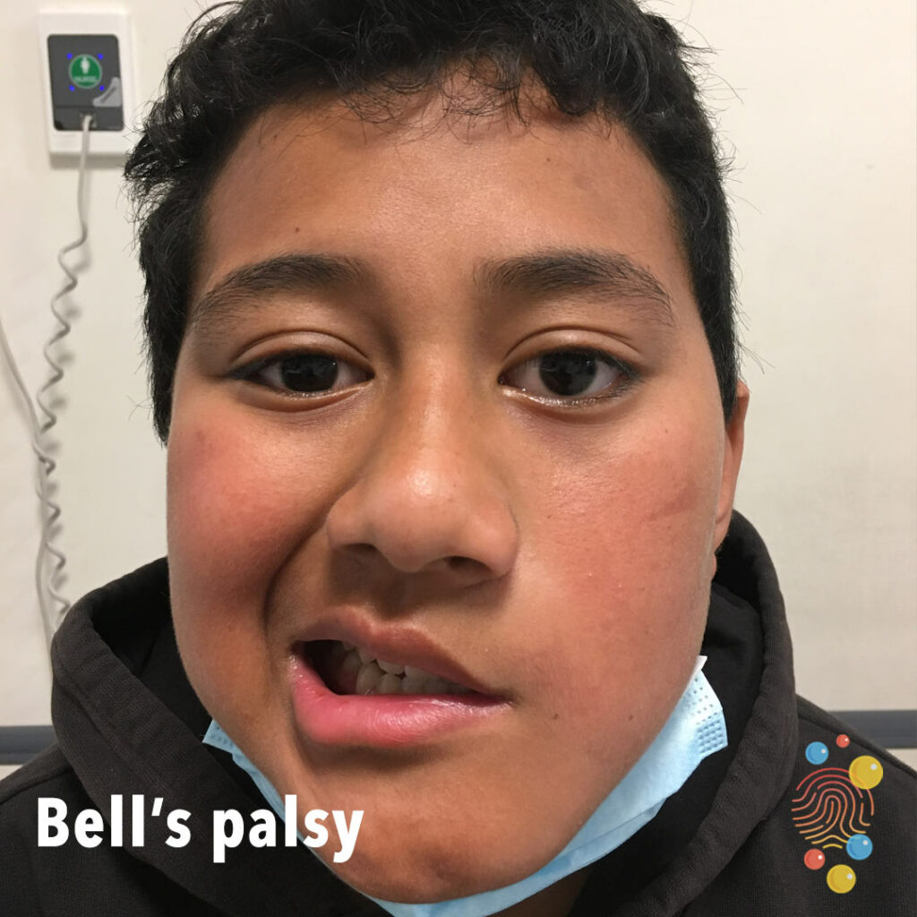
Bell’s Palsy
Learn more about Bell’s palsy
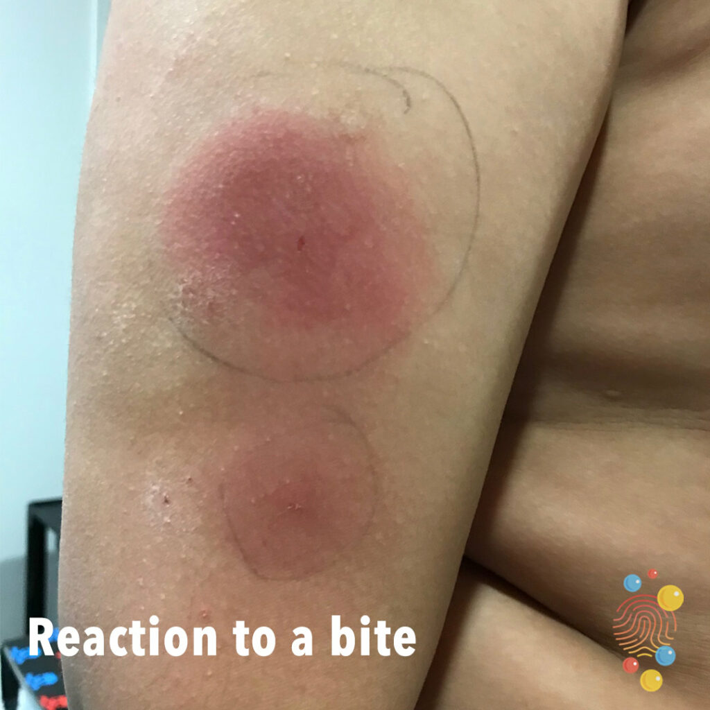
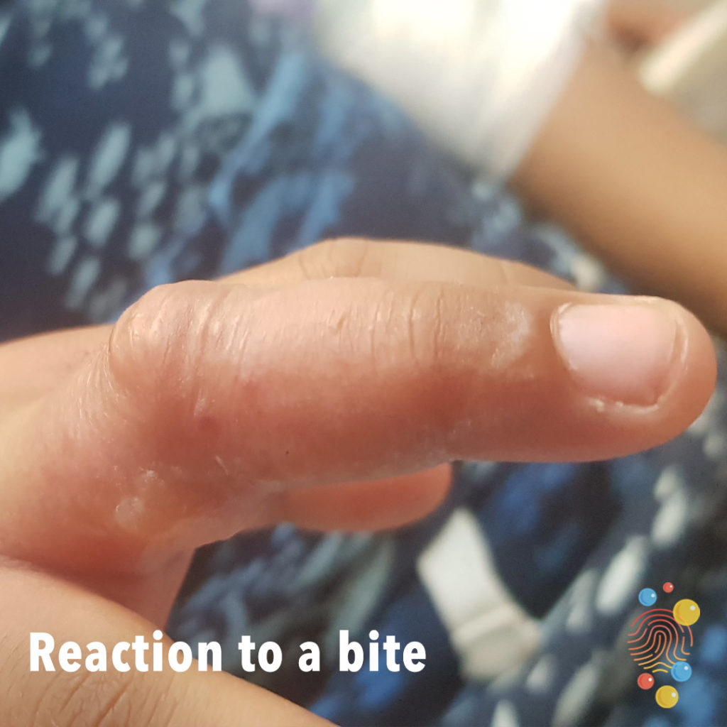
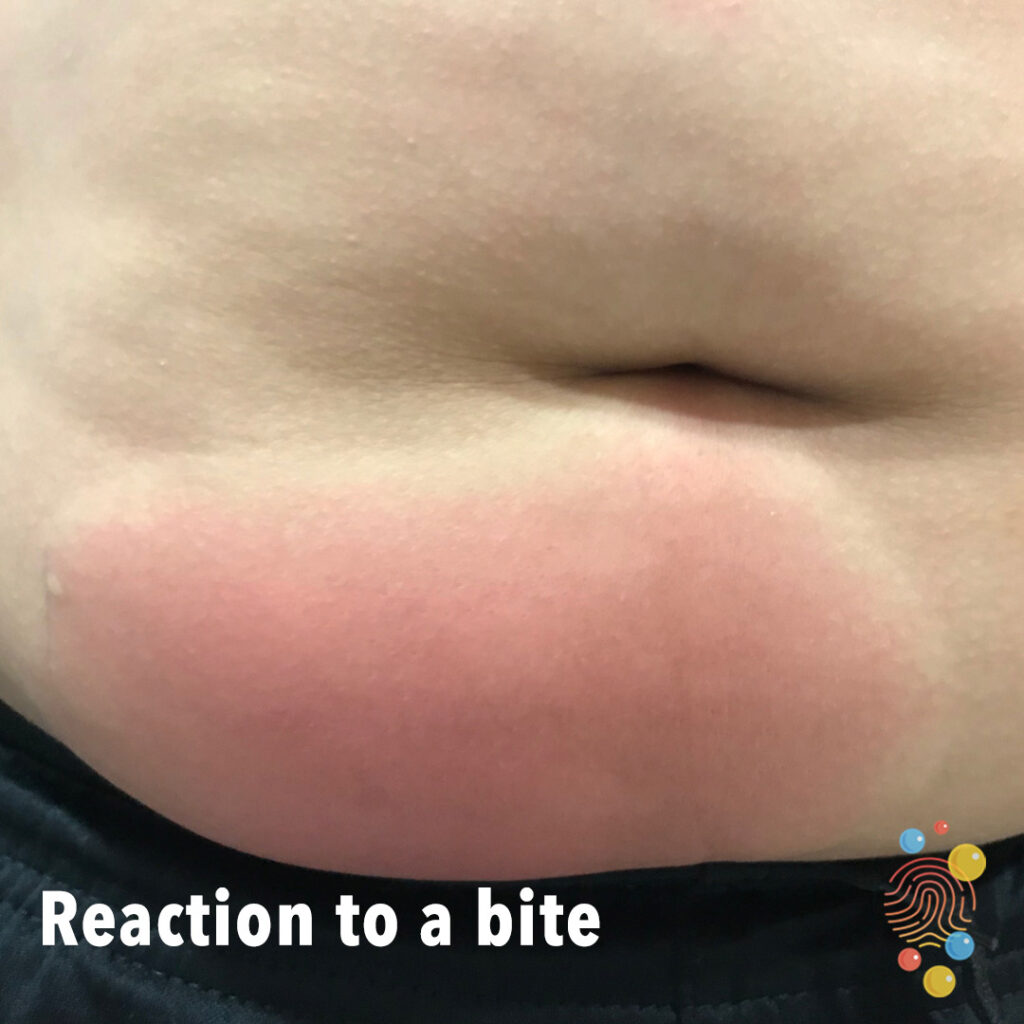
Reaction To A Bite
Learn more about bites
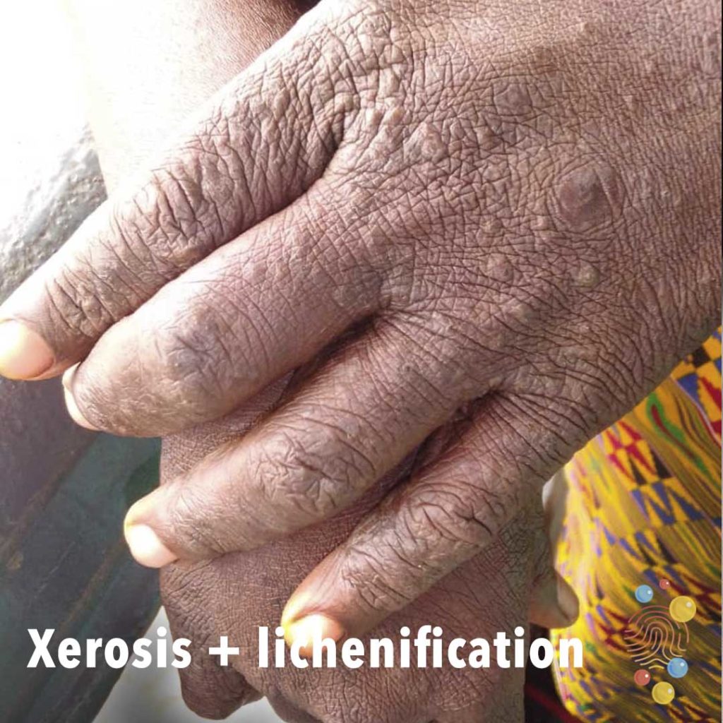
Xerosis + Lichenification
Learn more about xerosis lichenification
Urticaria
Learn more about urticaria
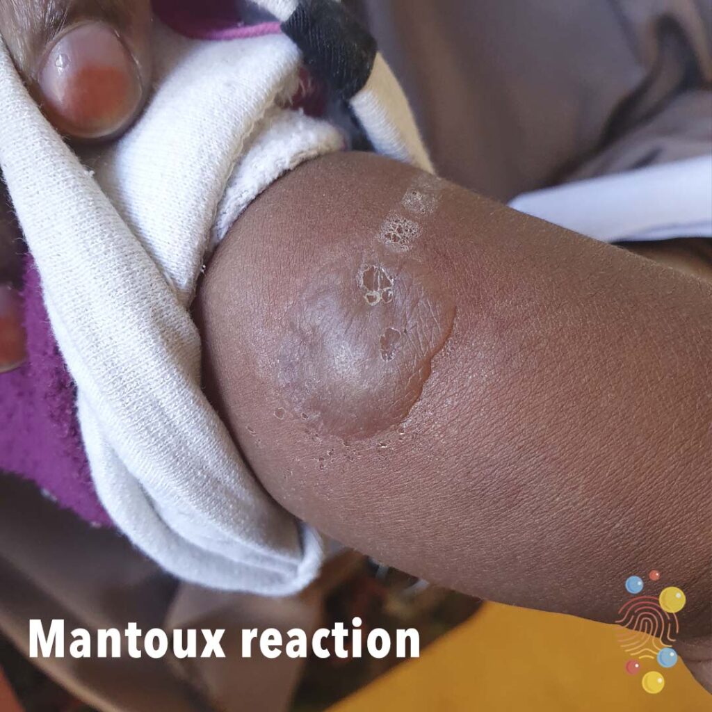
Mantoux Reaction
Learn more about the Mantoux test
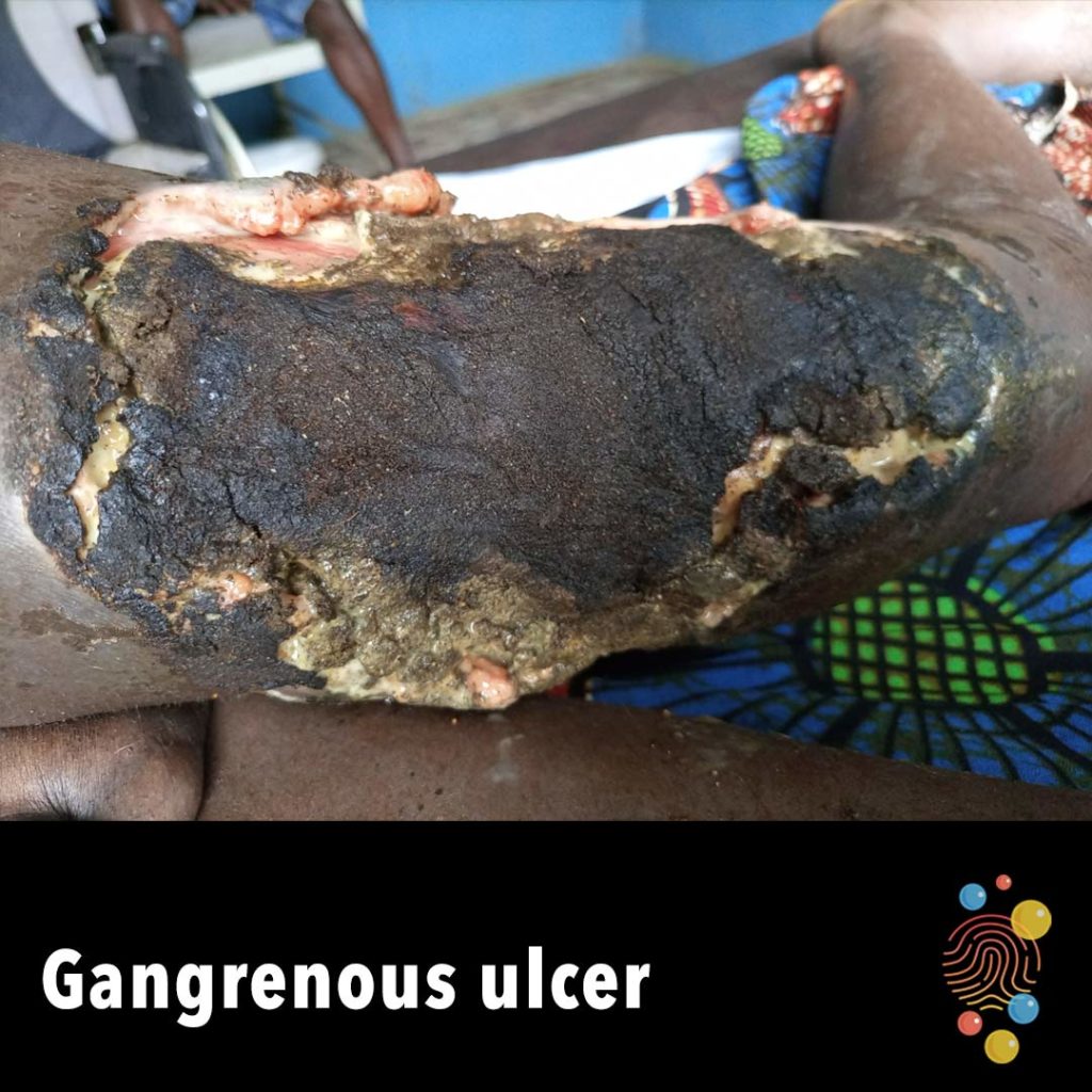
Gangrenous Ulcer
Deep ulceration of the thigh with necrotic tissue and eschar.
PIMS-TS
Erythematous papules with surrounding hazy erythema and follicular hyperkeratosis.
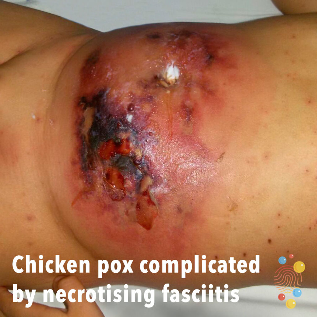
Chicken Pox Complicated By Necrotising Fasciitis
Learn more about chicken pox
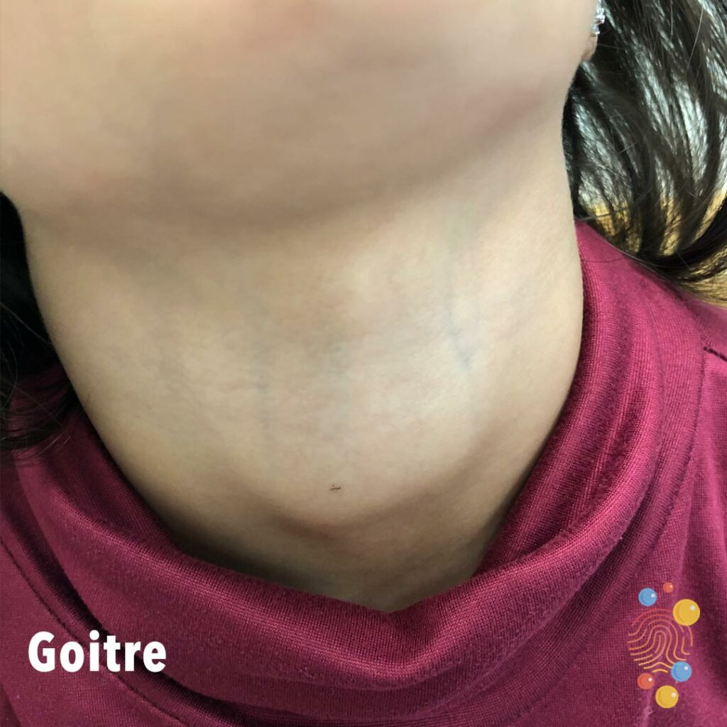
Goitre
Learn more about goitres

Accidental bruising to shin
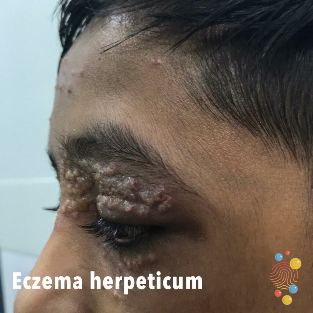
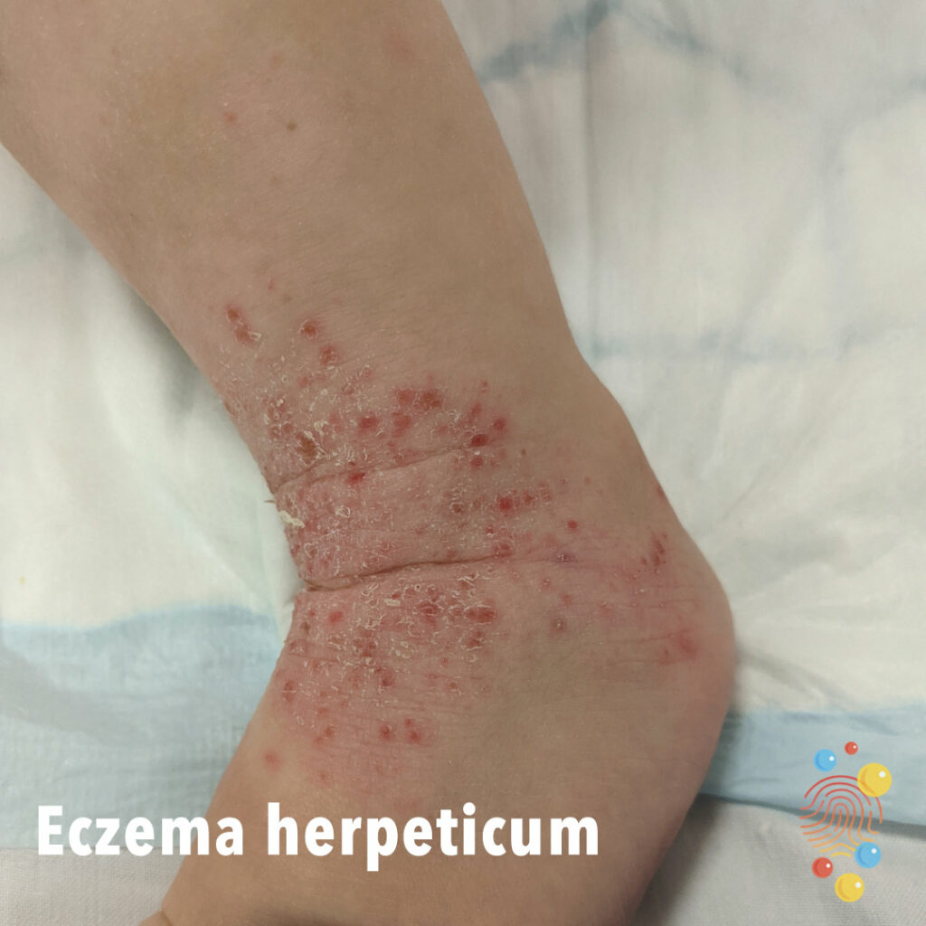
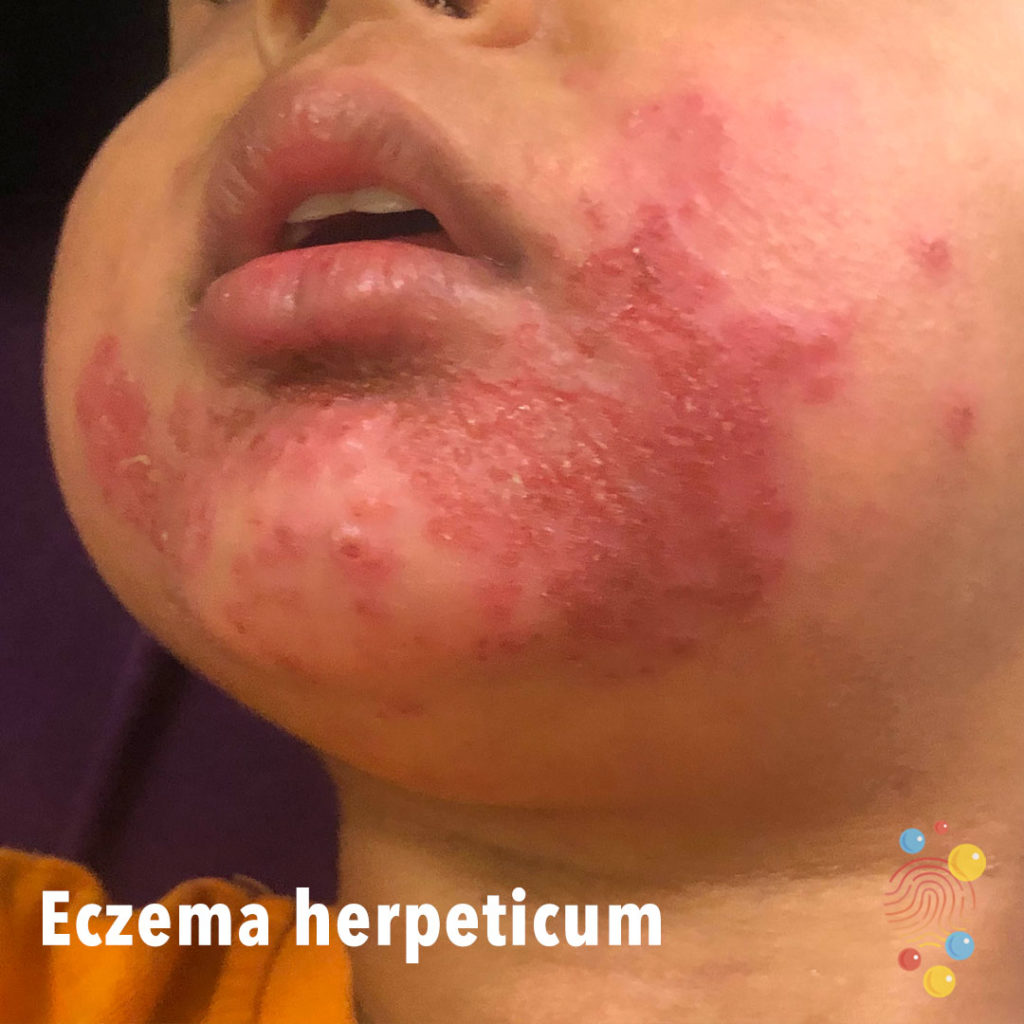
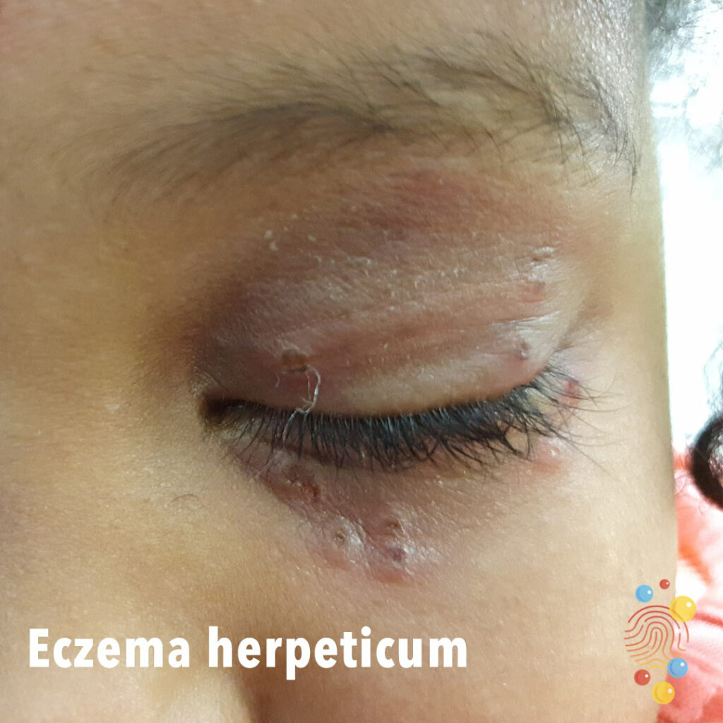
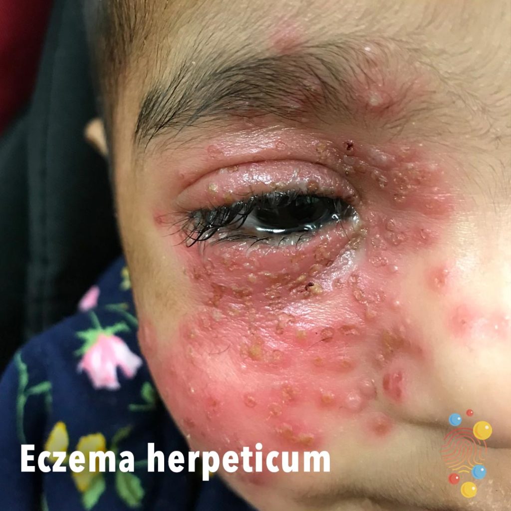
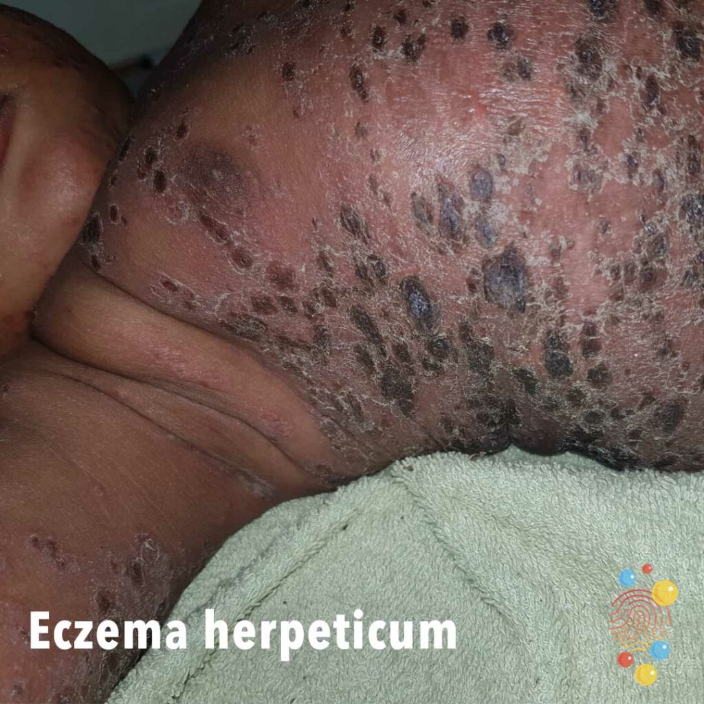
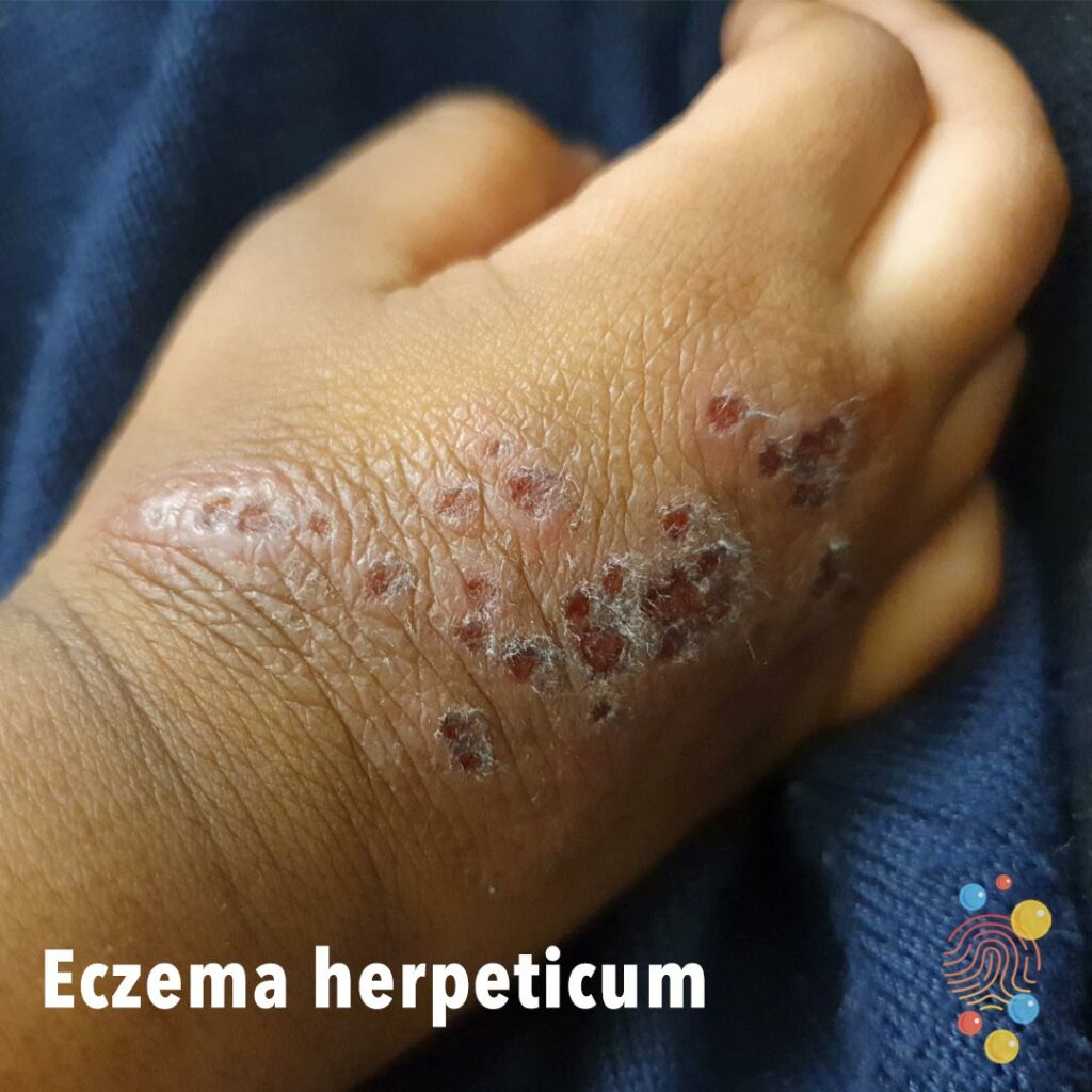
Eczema Herpeticum
Learn more about eczema herpeticum
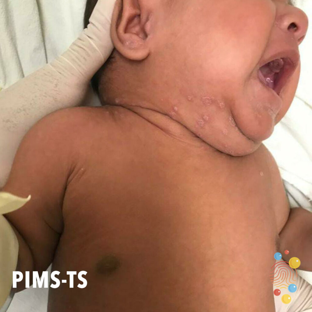
PIMS-TS
Learn more about PIMS-TS
Nummular Eczema
Learn more about eczema

Haemangioma
Learn more about haemangiomas
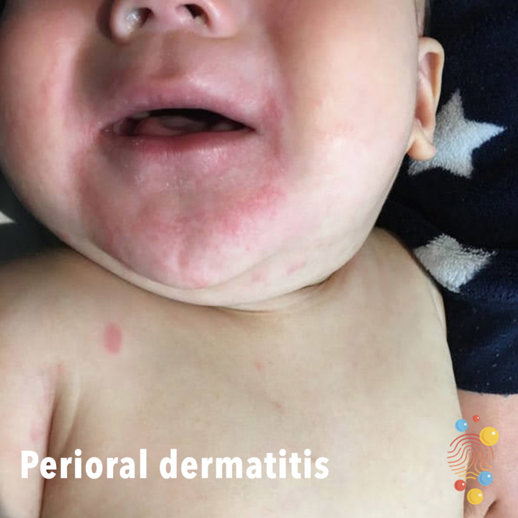
Perioral Dermatitis
Learn more about eczema
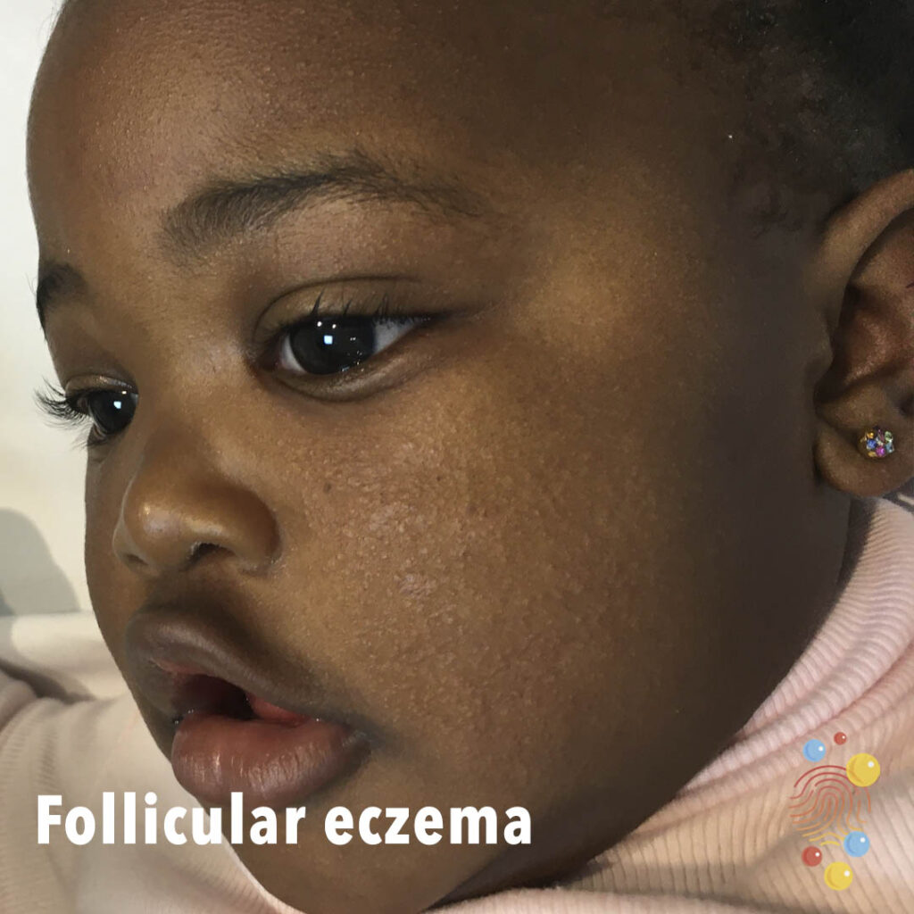
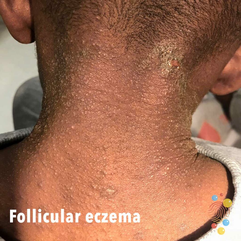
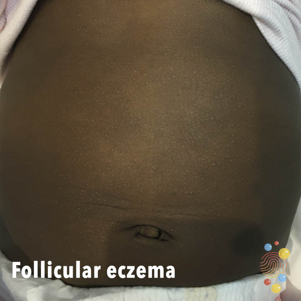
Follicular Eczema
Learn more about eczema
Post Scarlet Fever
Extensive desquamation on upper chest post scarlet fever.

Scabies
Learn more about scabies

Ranula
A ranula is a saliva-filled cyst that forms on the floor of the mouth under the tongue

Eczema
Lichenified hyperpigmented plaques on the abdomen with background follicular eczema.
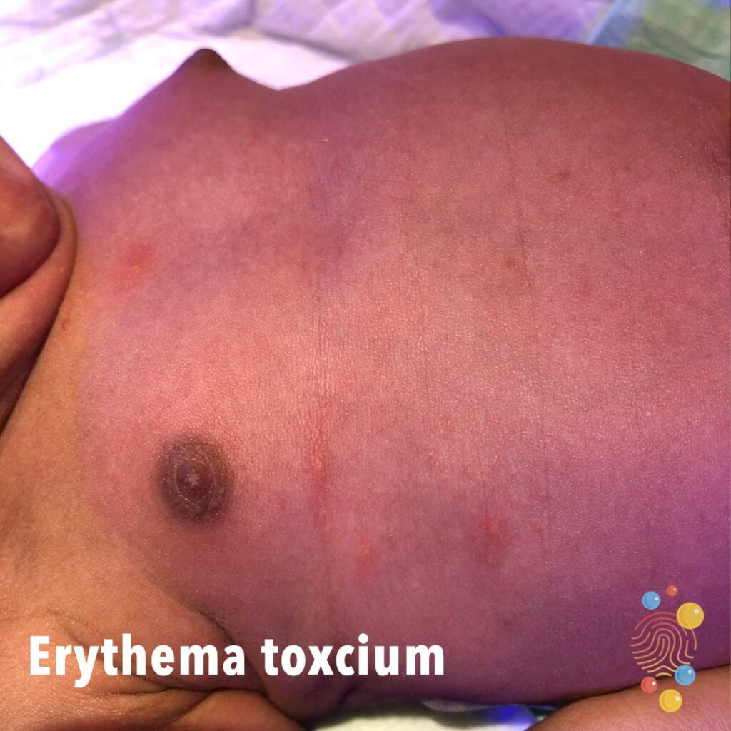
Erythema Toxicum
Learn more about erythema toxicum

Accessory Nipple
Learn more about accessory nipples

Vitiligo
Learn more about vitiligo
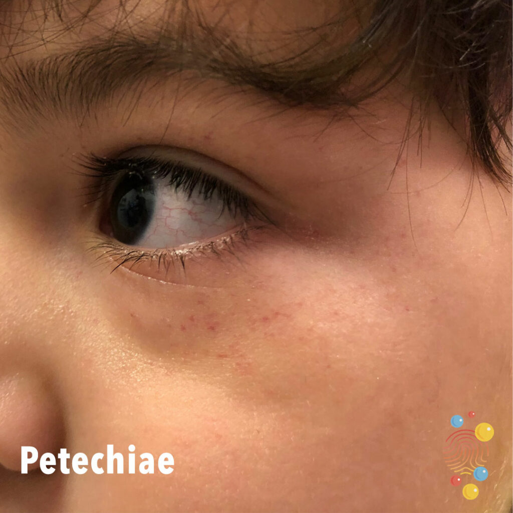
Petechiae
Petechiae around eyes – 4 year old male
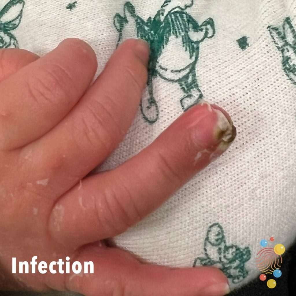
Infection
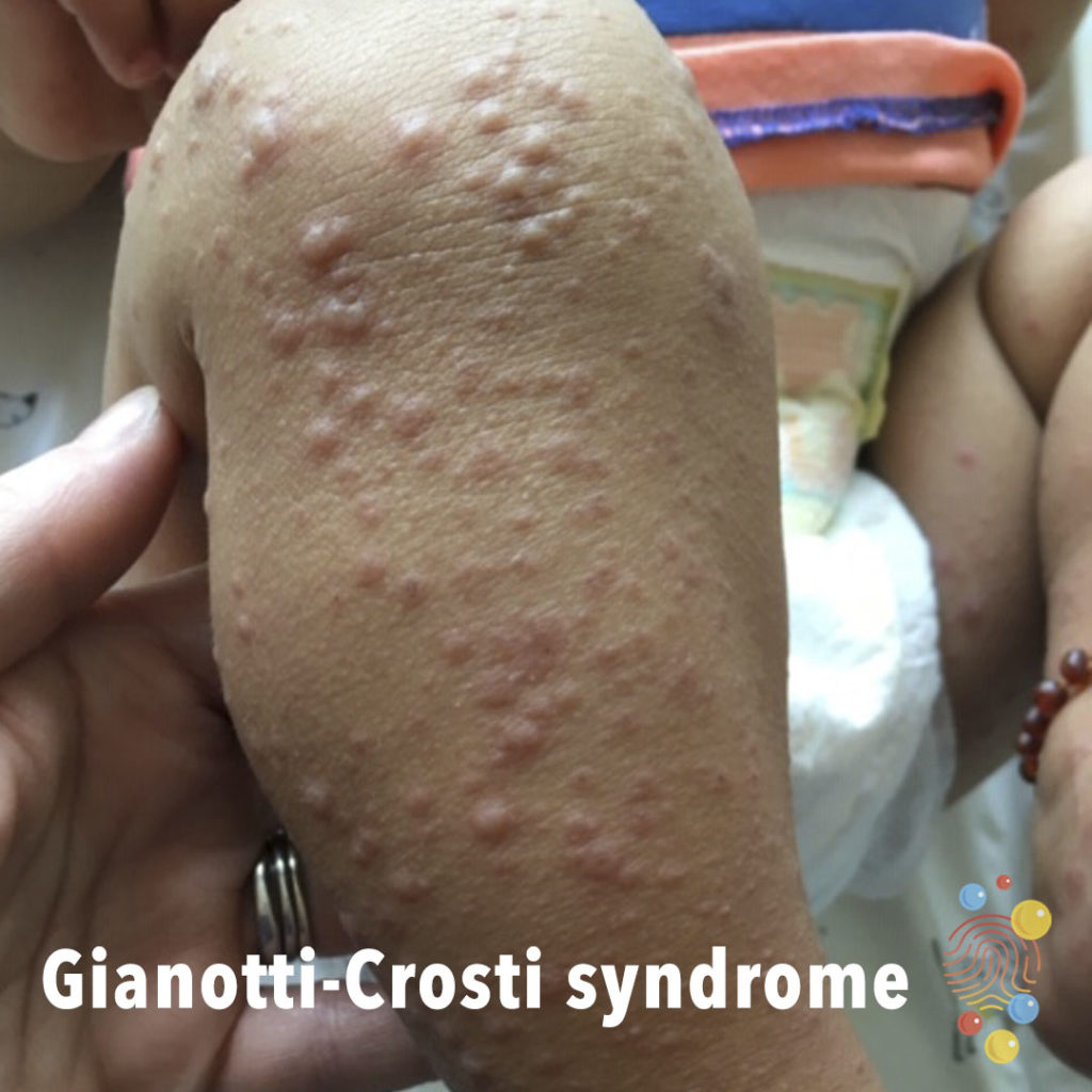
Gianotti-Crosti Syndrome
Learn more about Gianotti-Crosti syndrome
Chicken Pox
Learn more about chicken pox
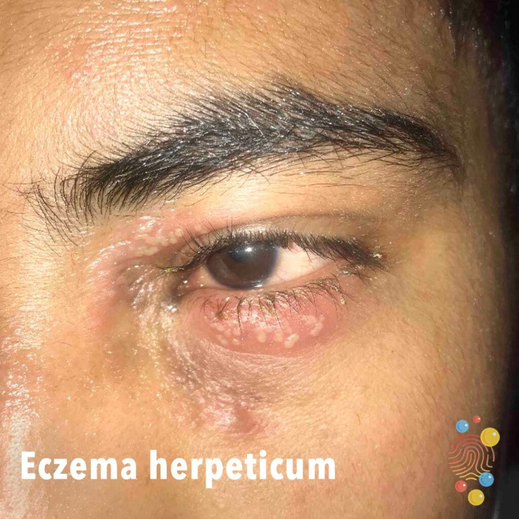
Eczema Herpeticum
Clusters of peri-ocular pustules on a background of erythematous patches. Numerous vesicles and erythematous changes across the face.
Learn more about eczema herpeticum
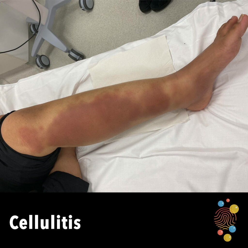
Cellulitis
Learn more about cellulitis

Petechiae
Learn more about petechiae
Chicken Pox
Multiple vesicles on an erythematous base.
Learn more about chicken pox
Neurofibromatosis
A 4-year-old girl with café-au-lait macula lesions on the chest, abdomen and extremities from birth. By maternal branch, all generations present the same type of café-au-lait mácula.
Pityriasis Alba
Learn more about pityriasis alba
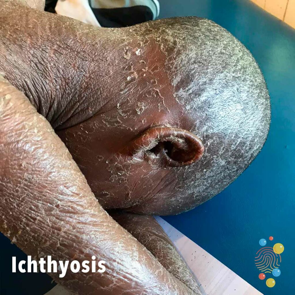
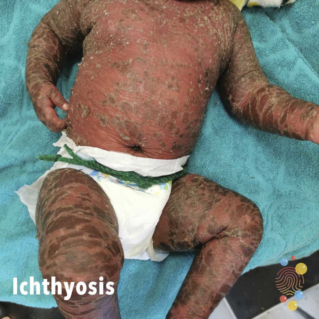
Ichthyosis
Learn more about ichthyosis
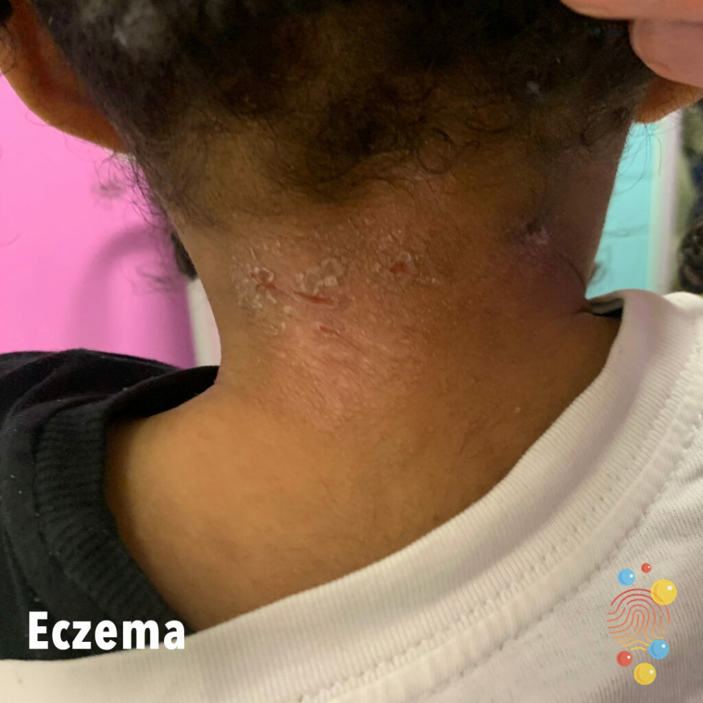
Eczema
Erythema, scale, and excorations on the posterior neck.
Eczema
Learn more about eczema

Clubbing
Learn more about clubbing

Bruise
Child ran into Ottoman bed.
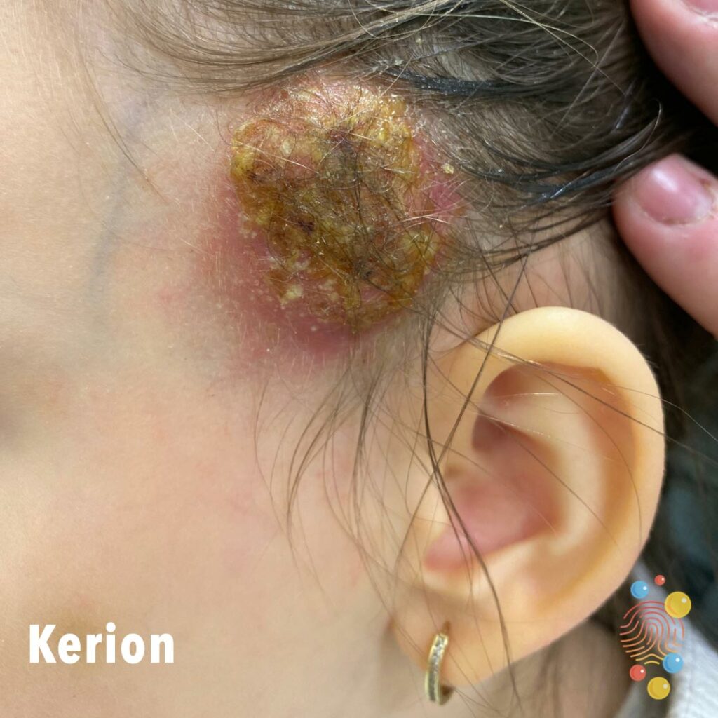
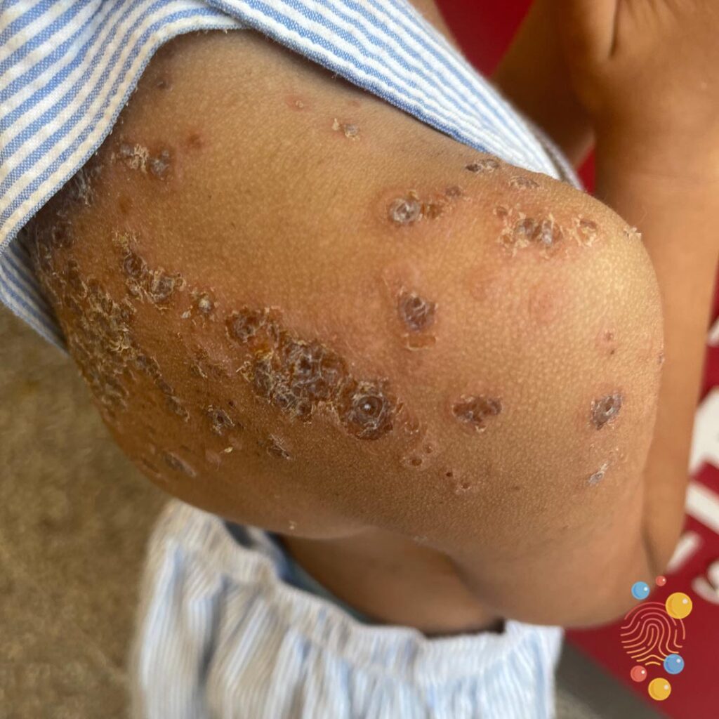
Kerion
4 year old with kerion
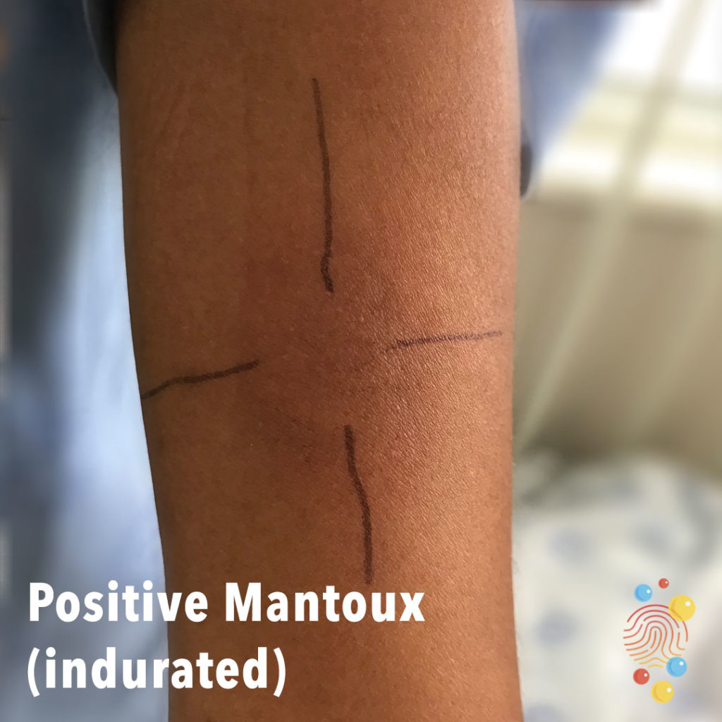
Positive Mantoux (Indurated)
Learn more about the Mantoux test

Periorbital Cellulitis
Learn more about cellulitis
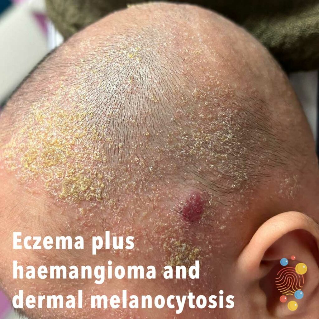
Eczema plus haemangioma and dermal melanocytosis
Eczema plus haemangioma and dermal melanocytosis
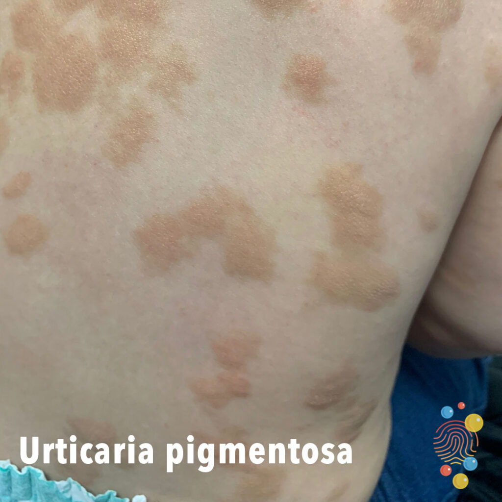
Urticaria Pigmentosa
Learn more about urticaria

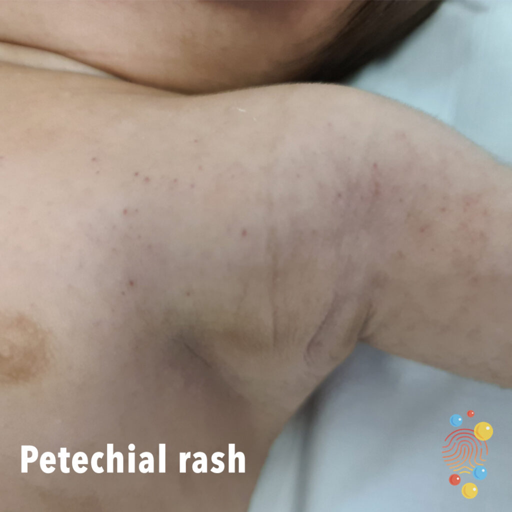
Petechial rash
Petechiae are tiny spots of bleeding under the skin. They can be caused by a simple injury, straining or more serious conditions. If you have pinpoint-sized red dots under your skin that spread quickly, or petechiae plus other symptoms, seek medical attention.
Eczema
Learn more about eczema
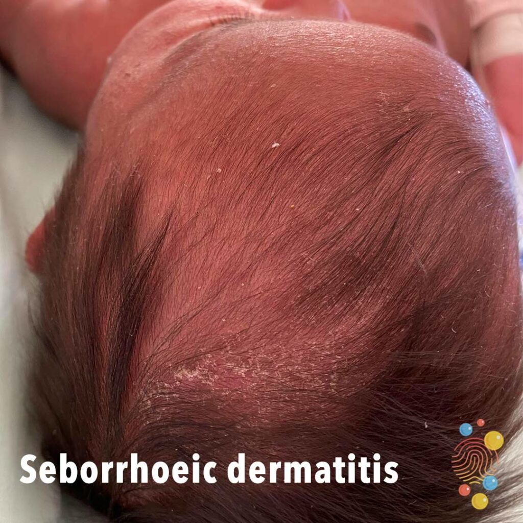
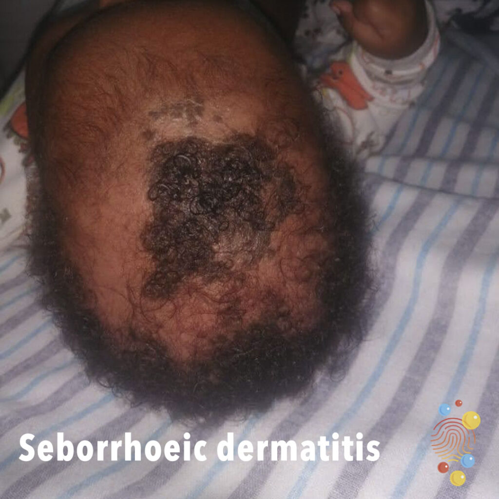
Seborrhoeic dermatitis
Learn more about seborrhoeic dermatitis
Dermal Melanocytosis
Learn more about dermal melanocytosis
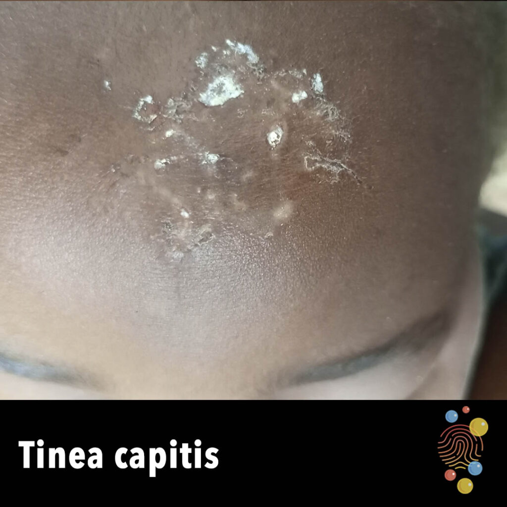
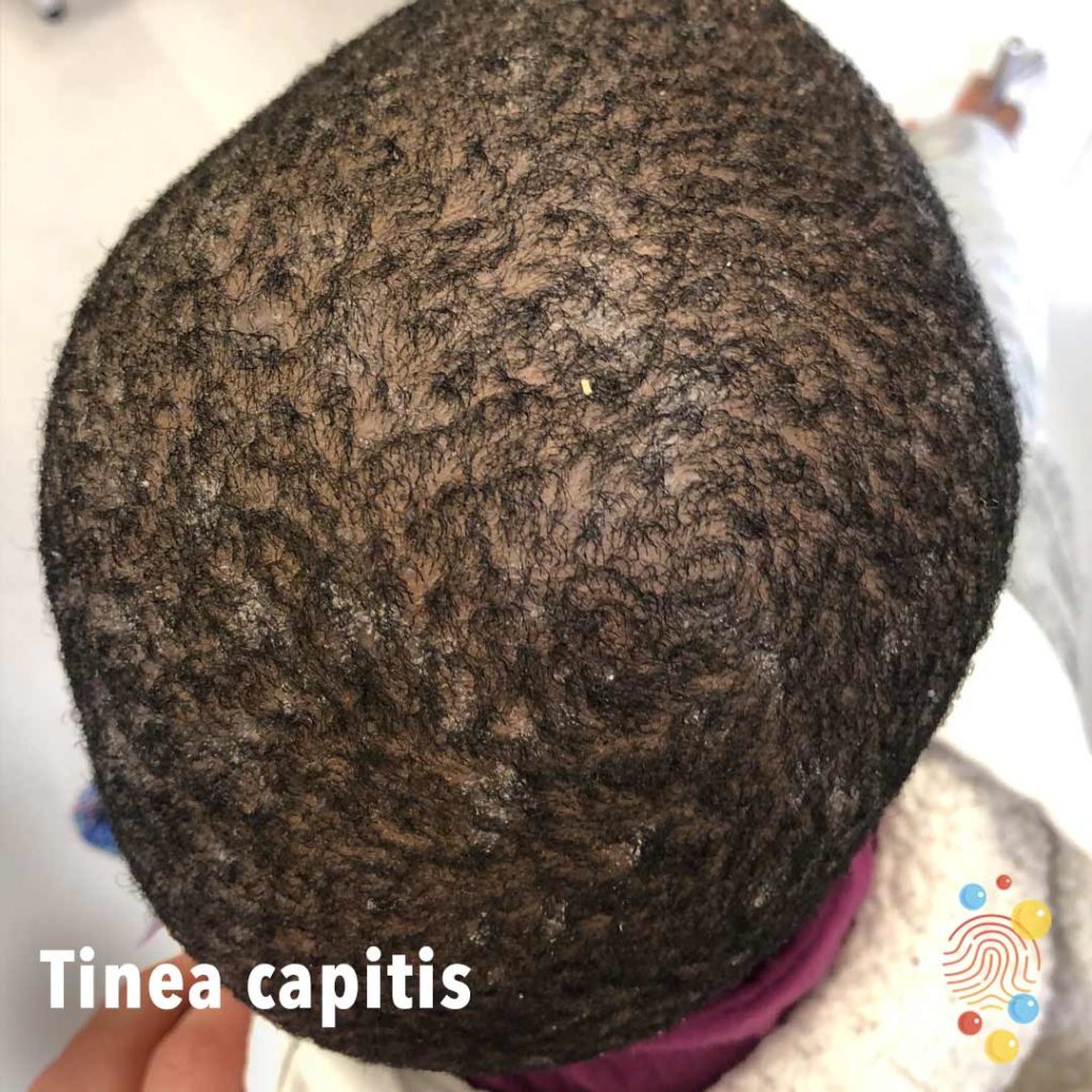
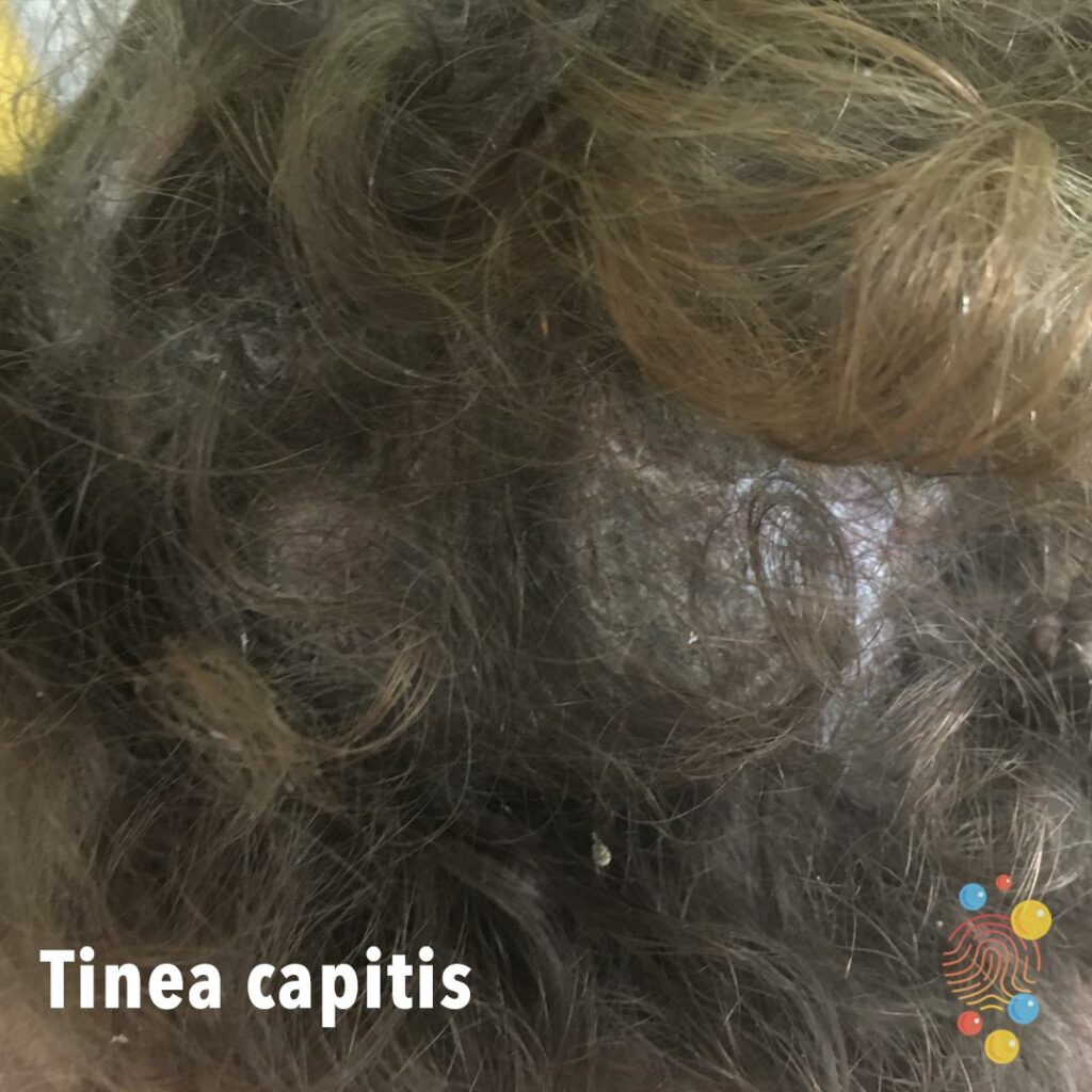
Tinea Capitis
Learn more about tinea capitis
Urticaria
Learn more about urticaria
Scabies
Learn more about scabies
Hand, Foot, + Mouth
Learn more about hand, foot and mouth
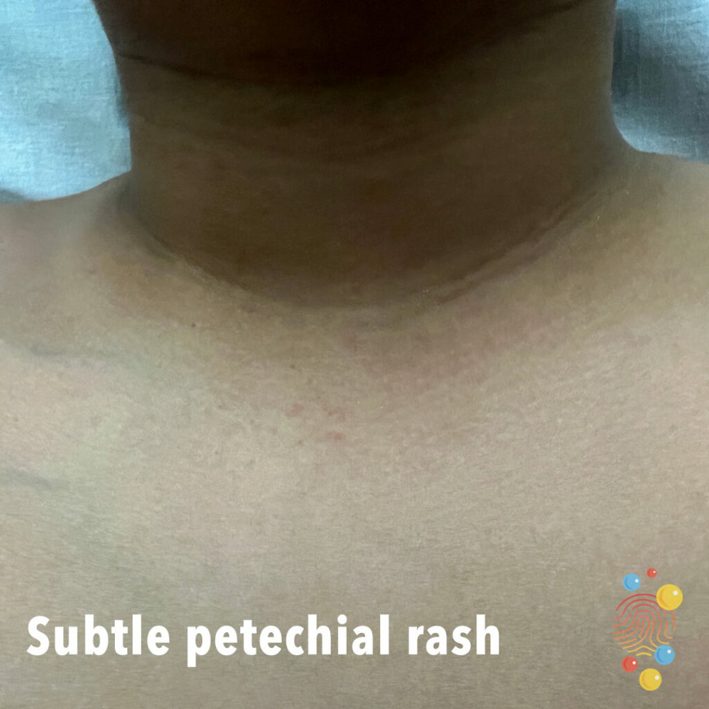
Subtle Petechial Rash

Eczema Coxsackium
Eruption of dark red macules, vesicles, and erosions distributed across areas previously affected by atopic dermatitis, with relative sparing of the trunk
Tinea corporis (ringworm)
Raised itchy dry skin with central sparing. Treatment daktacort.
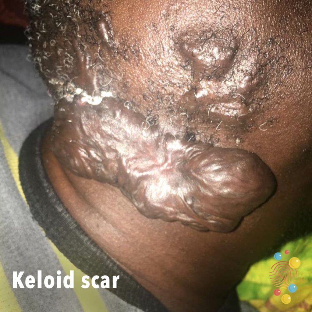
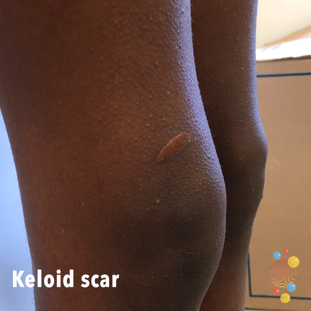
Keloid Scar
Learn more about keloid scars.
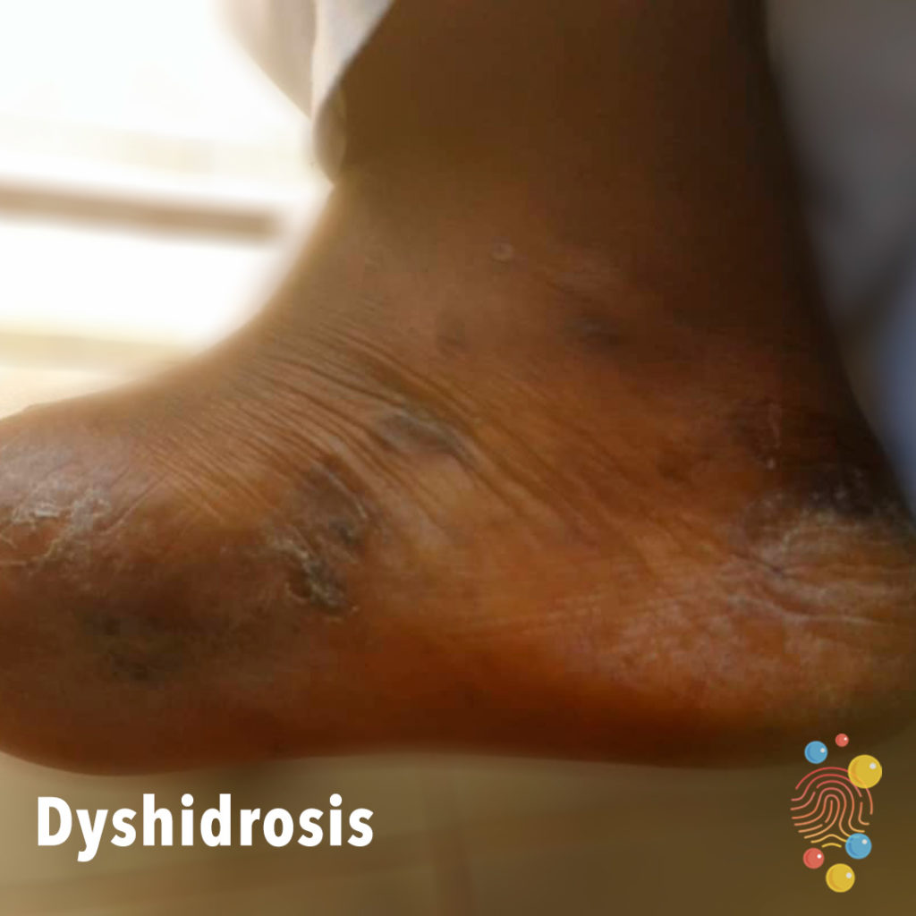
Dyshidrosis
Learn more about dyshidrosis

Nailbed Repair
Nailbed injury pre and post repair.
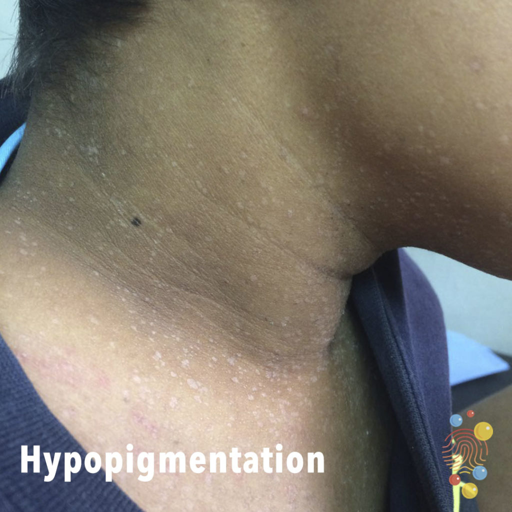
Hypopigmentation
Learn more about hypopigmentation

Eczema
Learn more about eczema

Cellulitis
Learn more about cellulitis
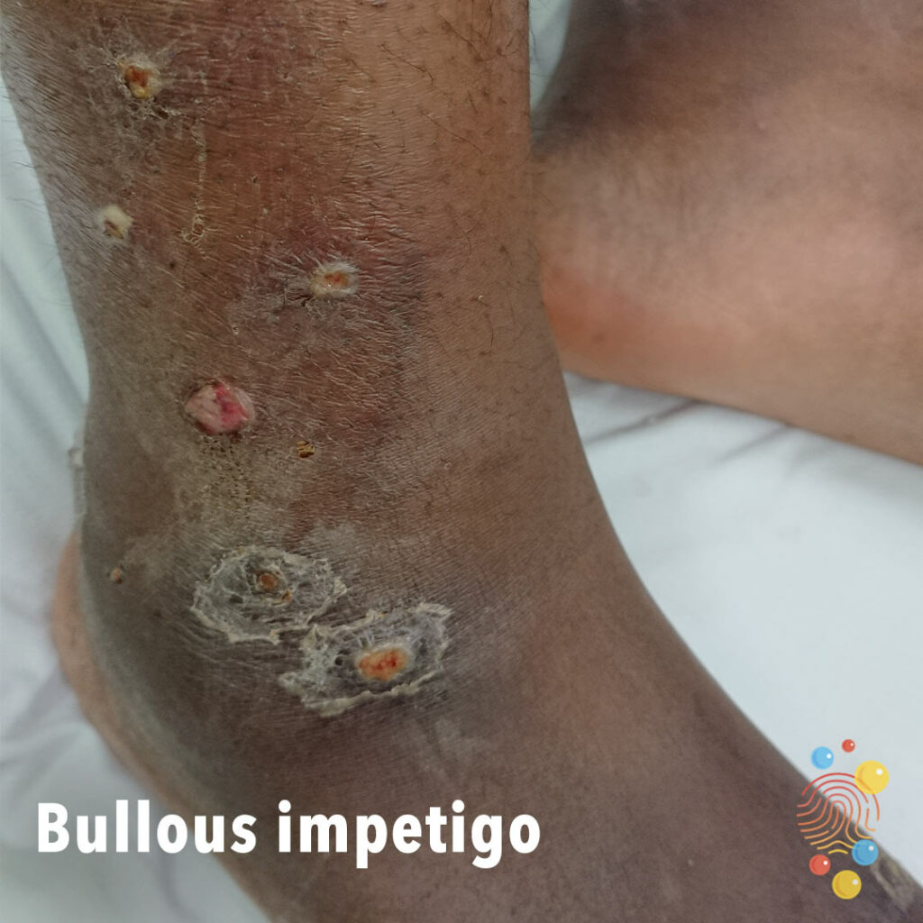
Bullous Impetigo
Learn more about bullous impetigo
Eczema
Learn more about eczema
PIMS-TS
Learn more about PIMS-TS
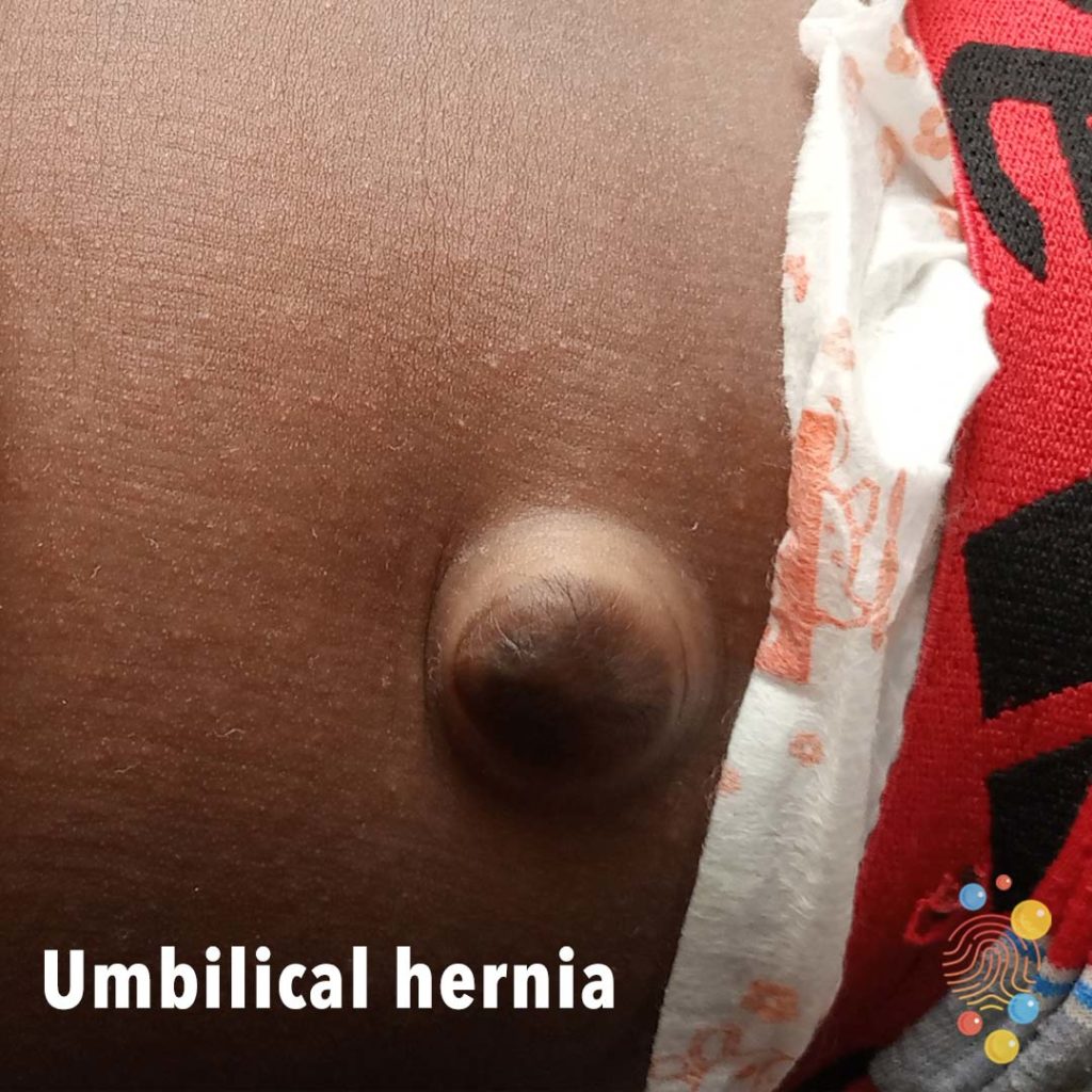
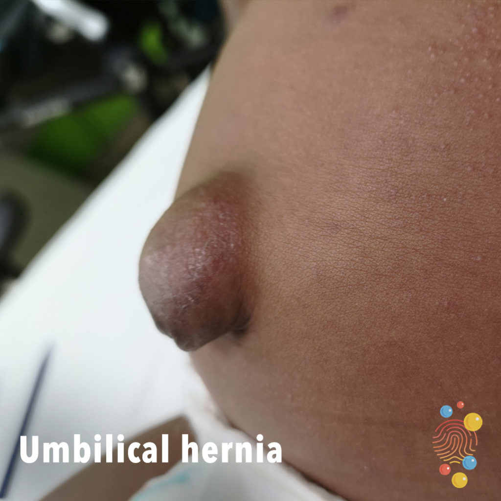
Umbilical Hernia
Learn more about umbilical hernias

Gianotti Crosti
Gianotti-Crosti syndrome (GCS) is a skin condition that usually affects children, but can also occur in adolescents and adults

Papular eczema
Learn more about eczema
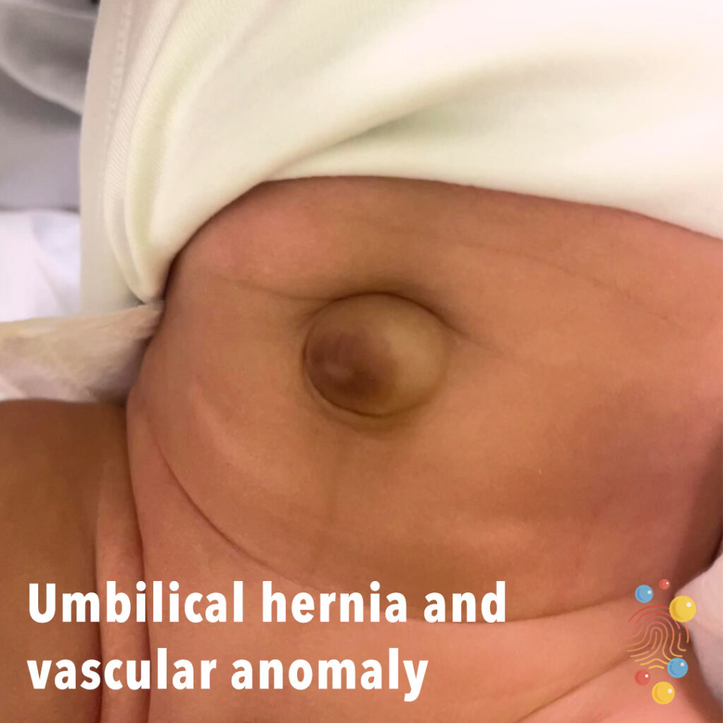
Umbilical hernia and vascular anomaly
Learn more about umbilical hernias

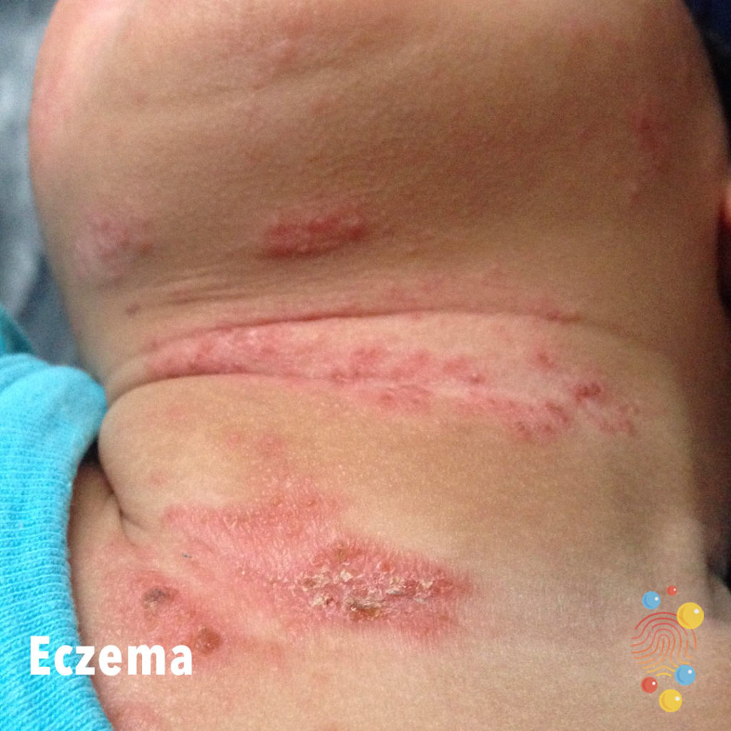
Eczema
Learn more about eczema
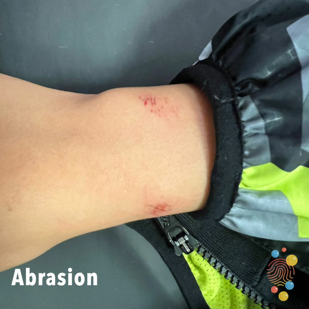
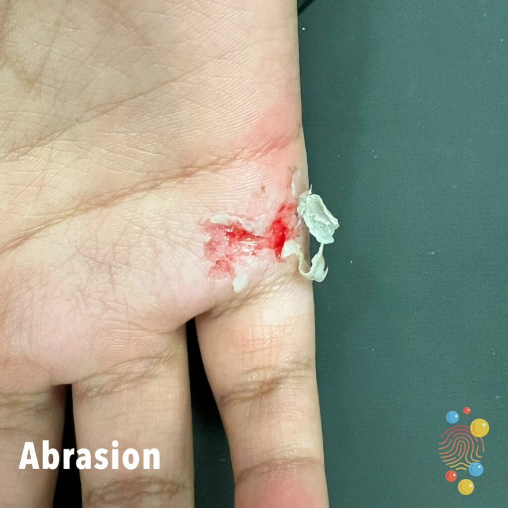
Abrasion
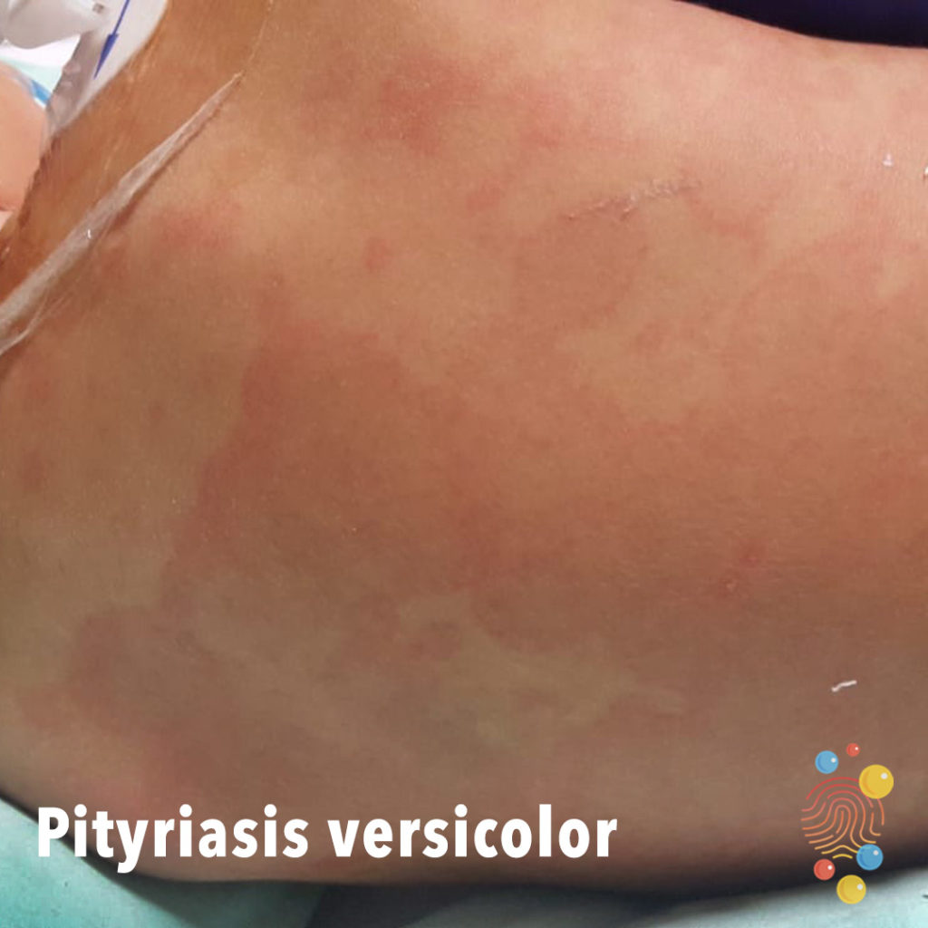
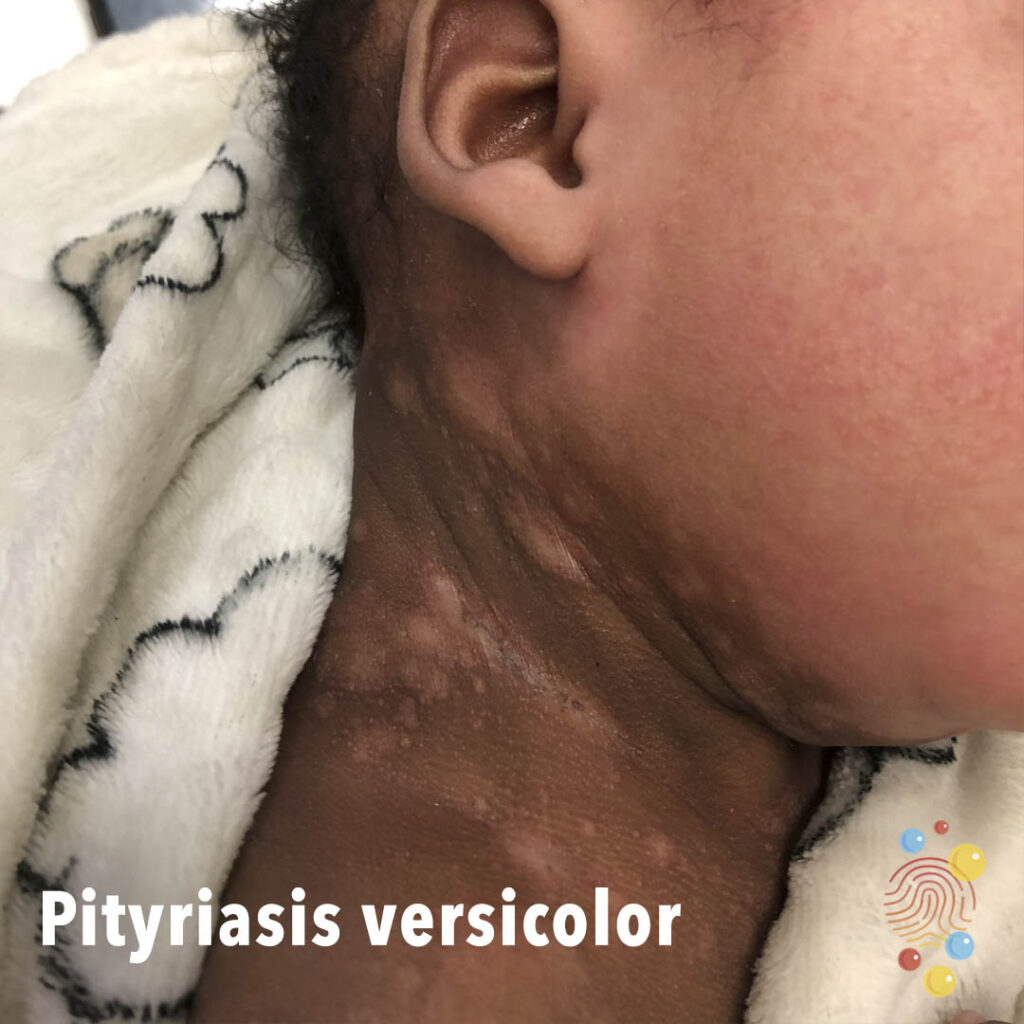
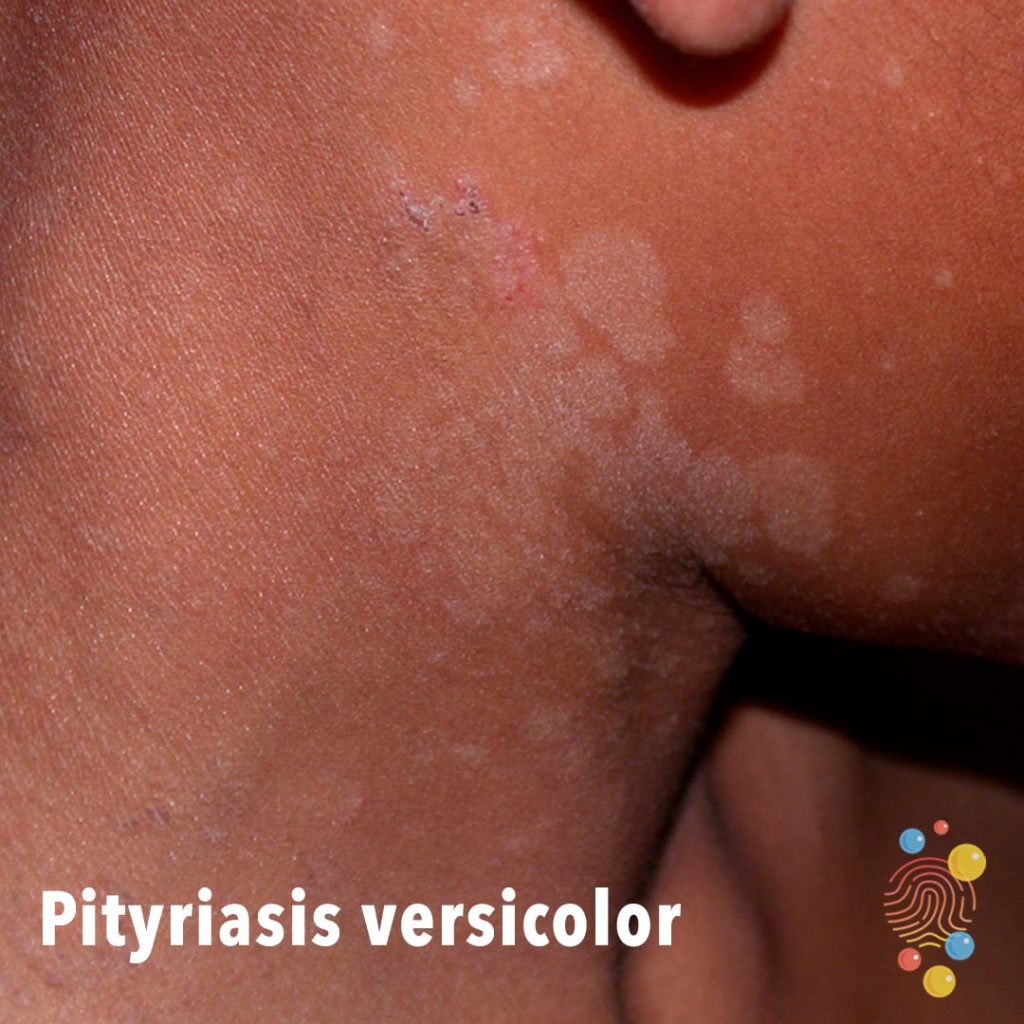
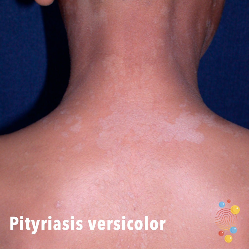
Pityriasis Versicolor
Learn more about pityriasis versicolor

Lymphoedema and hyperkeratosis
Symmetric swelling of lower limbs associated with hyperkeratosis, plantar keratoderma, and dystrophic toenails

Lymphoedema and hyperkeratosis
Symmetric swelling of lower limbs associated with hyperkeratosis, plantar keratoderma, and dystrophic toenails
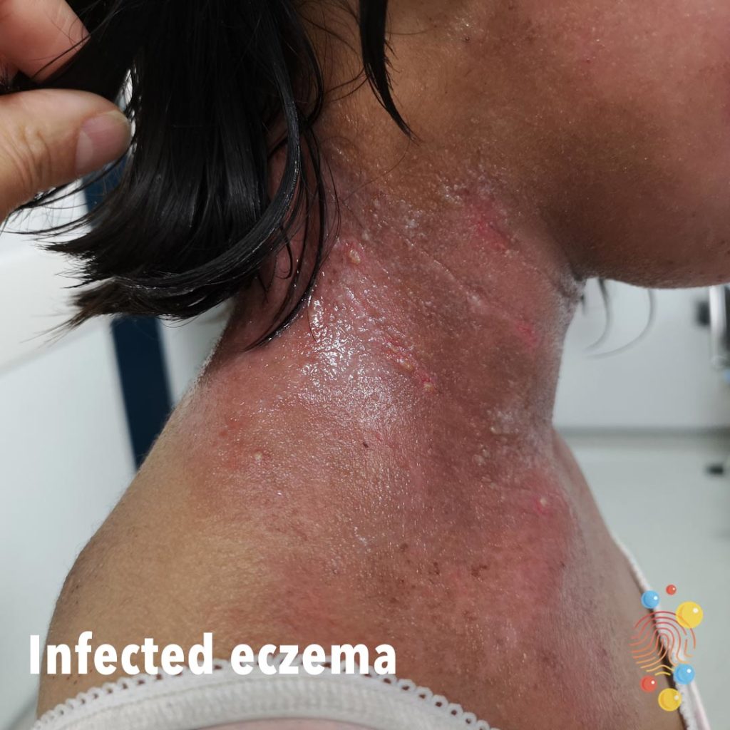
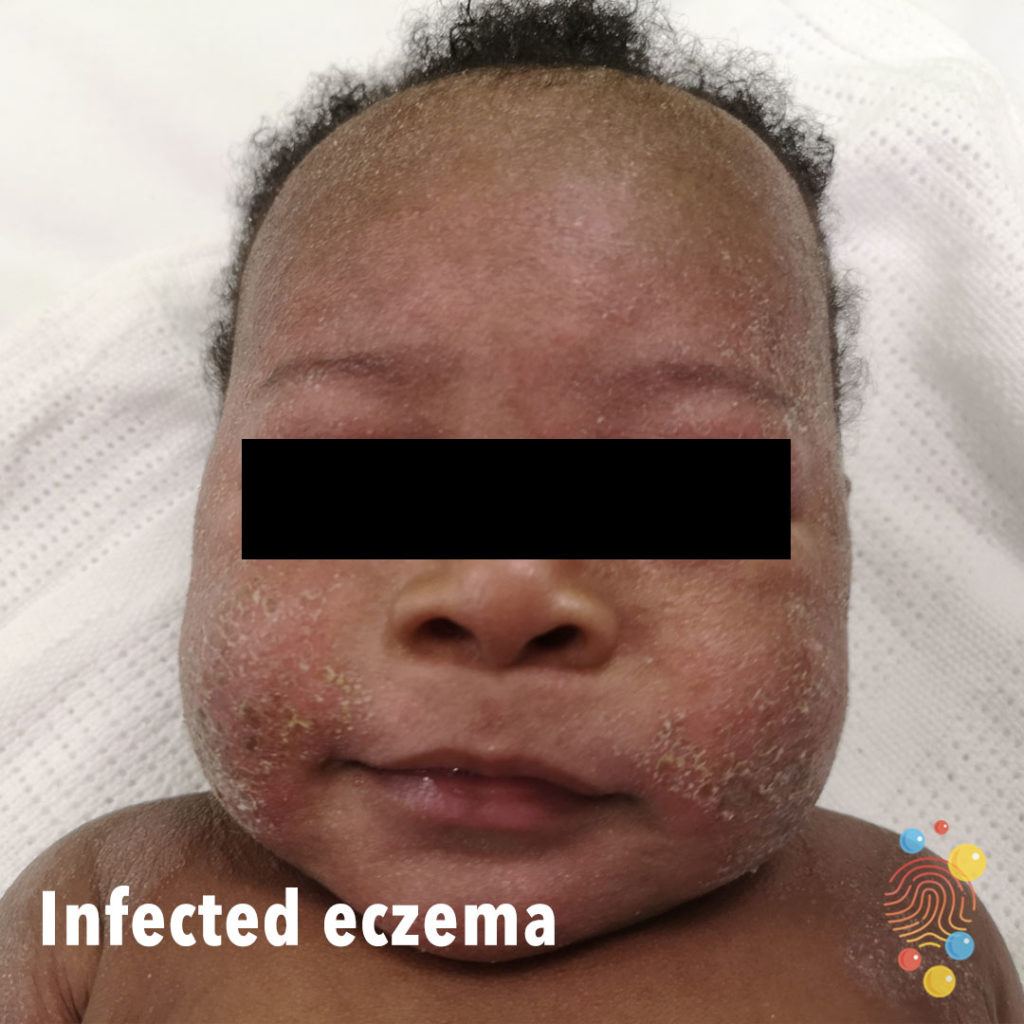
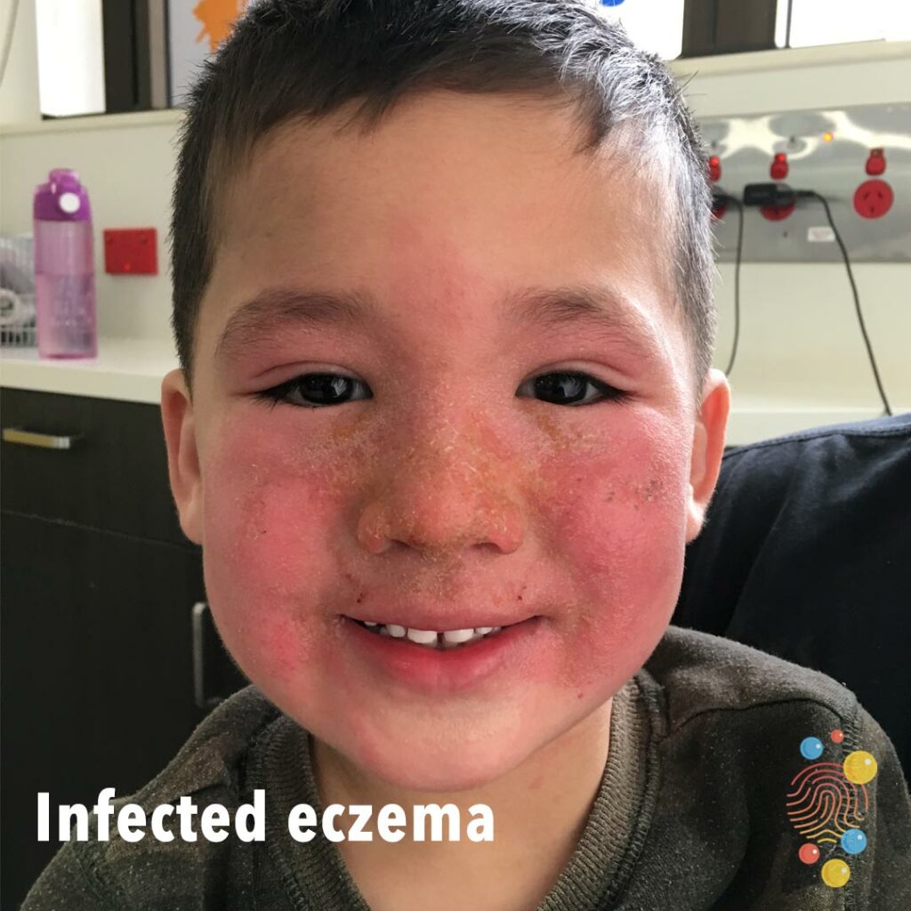
Infected Eczema
Learn more about eczema
Eczema
Learn more about eczema
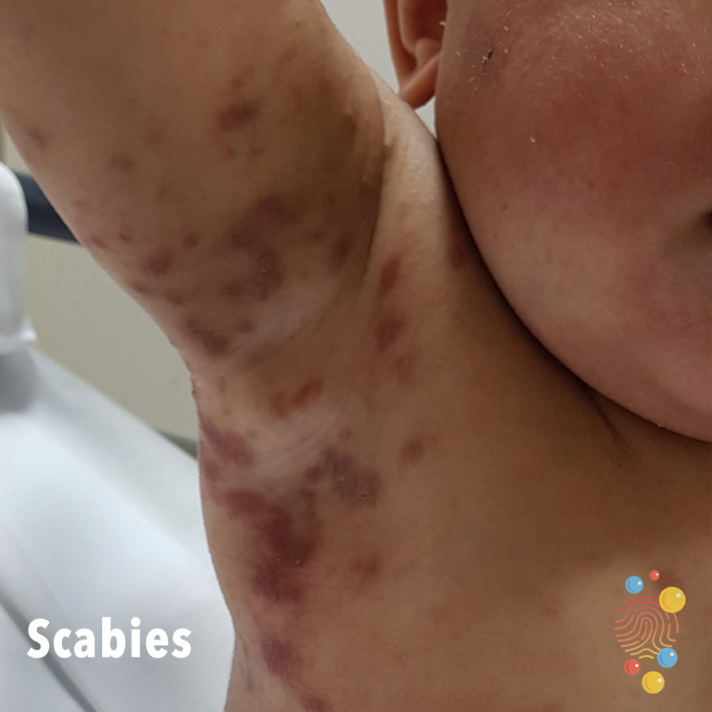
Scabies
Learn more about scabies
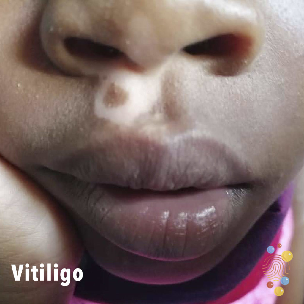
Vitiligo
Learn more about vitiligo

Post immunisation site
Post-immunisations (12 month imms)
Eczema Herpeticum
Eczema herpeticum (EH) is a rare, contagious, and severe skin infection that occurs when the human herpes simplex virus (HSV) infects inflamed skin
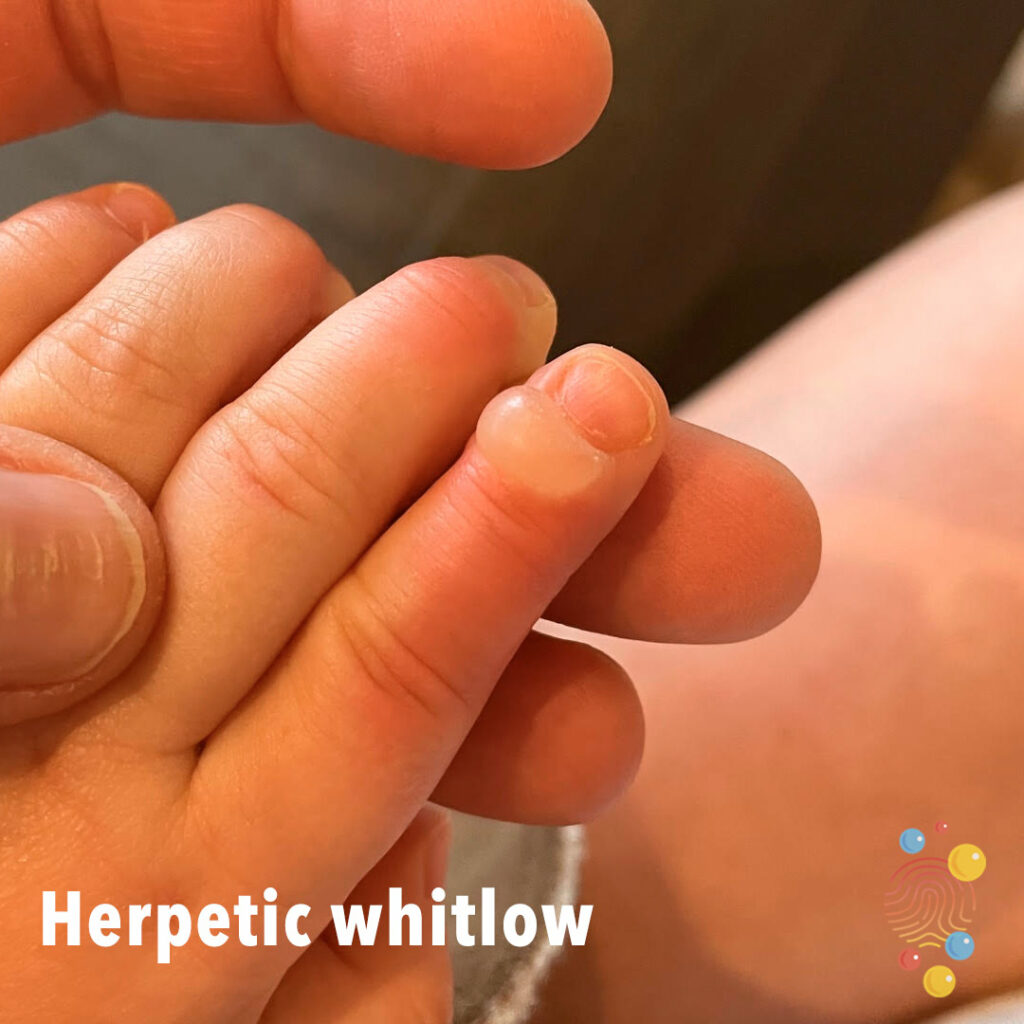
Herpetic whitlow
Learn more about herpes simplex virus
Urticaria
Learn more about urticaria
Paronychia
Small area of inflammation with surrounding pus on the skin surrounding the nail.
Learn more about paronychia

Eczema
Learn more about eczema
Pityriasis Alba
Learn more about pityriasis alba
Eczema
Learn more about eczema

Staphylococcal Skin Infection
Learn more about staphylococcal infection
Dermal Melanocytosis
Learn more about dermal melanocytosis
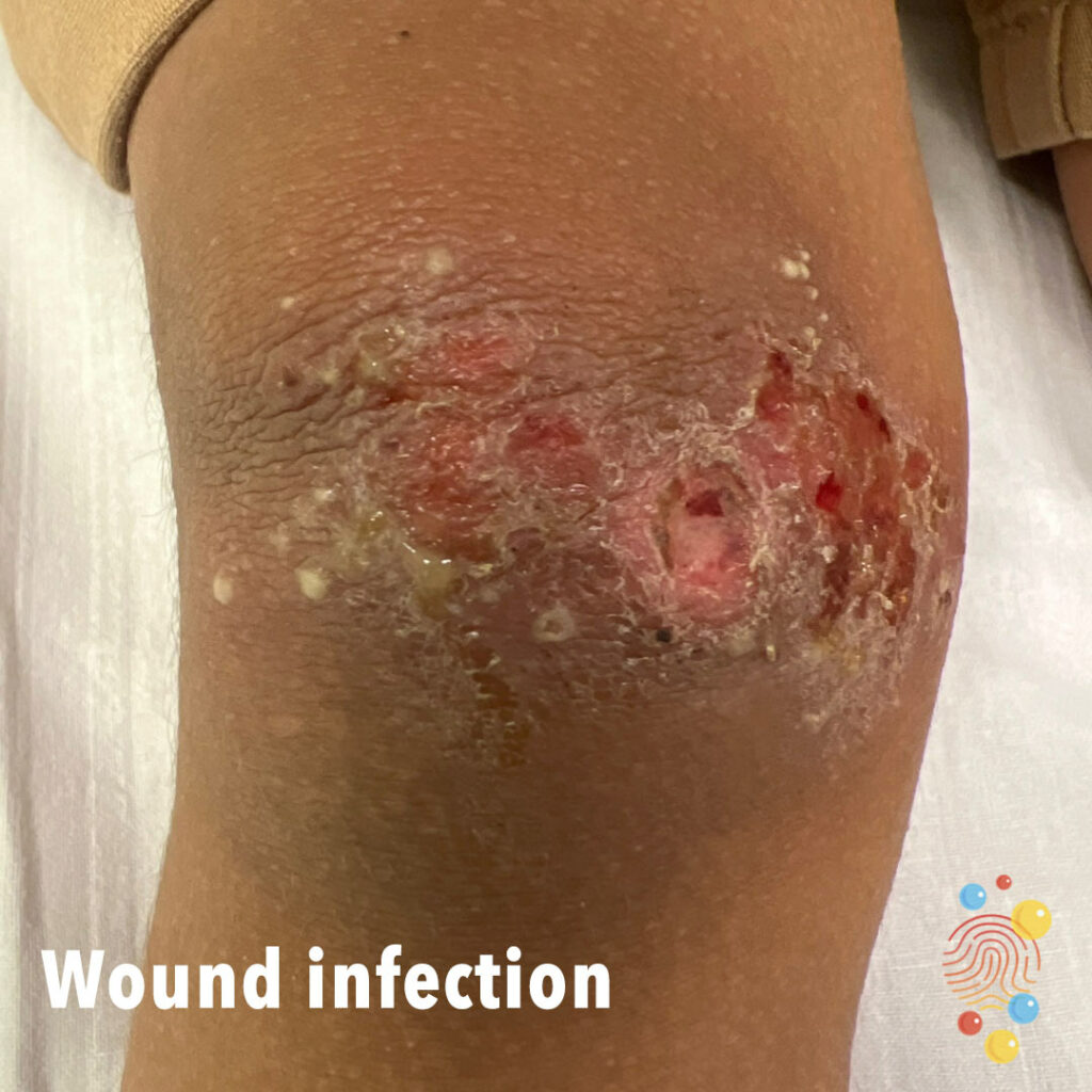
Wound Infection
3 year old boy. Tripped and fell twice in a week, a few days later noted to have pus in wound. Skin infection secondary to wound.
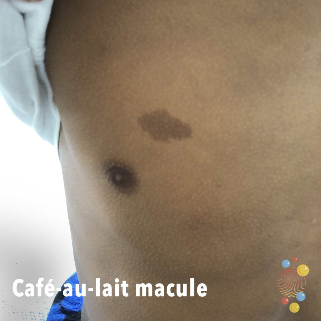
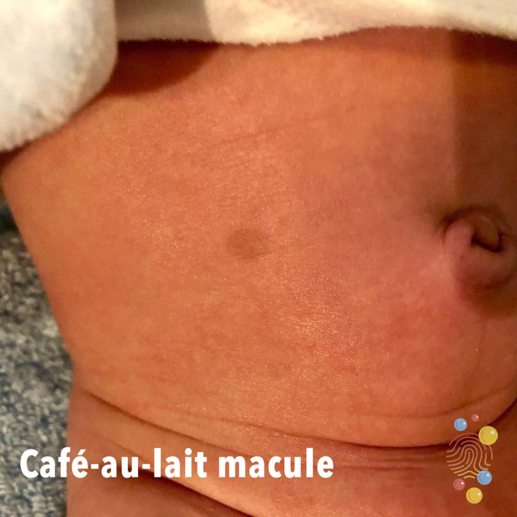
Café-Au-Lait Macule
Learn more about café-au-lait macules

Eczema Coxsackium
Eruption of dark red macules, vesicles, and erosions distributed across areas previously affected by atopic dermatitis, with relative sparing of the trunk
Normal Bruising Pattern
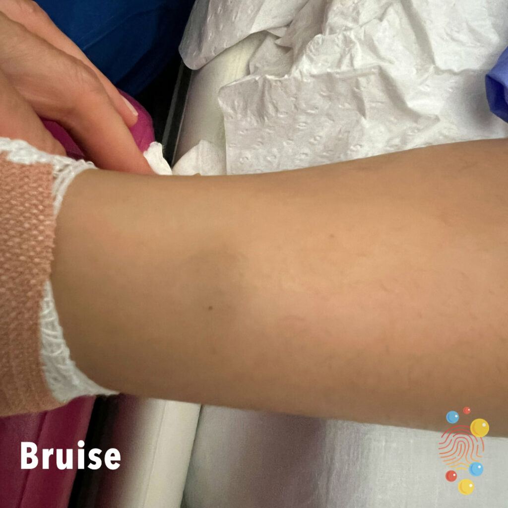
Bruise
Bruise to shin

Bullous Impetigo
Extensive healing erosions with haemorrhagic crust and a collarette of scale
Pityriasis Versicolor
Learn more about pityriasis versicolor
Henoch-Schonlein Purpura
Learn more about Henoch-Schonlein purpura

Scabies
Learn more about scabies
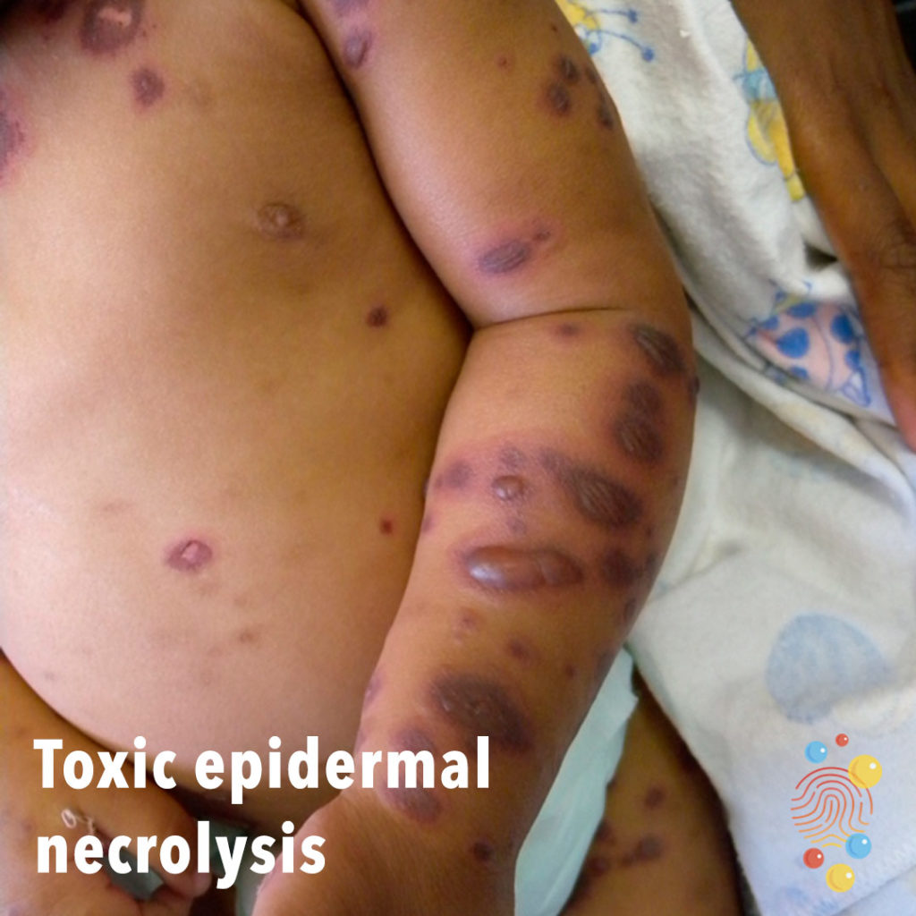
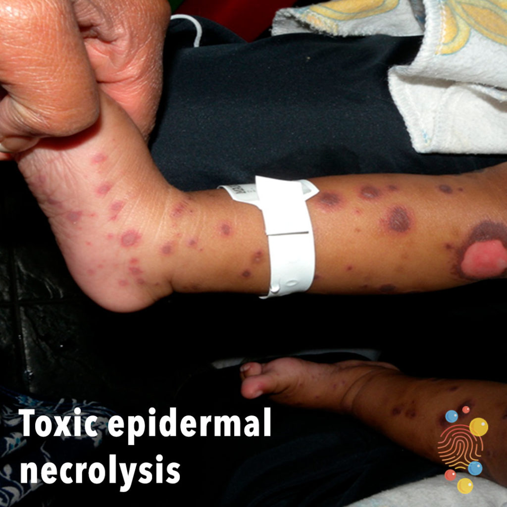
Toxic Epidermal Necrolysis
Learn more about toxic epidermal necrylosis

Umbilical Hernia
Learn more about umbilical hernias
Tick Bite
Learn more about tick bites
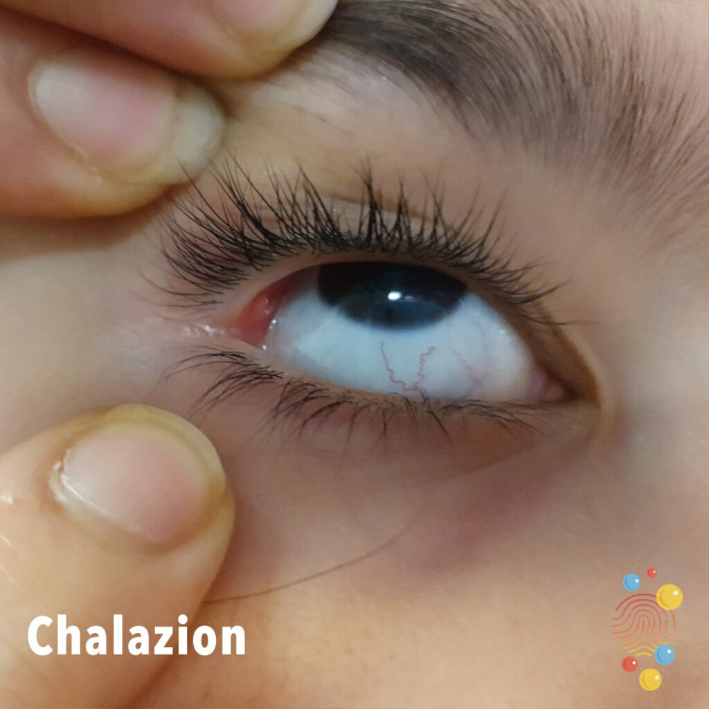
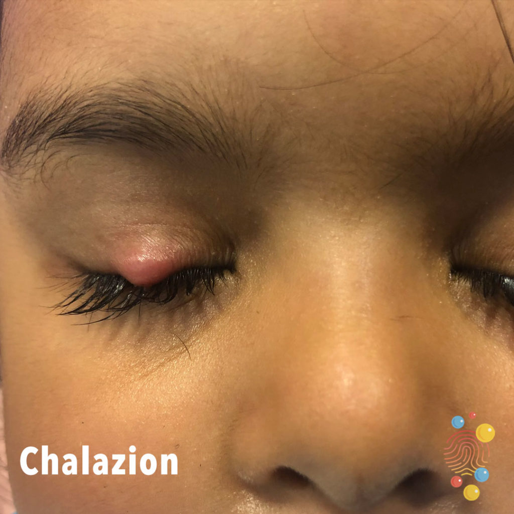
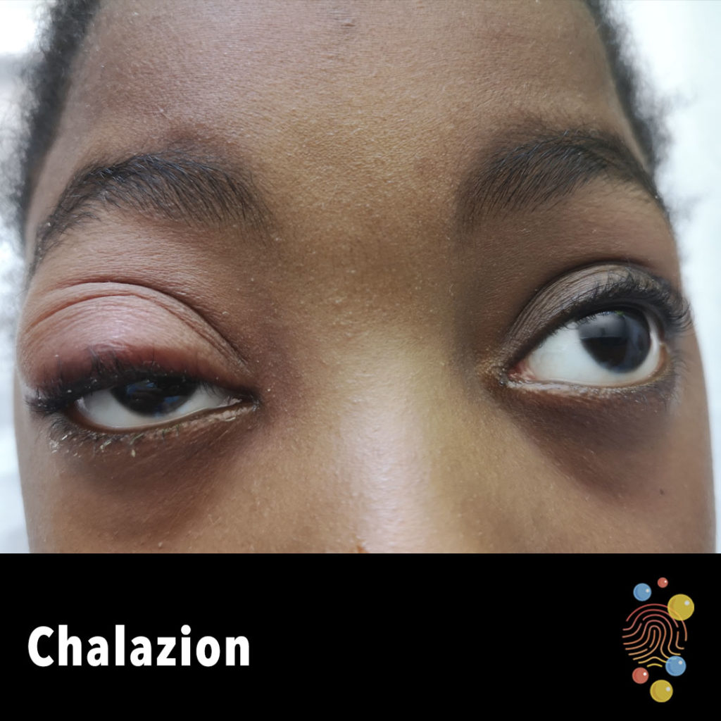
Chalazion
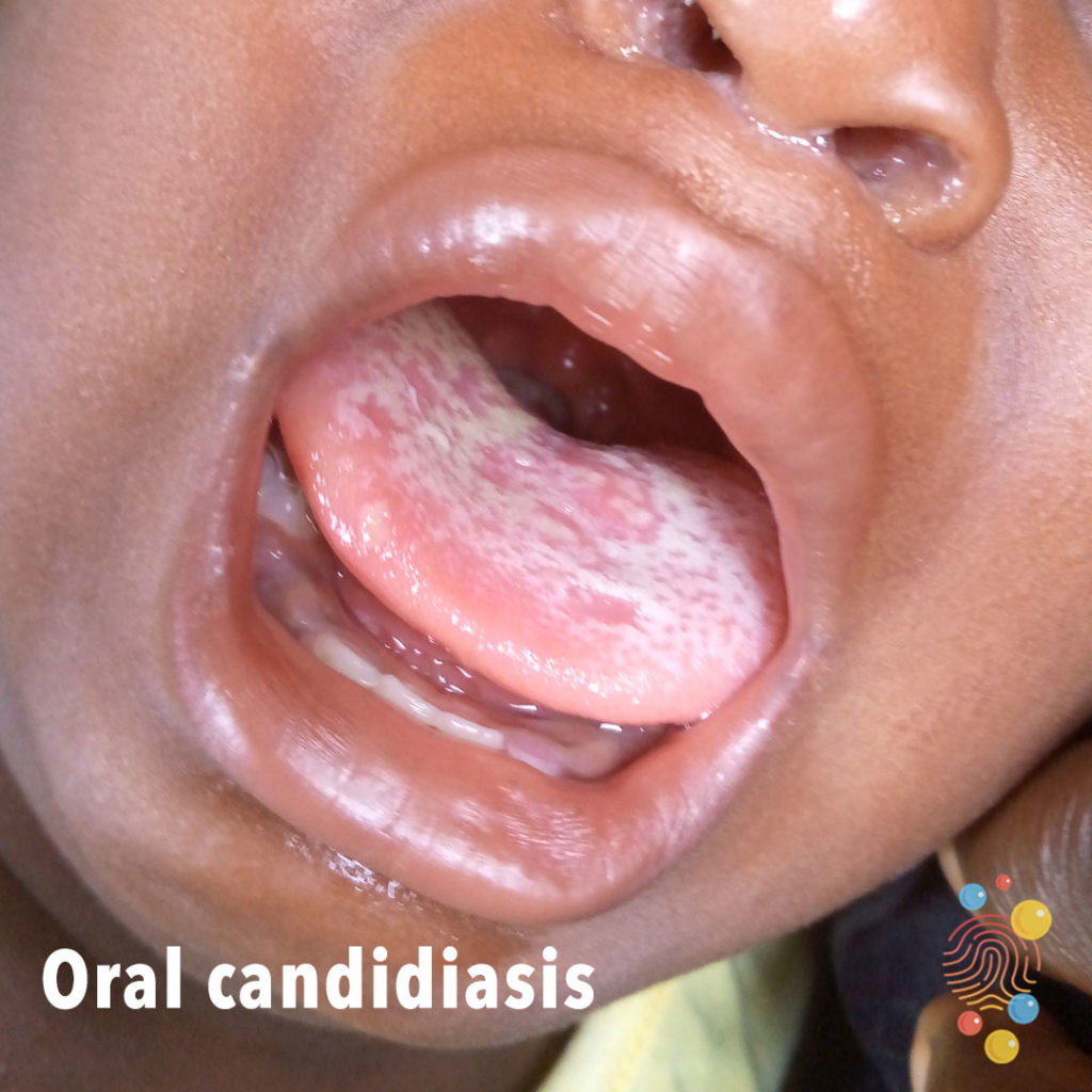
Oral Candidiasis
Learn more about neonatal thrush
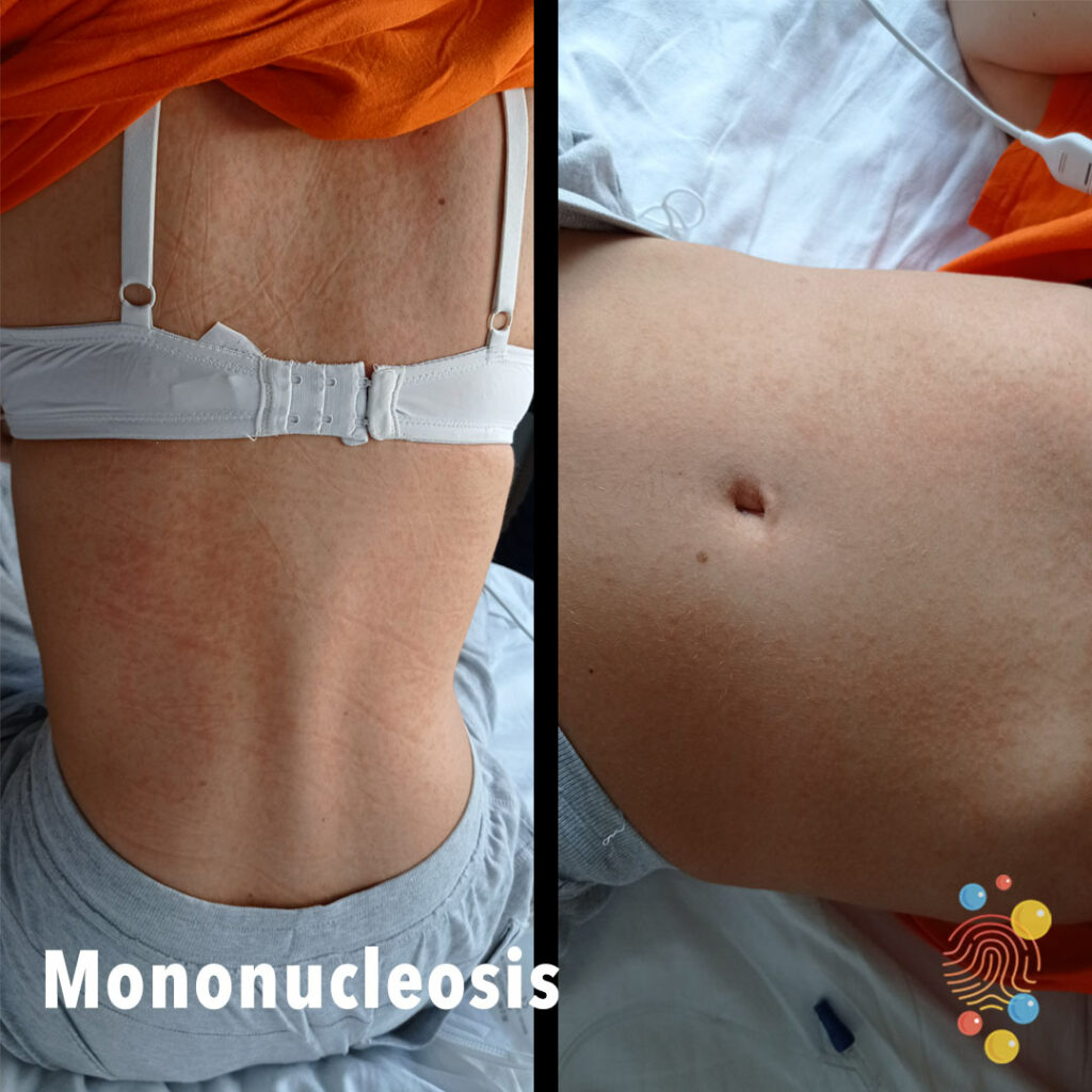
Mononucleosis
Learn more about infectious mononucleosis
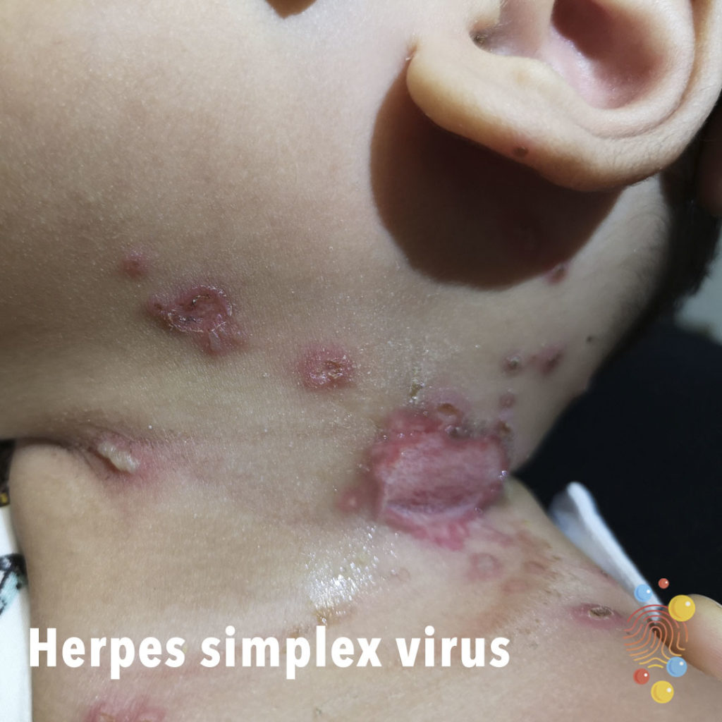
Herpes Simplex Virus
Learn more about herpes simplex virus
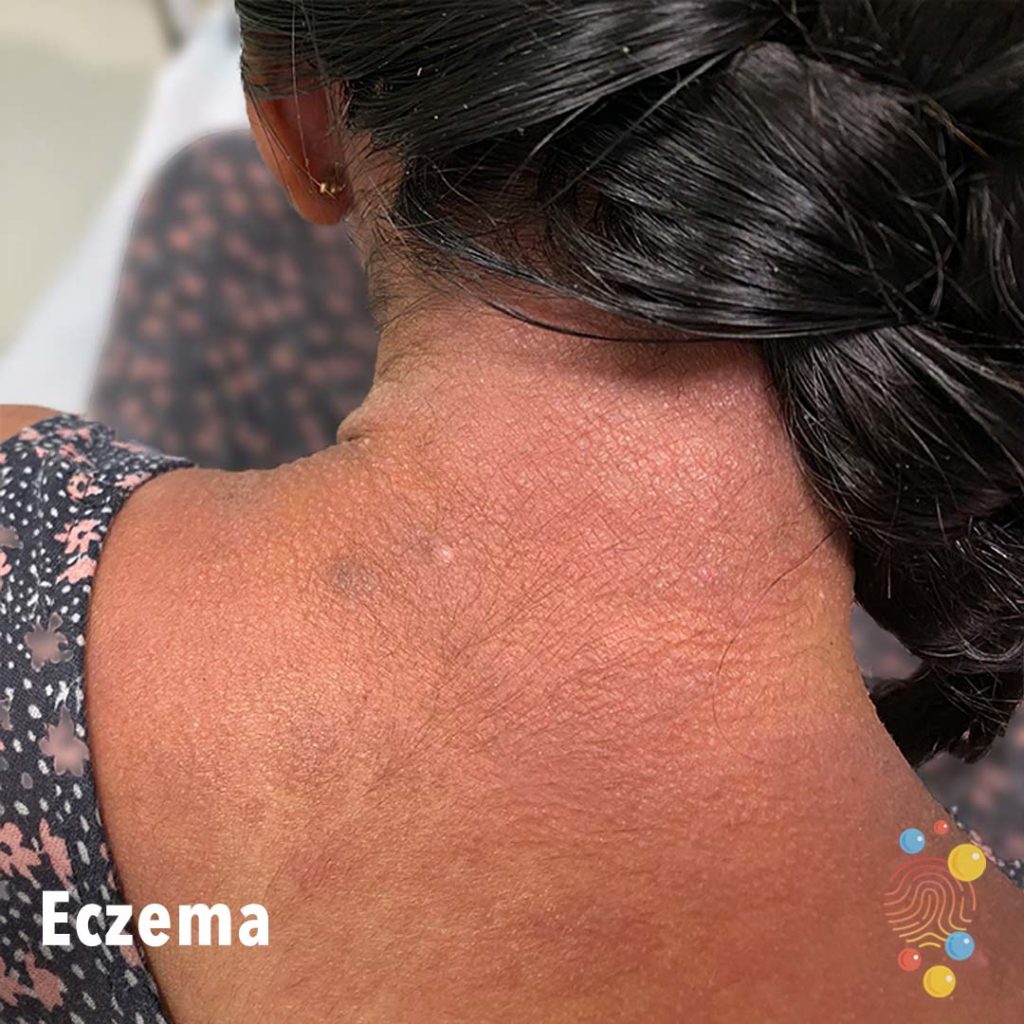
Eczema
Learn more about eczema

Hidradenitis Suppurativa
Learn more about hidradenitis suppurativa

Herpes Simplex Virus
Learn more about herpes simplex virus
PIMS-TS
Learn more about PIMS-TS
Leprosy
Henoch-Schonlein Purpura
Learn more about Henoch-Schonlein purpura
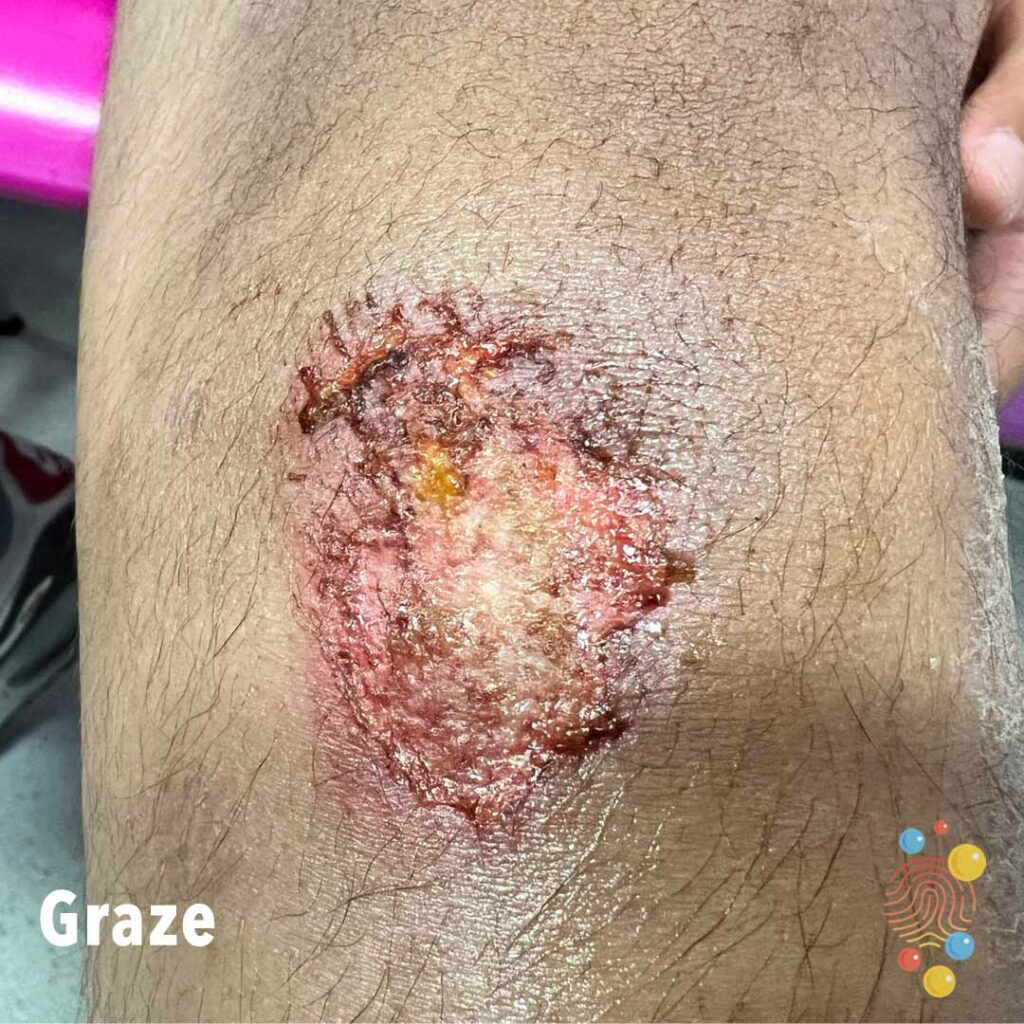
Grazed Knee
Grazed Knee – 13 year old boy
Dermal Melanocytosis
Learn more about dermal melanocytosis
Pyogenic Granuloma
Learn more about pyogenic granulomas
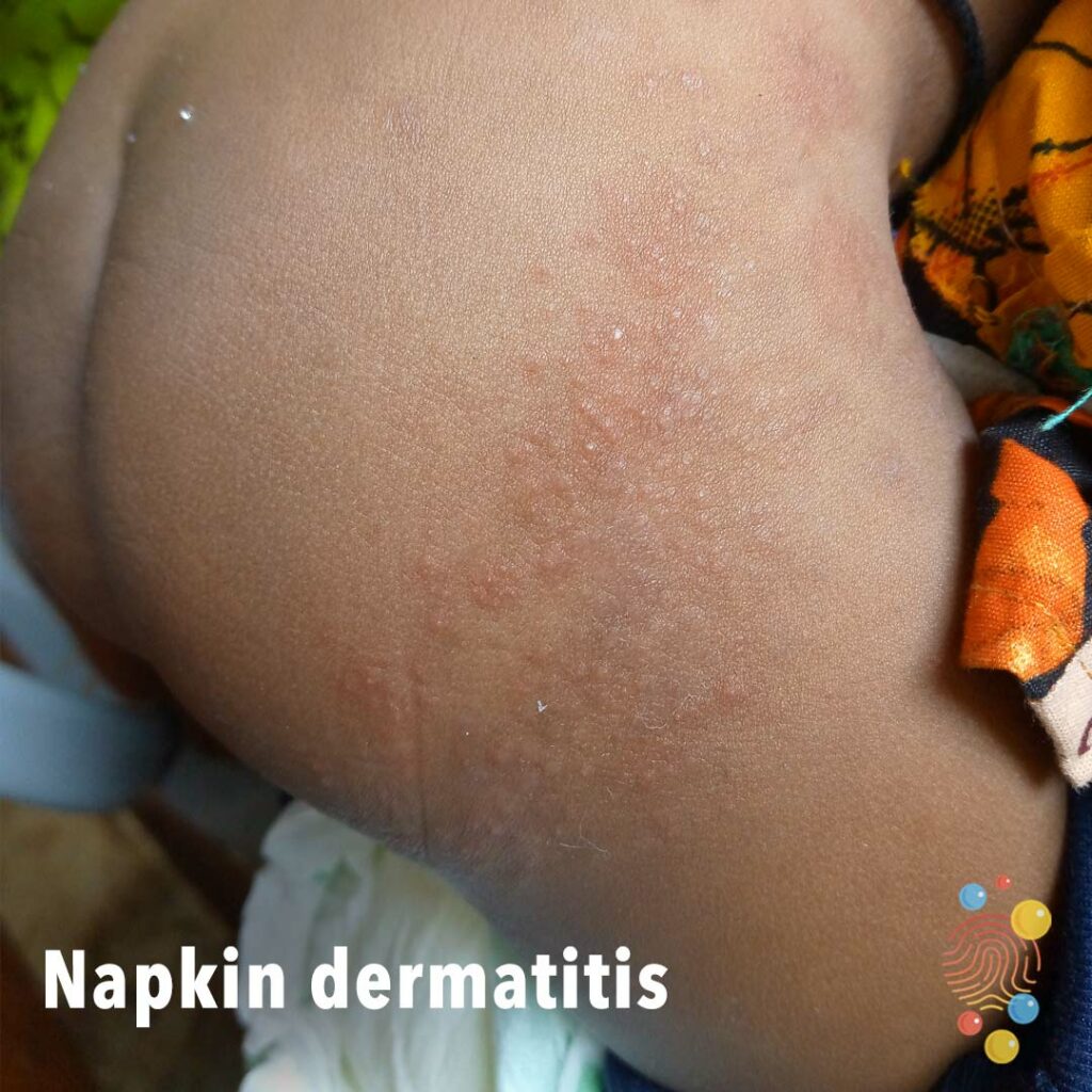
Napkin Dermatitis
Learn more about napkin dermatitis

Mantoux Blister
Learn more about the Mantoux test
Eczema
Learn more about eczema
Eczema Coxsackium

Eczema
Severe lichenified eczema with induration and impetiginisation
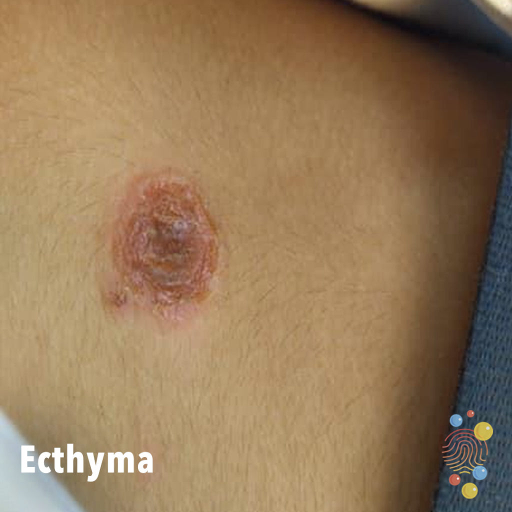
Ecthyma
Learn more about ecthymas
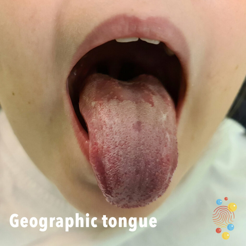
Geographic Tongue
Learn more about geographic tongue
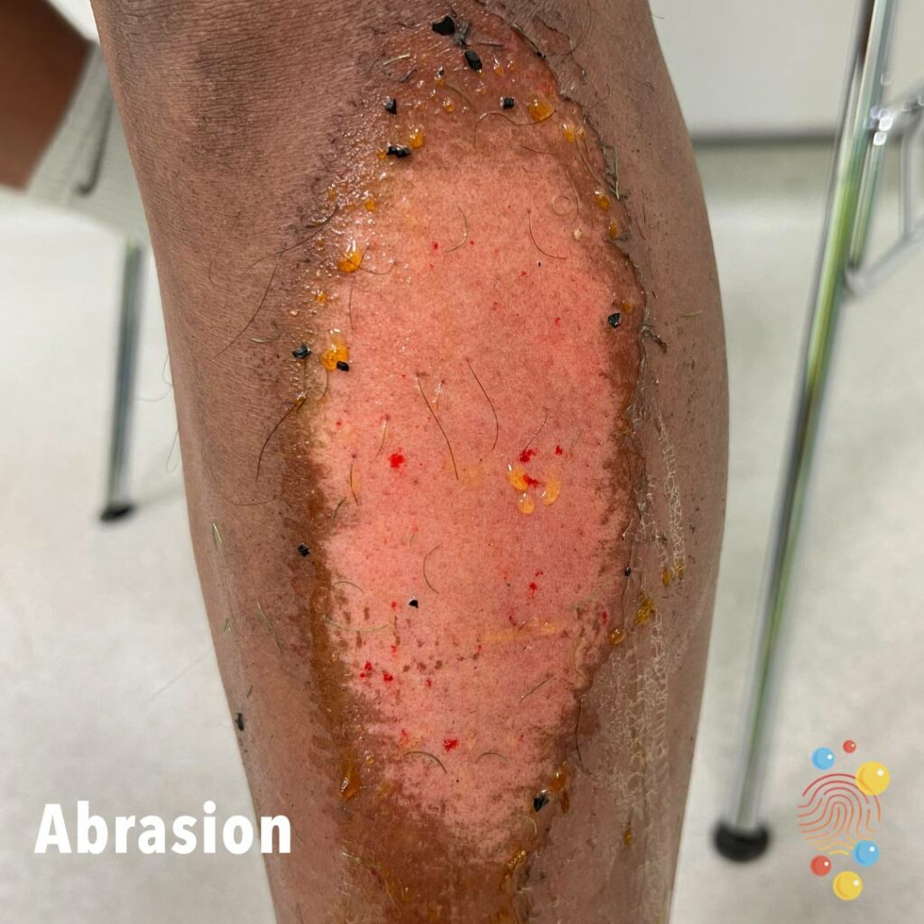
Abrasion
Abrasion to lower leg from AstroTurf – 17 year old male
Scarlet Fever
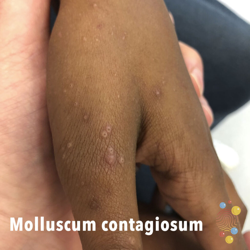
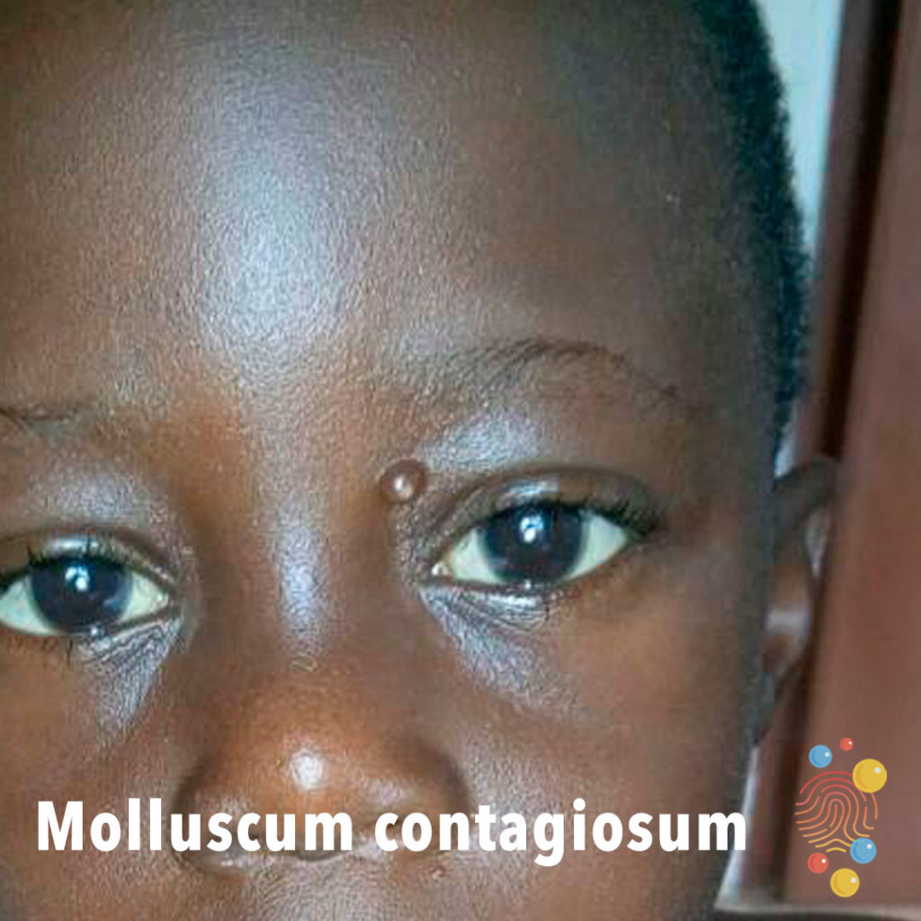
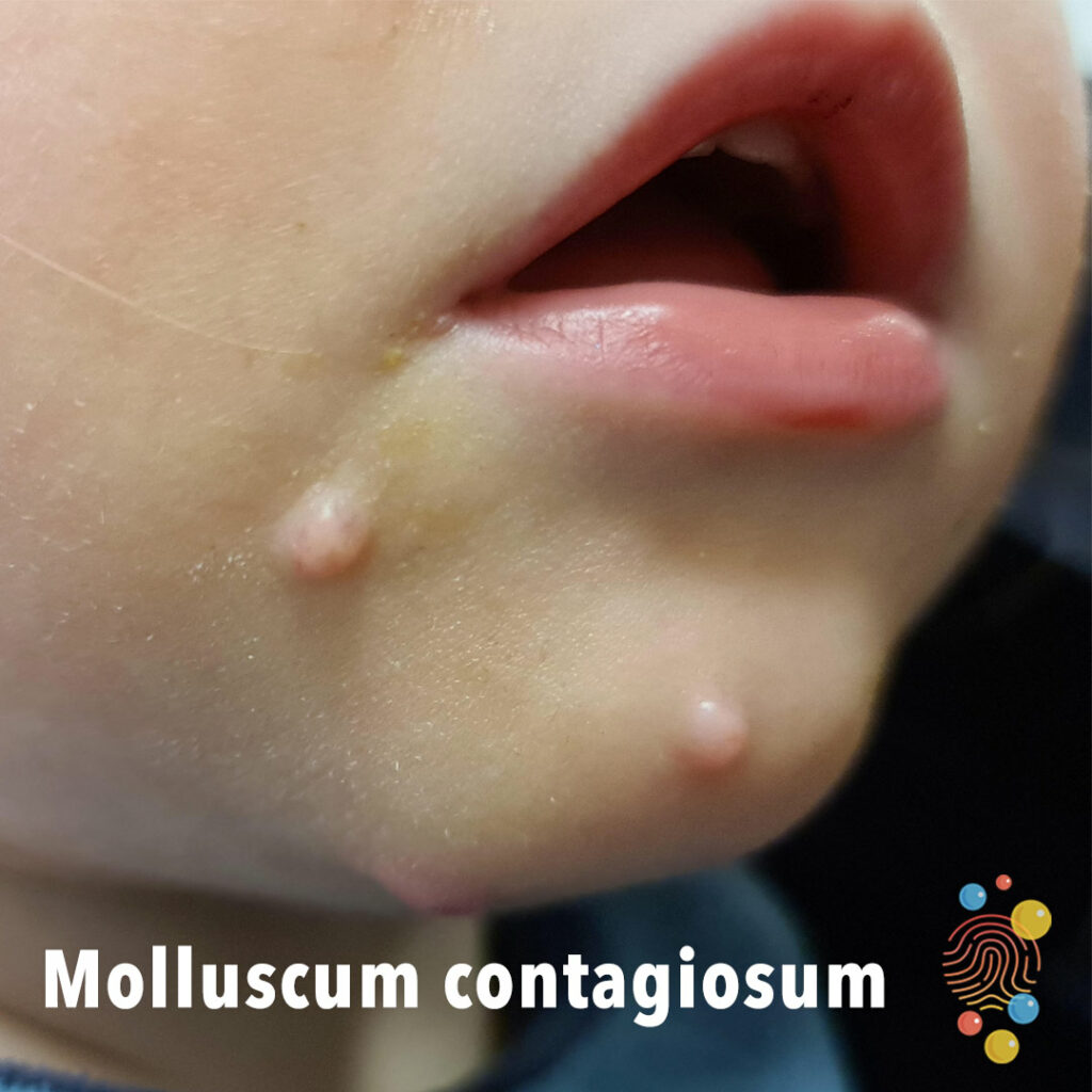
Molluscum Contagiosum
Learn more about molluscum contagiosum
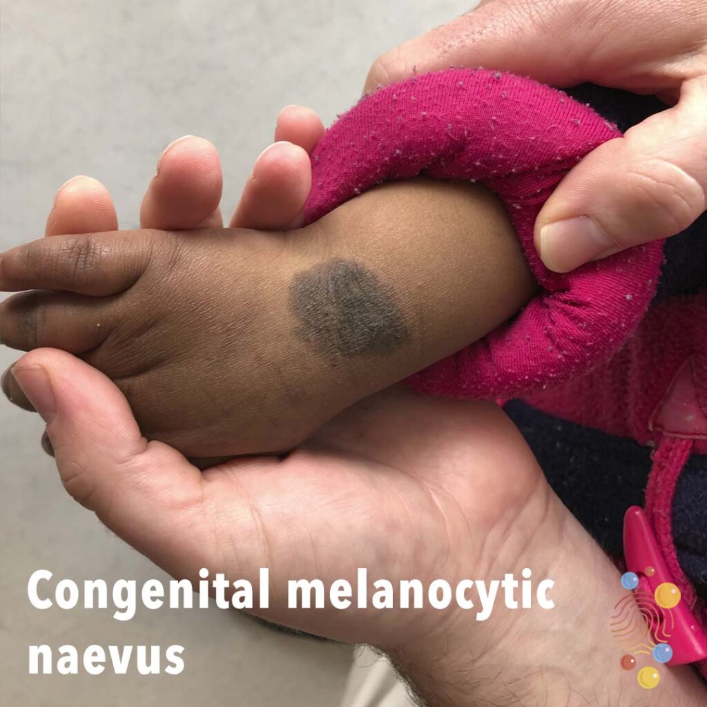
Congenital Melanocytic Naevus
Learn more about congenital melancytic naevi
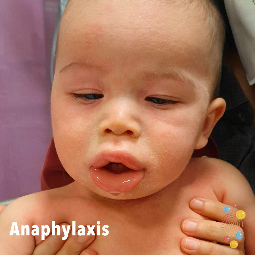
Anaphylaxis
Learn more about anaphylaxis

Hidradenitis Suppurativa
Learn more about hidradenitis suppurativa

Steven’s Johnson syndrome
Eczema
Learn more about eczema

Eczema
Severe lichenified eczema with induration and impetiginisation

Eczema
Learn more about eczema
Eczema Herpeticum
Learn more about eczema herpeticum
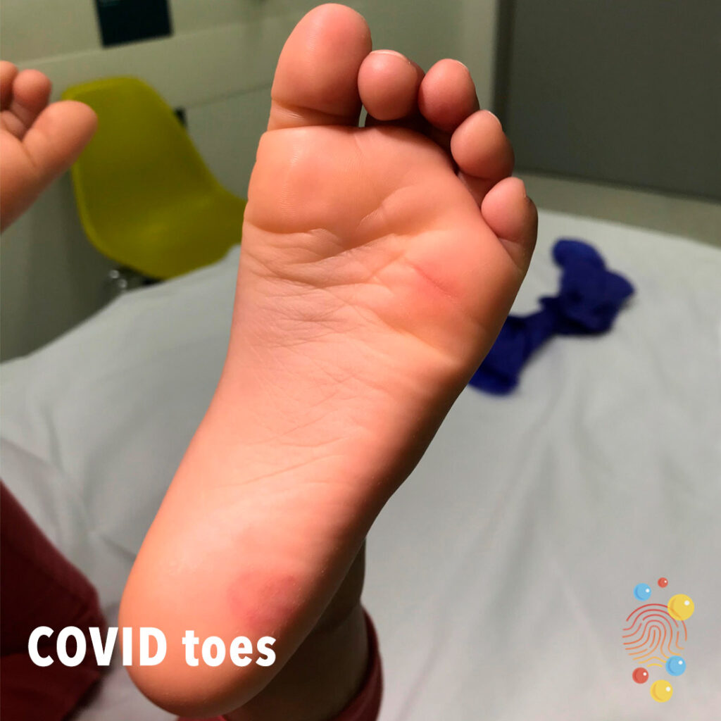
COVID toes
Learn more about COVID
Eczema
Severe erythema, lichenification, and bleeding of the lower limbs.
Eczema Herpeticum
Eczema herpeticum (EH) is a rare but severe skin infection that occurs when the human herpes simplex virus (HSV) infects inflamed skin
Eczema
Learn more about eczema

Bullous Impetigo
Multiple clustered erosions with central ulceration on the back
Staphylococcal Infection
Learn more about staphylococcal infection
Pustular psoriasis
Learn more about psoriasis
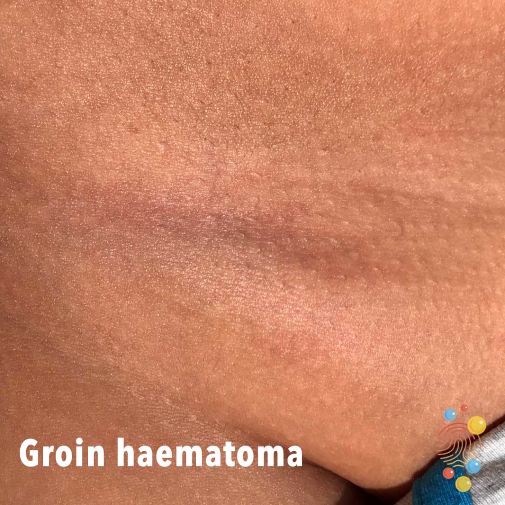
Groin Haematoma
Non blanching patch of erythema.
Eczema
Learn more about eczema
Molluscum Contagiosum
Learn more about molluscum contagiosum
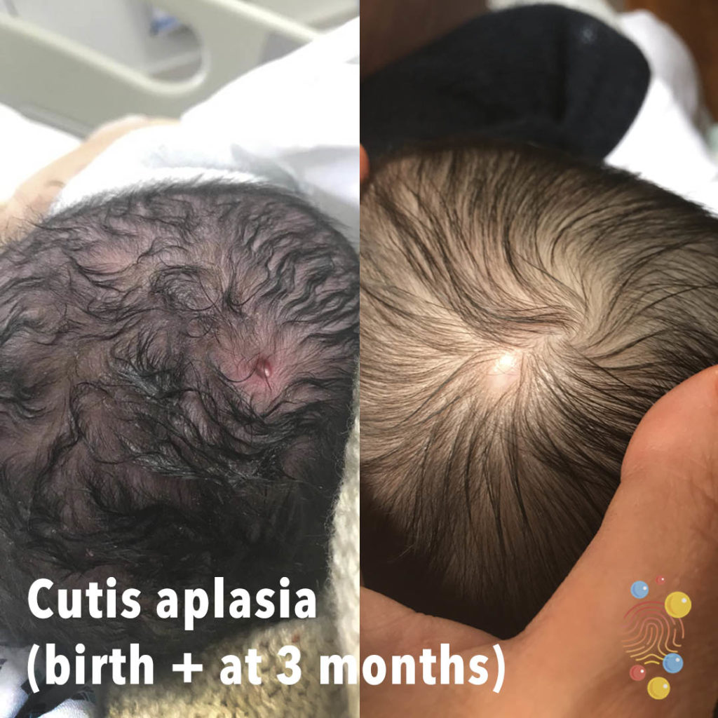
Cutis Aplasia
Learn more about cutis aplasia
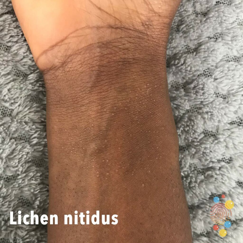
Lichen Nitidus
Learn more about lichen nitidus
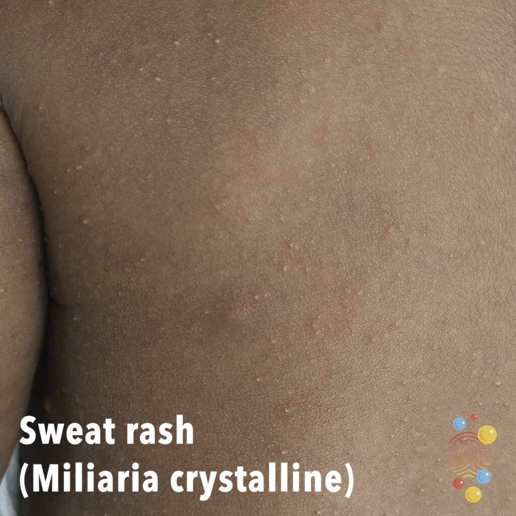
Sweat Rash (Miliaria Crystalline)
Learn more about miliaria
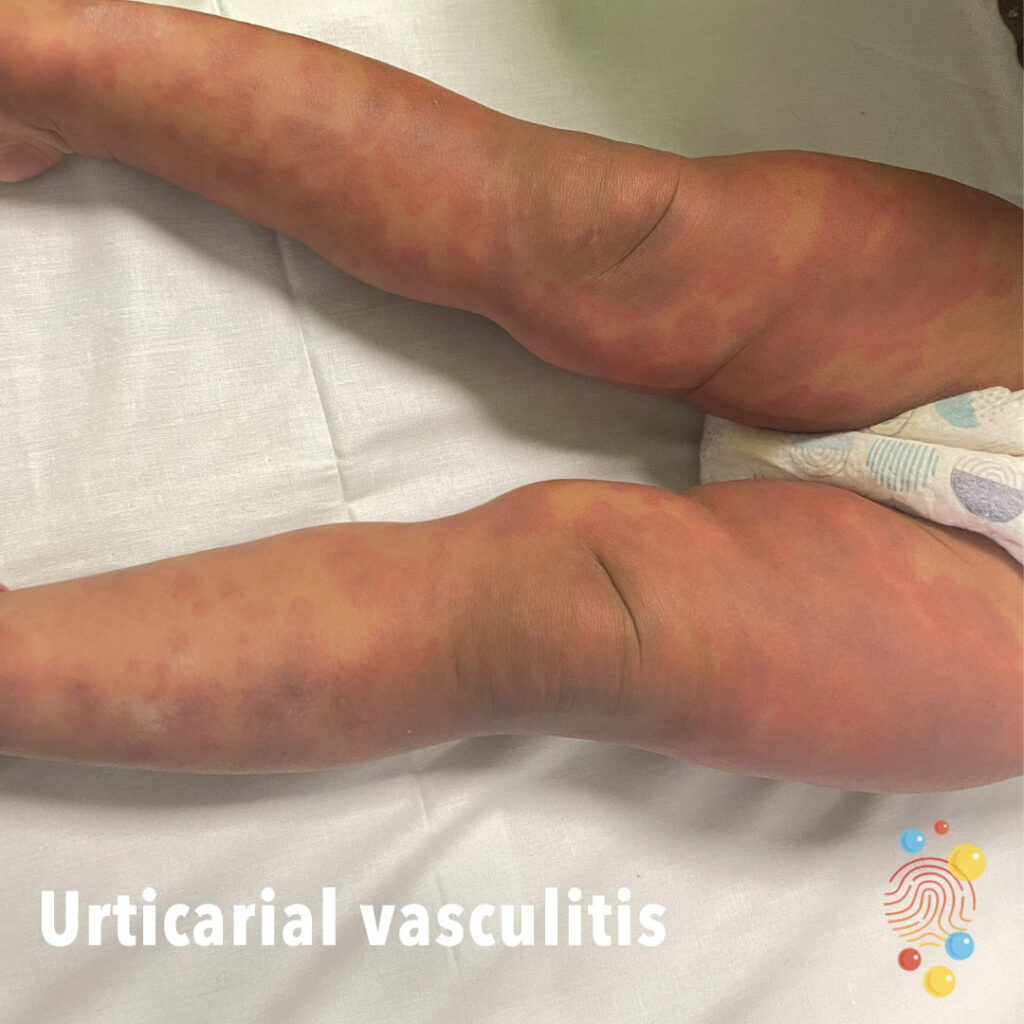
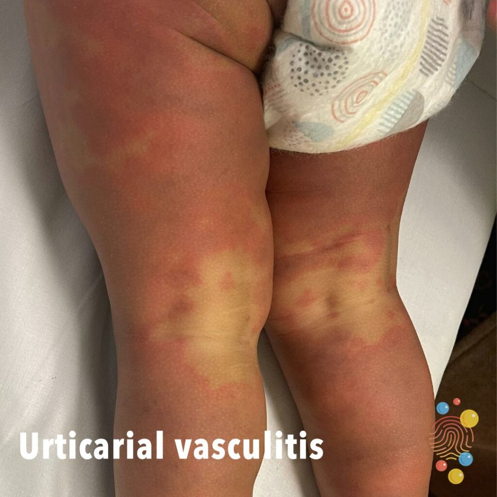
Urticarial Vasculitis
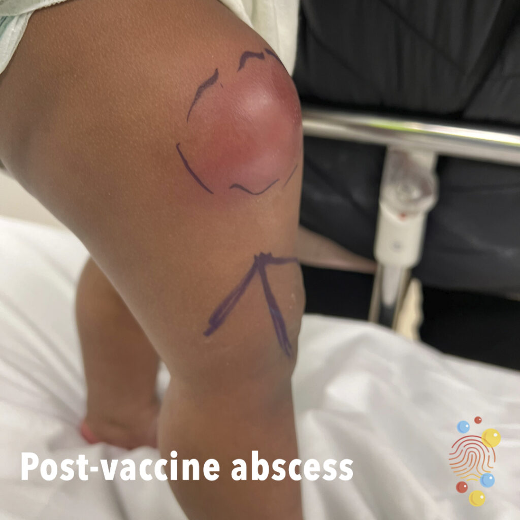
Post Vaccine Abscess
Thigh abscess post men c vaccine

Cephalhaematoma
Learn more about cephalhaematoma
Urticaria

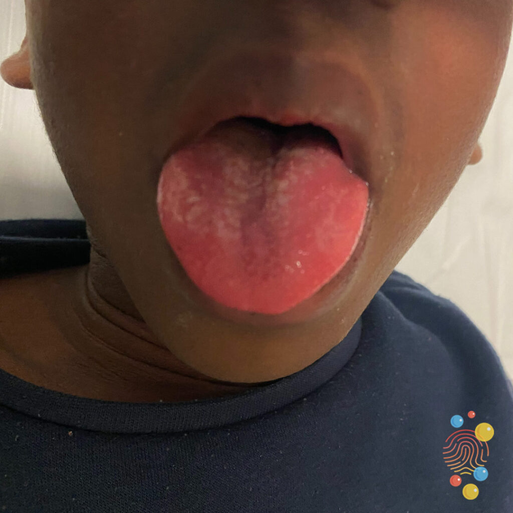
Strawberry Tongue
Learn more about strawberry tongues
Bullous Impetigo
Bullous impetigo is a bacterial skin infection that causes large, fluid-filled blisters to appear on the body
Paronychia
Paronychia (pahr-uh-NIK-ee-uh) is an infection of the skin around a fingernail or toenail.
Dermal Melanocytosis
Learn more about dermal melanocytosis

Head Injury

PIMS-TS
Learn more about PIMS-TS
Blue Sclerae In Osteogenesis Imperfecta
Learn more about blue sclerae
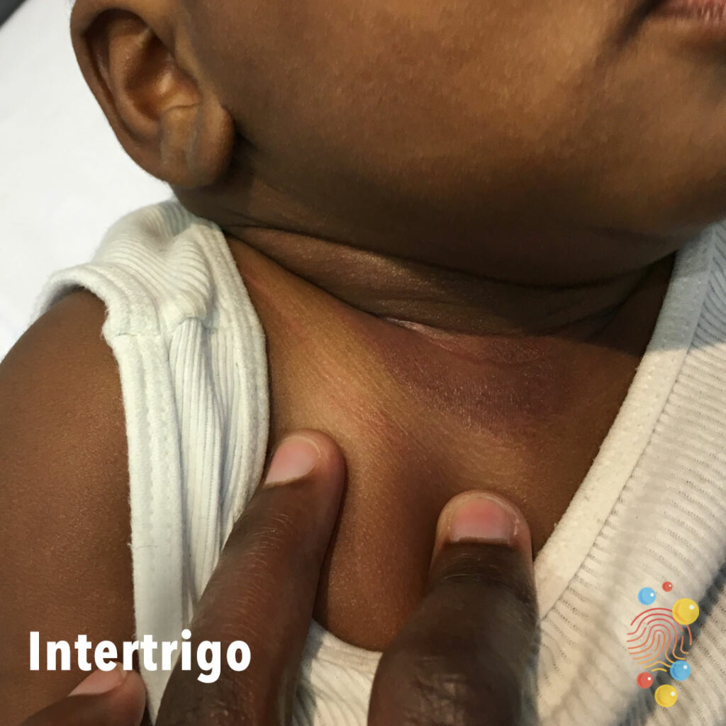
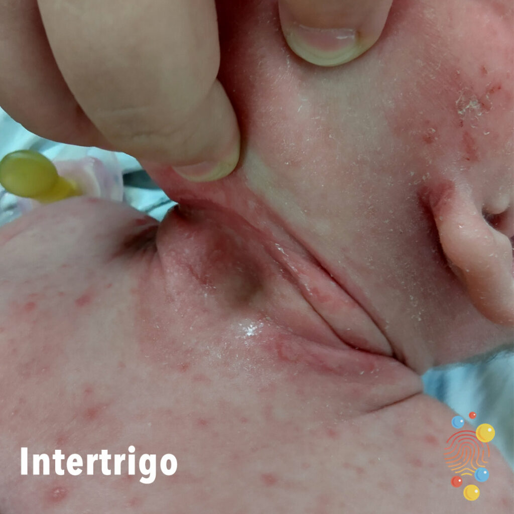
Intertrigo
Learn more about intertrigo

Burn – Pre & Post Deroofing

Hypopigmentation
Learn more about hypopigmentation
Eczema Herpeticum
Learn more about eczema herpeticum

Strawberry Tongue
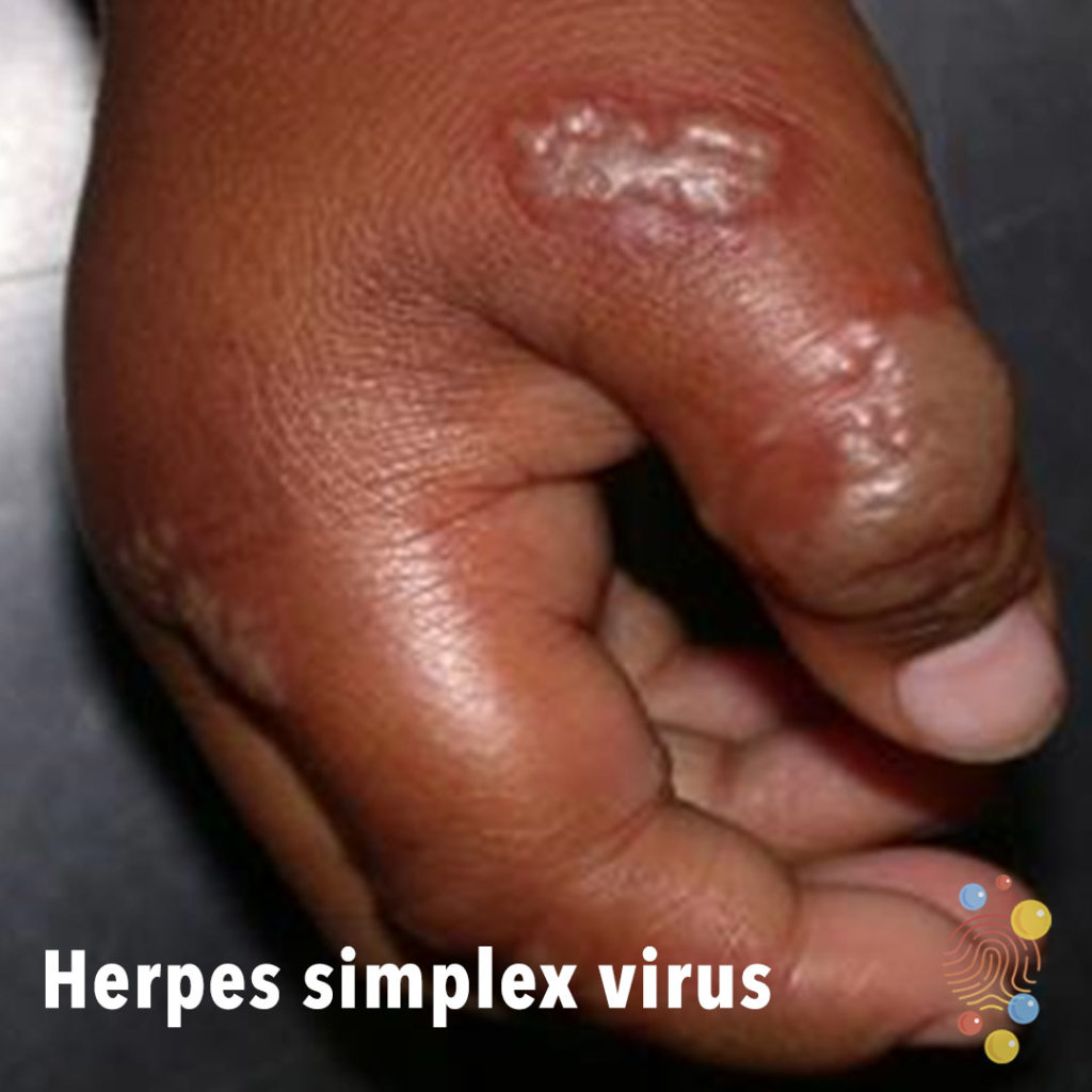
Herpes Simplex Virus
Learn more about herpes simplex virus
Folliculitis
Widespread follicular rash upper chest, with papules and some small pustules.
Learn more about folliculitis

Eczema Herpeticum
Urticaria
Learn more about urticaria

Becker’s Naevus
Learn more about beckers naevus

Abscess
Learn more about abscesses
Eczema herpeticum
Learn more about eczema herpeticum
Umbilical Hernia
Learn more about umbilical hernia
Eczema
Learn more about eczema
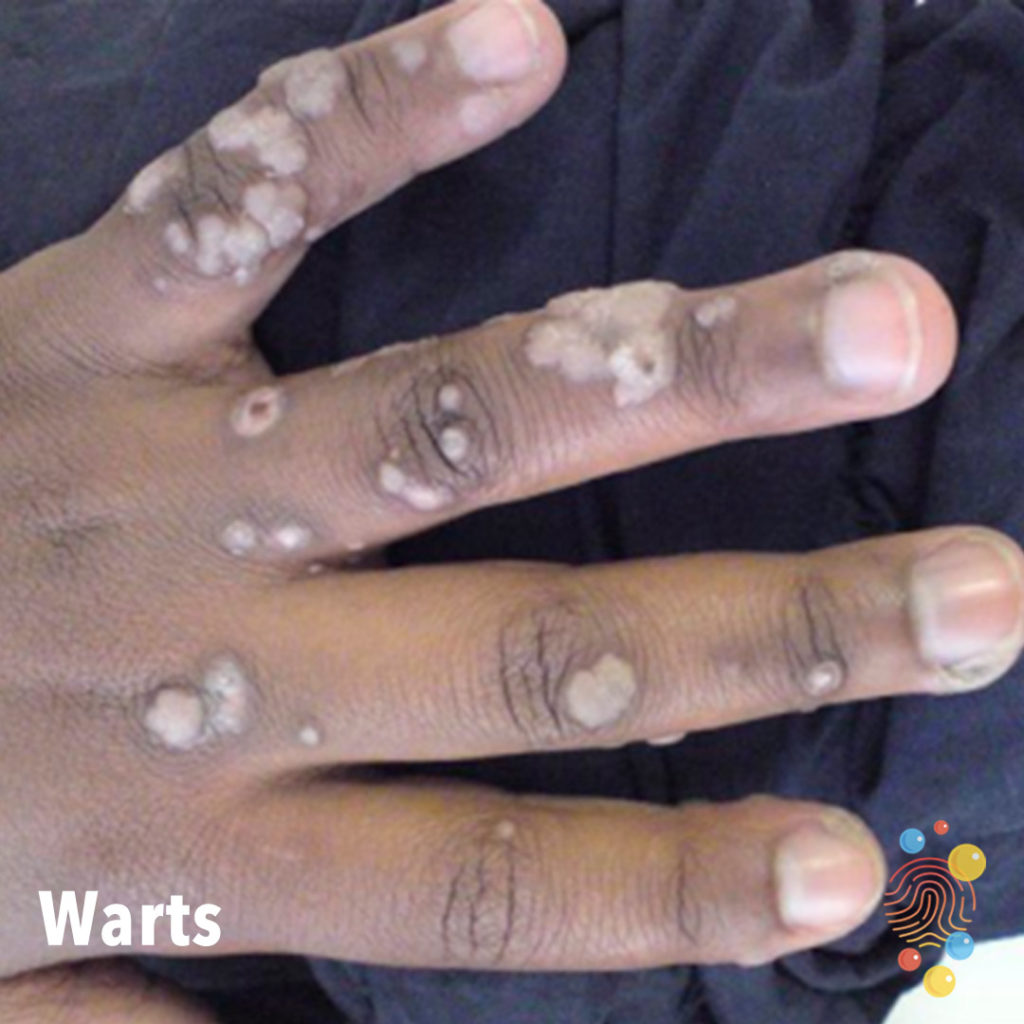
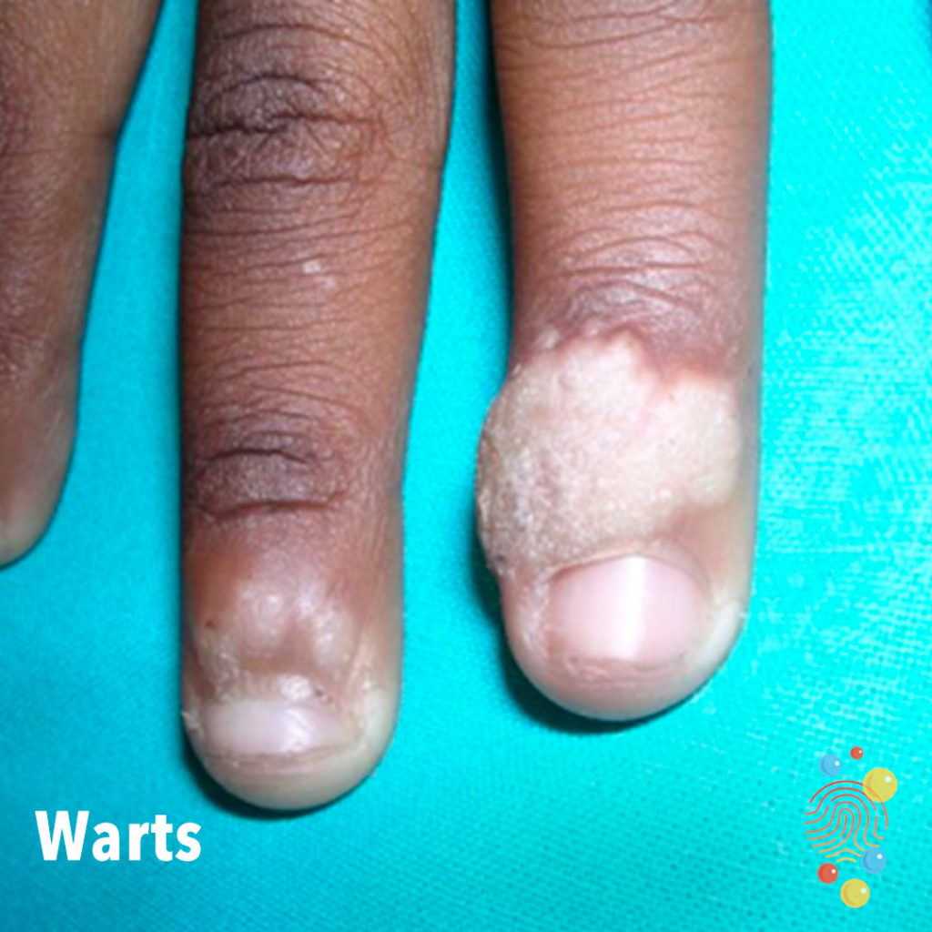
Warts
Learn more about warts
Urticaria
Learn more about urticaria

Bullous Impetigo
Extensive healing erosions with haemorrhagic crust and a collarette of scale
Epidermal Naevus
Learn more about epidermal naevus
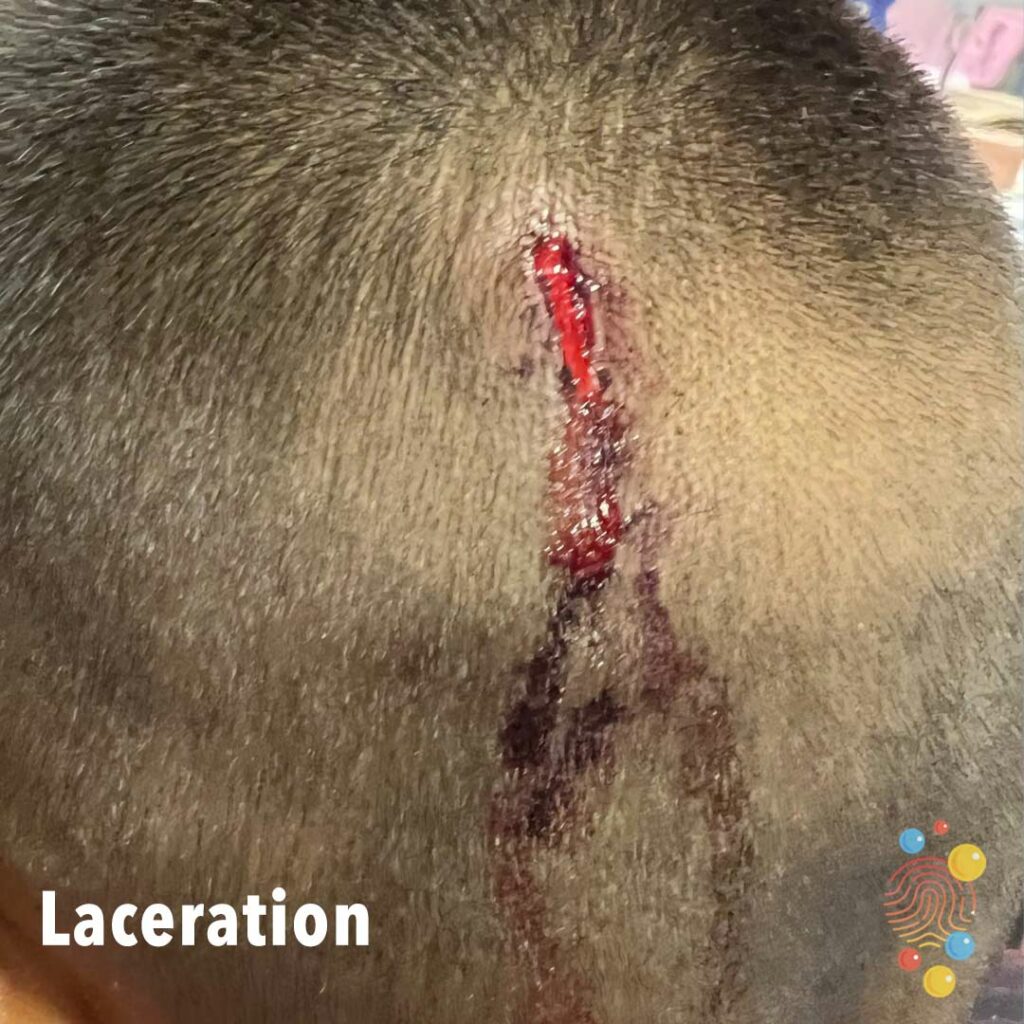
Laceration
Head Laceration

Shingles
Shingles, also known as herpes zoster or zona, is a viral disease characterized by a painful skin rash with blisters in a localized area. Typically the rash occurs in a single, wide mark either on the left or right side of the body or face.
Herpes Simplex Virus
Learn more about herpes simplex virus
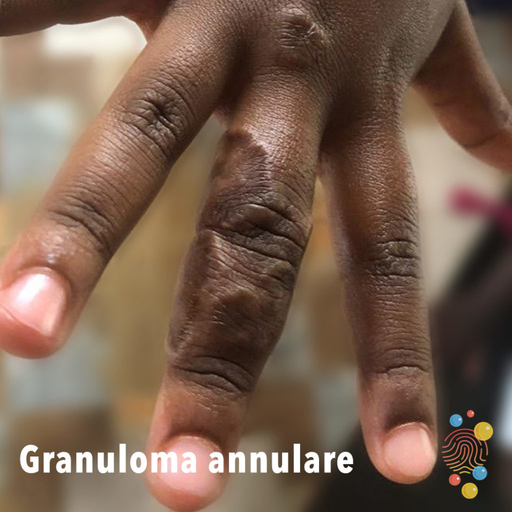
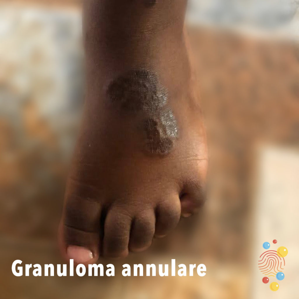
Granuloma Annulare
Learn more about granuloma annulare
Café-Au-Lait Macule
Learn more about café-au-lait macules

Stomatitis
Stomatitis in child with bilateral pneumonia, urticaria rash and cardiovascular instability requiring >40ml/kg fluid + inotropes.
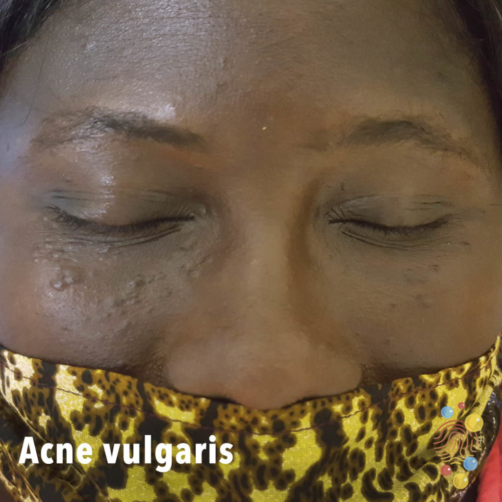
Acne Vulgaris
Learn more about acne vulgaris
Discoid Eczema
Learn more about eczema
Bullous Impetigo
Bullous impetigo is a bacterial skin infection that causes large, fluid-filled blisters called bullae
Impetigo
Learn more about bullous impetigo

BCG Abscess
Learn more about BCGs

Folliculitis
Learn more about folliculitis
Urticarial Vasculitis
Peri-Orbital Cellulitis
Eczema
Learn more about eczema
Urticaria
Learn more about urticaria
Eczema
Learn more about eczema
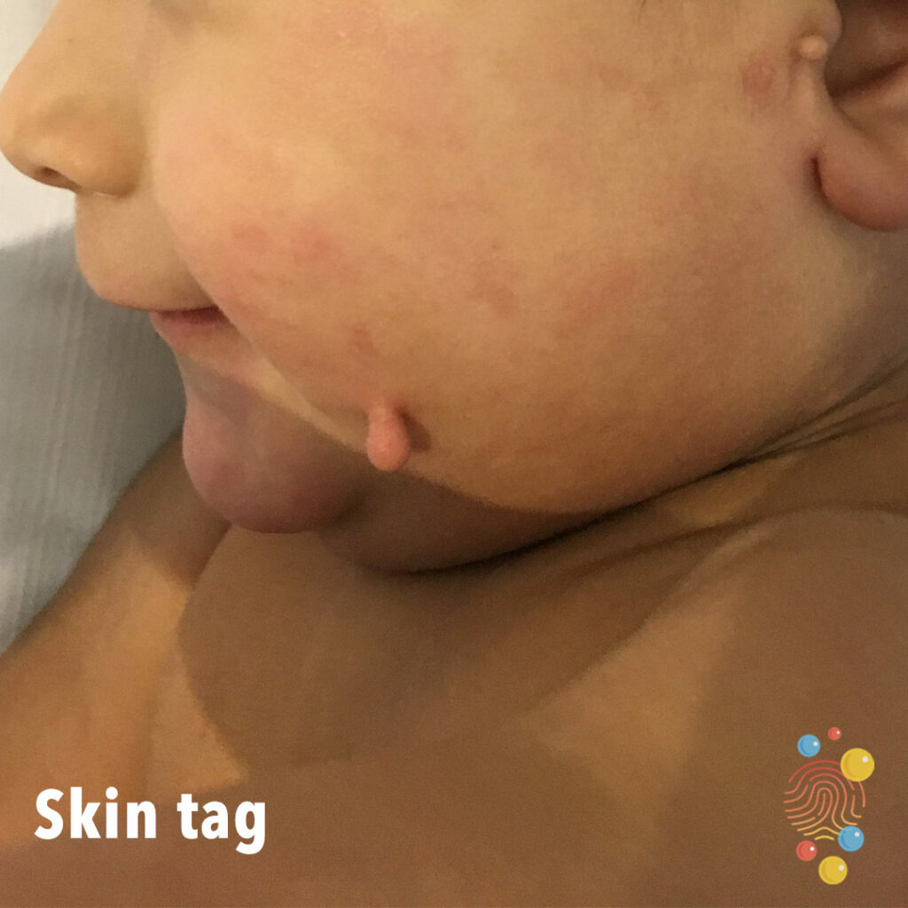
Skin Tag
Learn more about skin tags
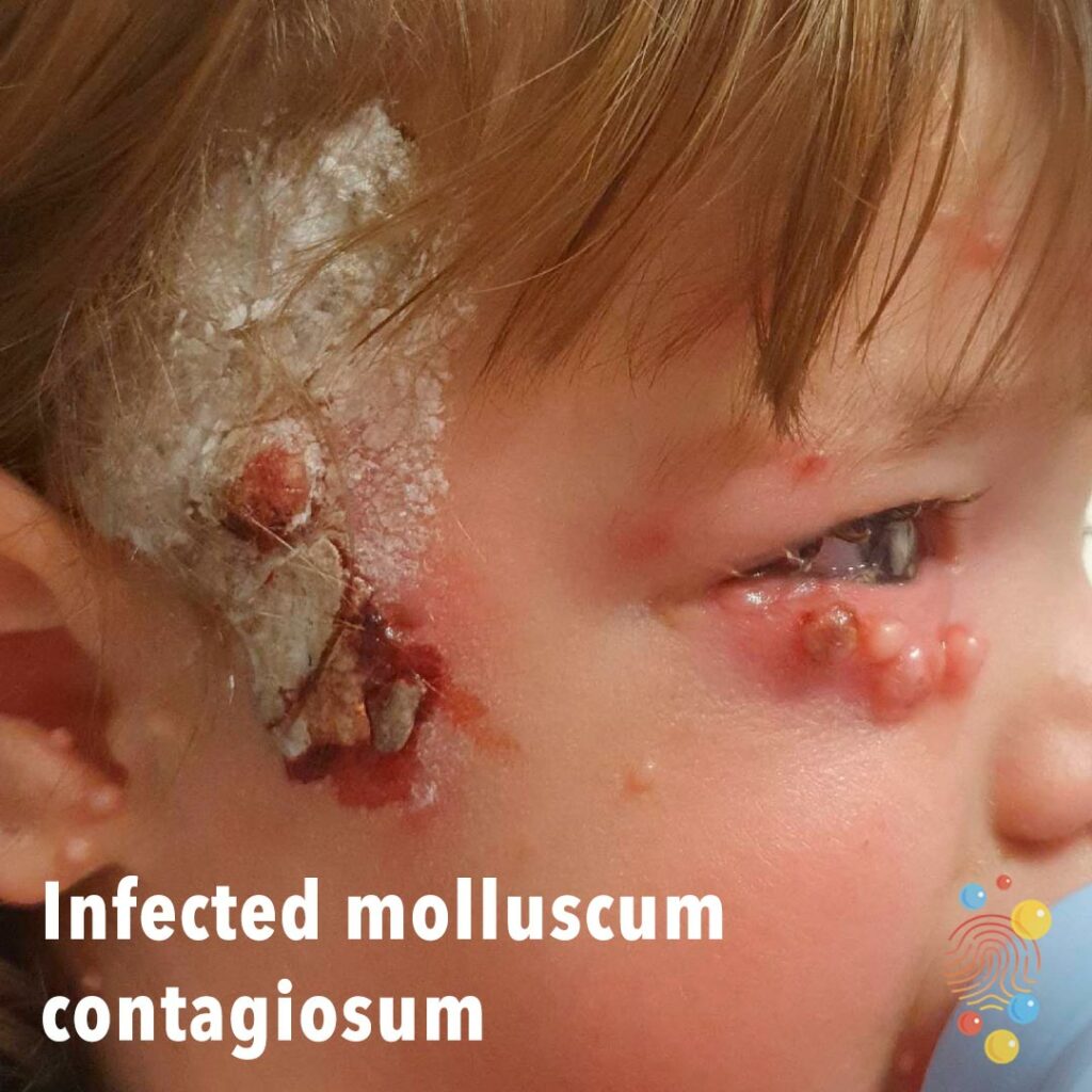
Infected Molluscum Contagiosum
Learn more about molluscum contagiosum

Miliaria
Learn more about miliaria
Infantile haemangioma
Superficial infantile haemangioma on the anterior neck.
Abscess
Learn more about abscesses
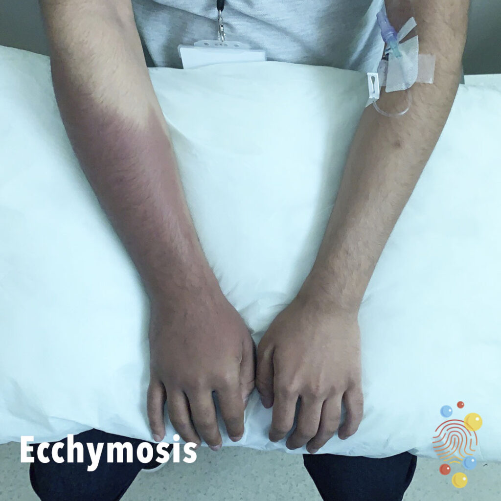
Ecchymosis
Learn more about ecchymosis

Gianotti Crosti
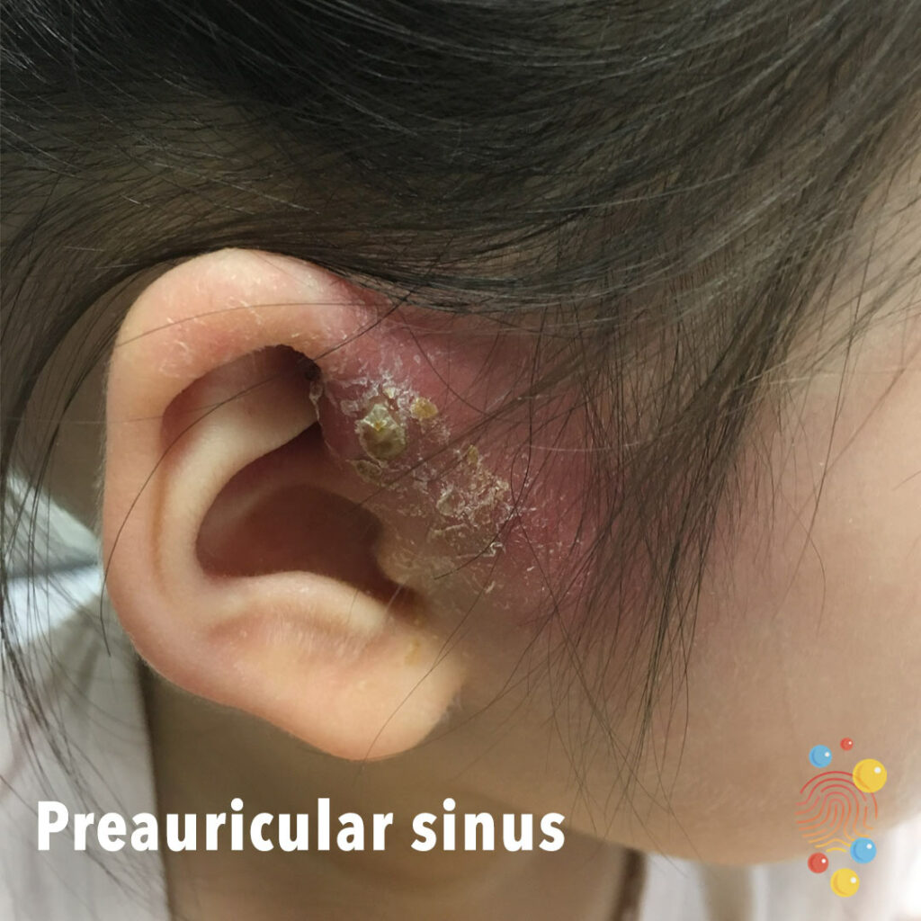
Pre-Auricular Sinus
Learn more about sinuses

Parvovirus
Bright red rash in symmetrical distribution on cheeks
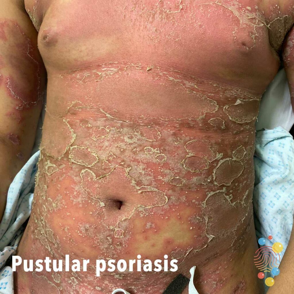
Pustular psoriasis
Learn more about psoriasis
Follicular eczema
Learn more about eczema
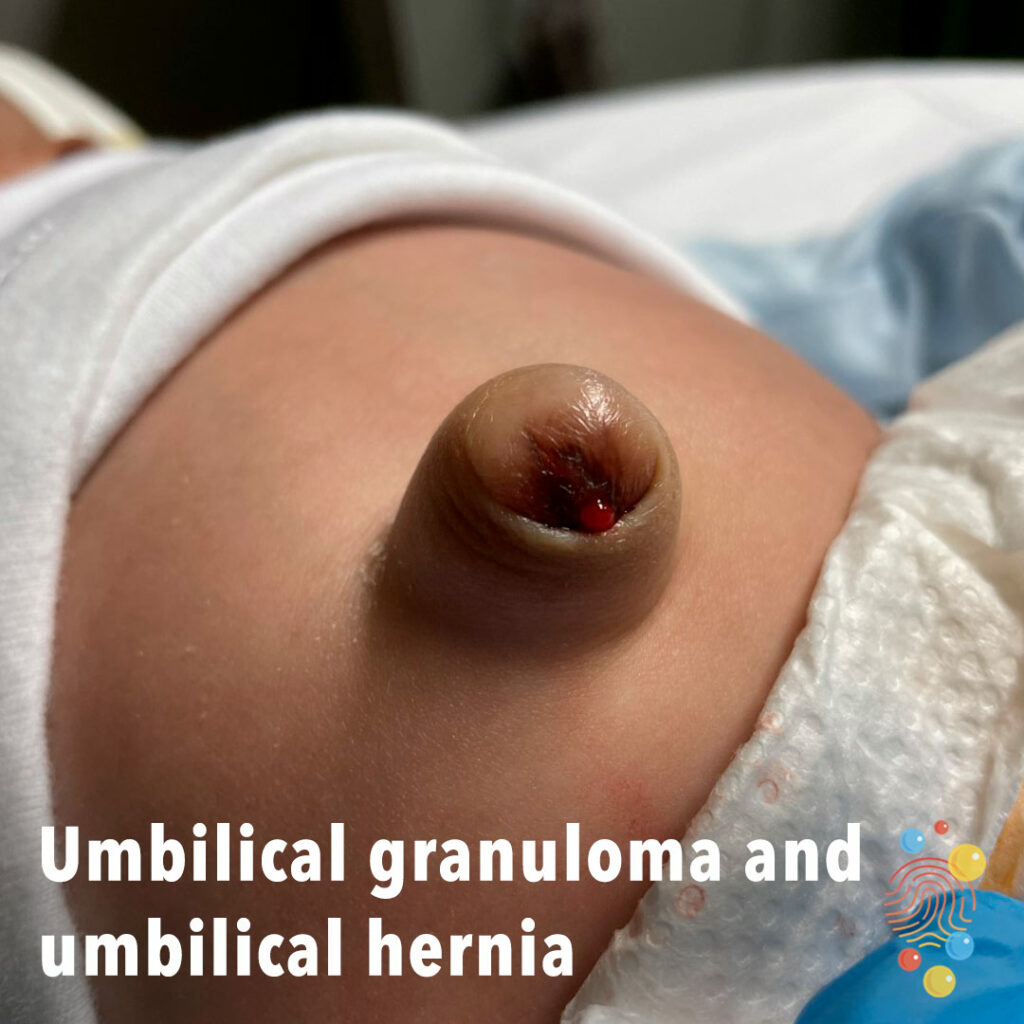
Umbilical Granuloma And Umbilical Hernia
Learn more about umbilical hernias

Bullous Impetigo
Extensive healing erosions with haemorrhagic crust and a collarette of scale
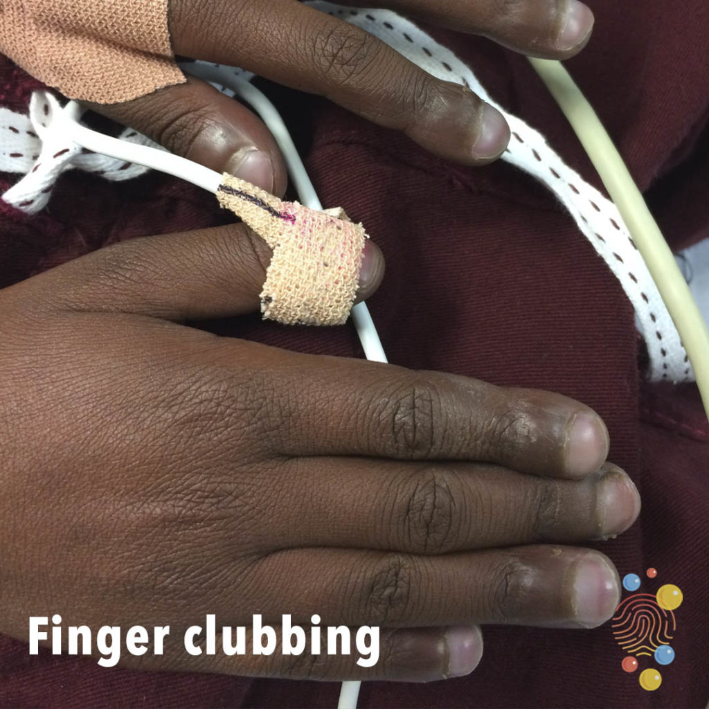
Finger Clubbing
Learn more about clubbing
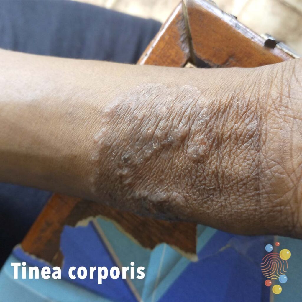
Tinea Corporis
Learn more about tinea corporis
Eczema

Epidermoid Cyst
Learn more about epidermoid cysts

Tracking Cellulitis
Tracking cellulitis is a term used to describe when a skin infection spreads, or “tracks,” from the initial area of infection. Cellulitis is a bacterial infection that occurs when bacteria enters the skin through a break, such as an injury or insect bite. It often affects the lower legs but can also occur on the arms, face, and other areas.

Hand, foot & mouth
Learn more about hand, foot and mouth

Pityriasis Alba
Learn more about pityriasis alba

Jellyfish sting
Learn more about bites
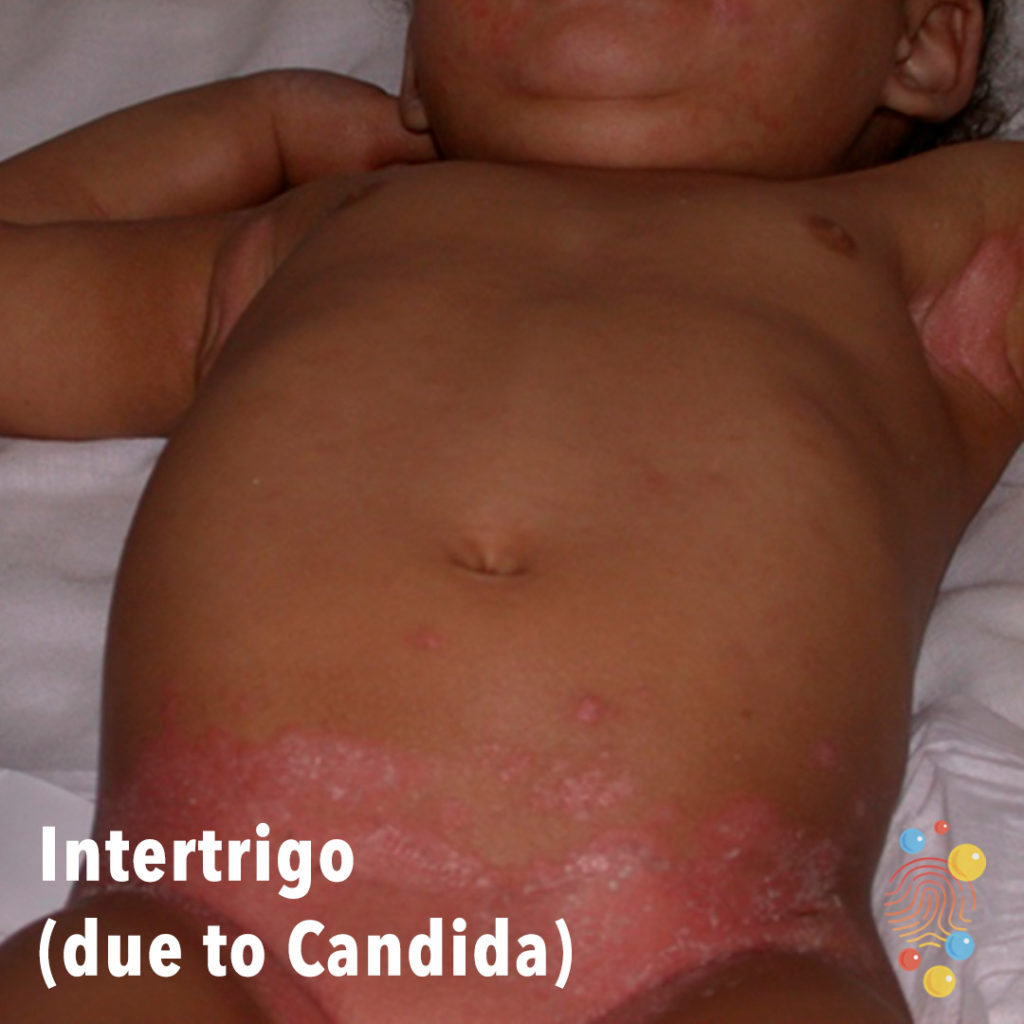
Intertrigo (Due To Candida)
Learn more about intertrigo

Ezcema
Learn more about eczema

Measles
Learn more about measles

Impetigo
Eczema
Learn more about eczema

Herpes Stomatitis
Vesiculopustular eruption of lips with crust and ulceration.
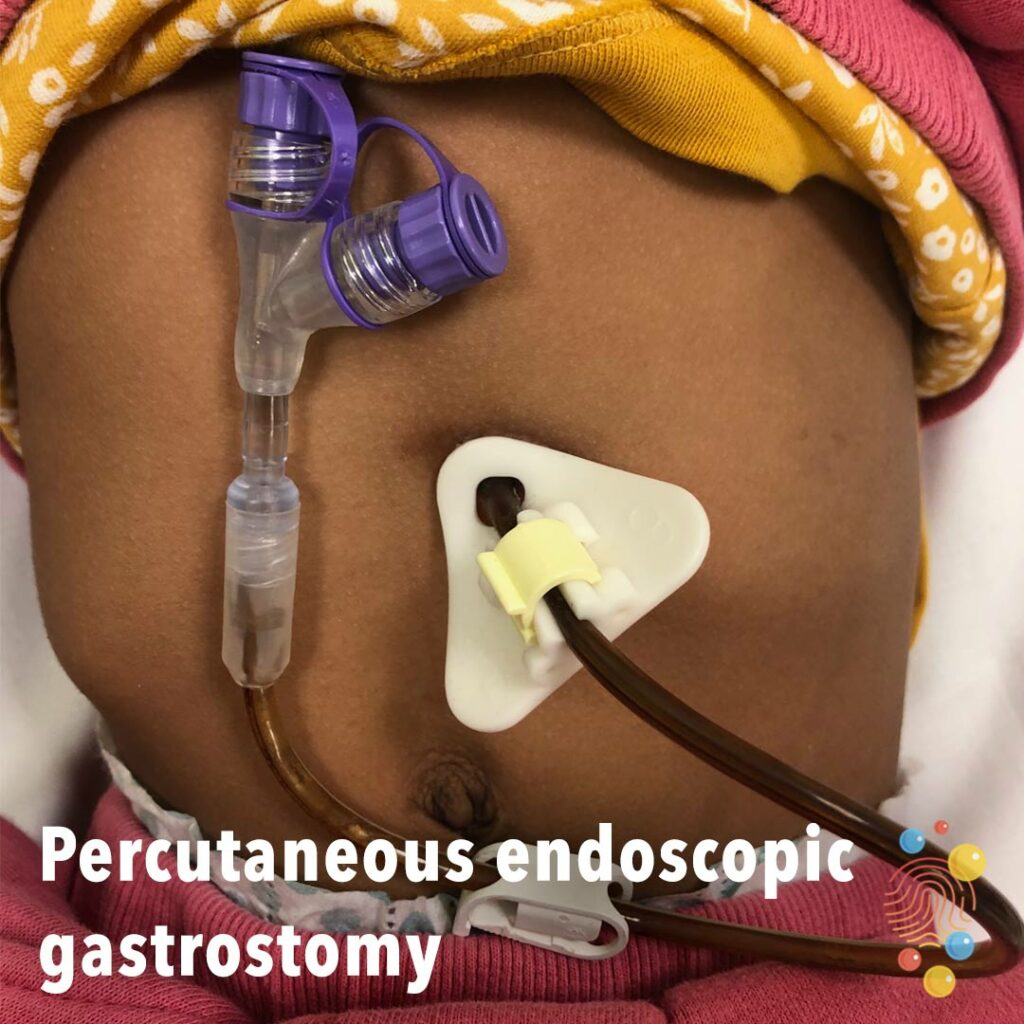
Percutaneous Endoscopic Gastrostomy
Learn more about gastrostomies
Infected Eczema
Learn more about eczema

Erythema Associated With Scombroid Poisoning
Learn more about scombroid poisoning

Impetigo
Learn more about bullous impetigo
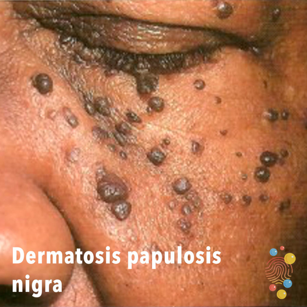
Dermatosis Papulosis Nigra
Learn more about dermatosis papulosis nigra
Paronychia
Paronychia (pahr-uh-NIK-ee-uh) is an infection of the skin around a fingernail or toenail.
Ichthyosis
Learn more about ichthyosis
Seborrhoeic Dermatitis
Learn more about seborrhoeic dermatitis
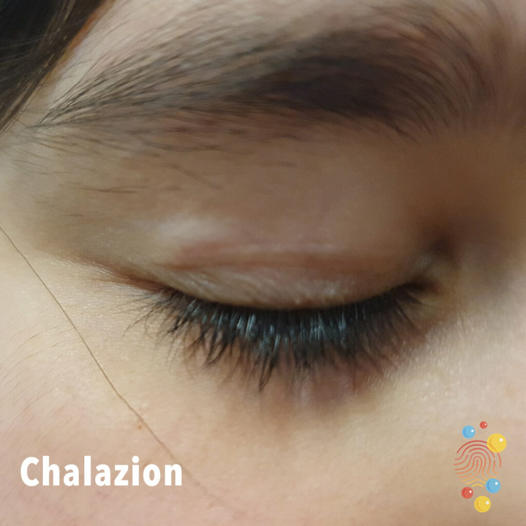
Chalazion
Eczema
Learn more about eczema
Reaction To A Bite
Learn more about bites

Periorbital Cellulitis
Learn more about periorbital cellulitis
Abscess
Learn more about abscesses

Normal umbilical cord
4 day baby with normal dry cord

Mouth Injury

Steven’s Johnson syndrome
Stevens–Johnson syndrome is a type of severe skin reaction. Together with toxic epidermal necrolysis and Stevens–Johnson/toxic epidermal necrolysis overlap, they are considered febrile mucocutaneous drug reactions and probably part of the same spectrum of disease, with SJS being less severe.

Avulsed Nail
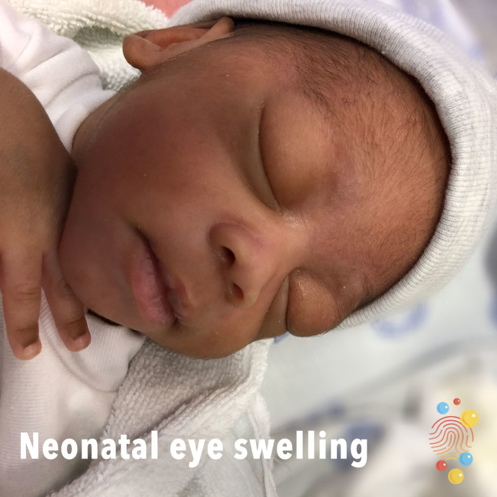
Neonatal Eye Swelling
Bilateral eye swelling.
Scabies
Learn more about scabies

Acute haemorrhagic oedema of infancy
Multiple urticated bruises, some of which have a targetoid appearance
Eczema
Learn more about eczema
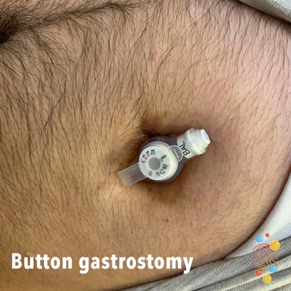
Button gastrostomy
Learn more about gastrostomies
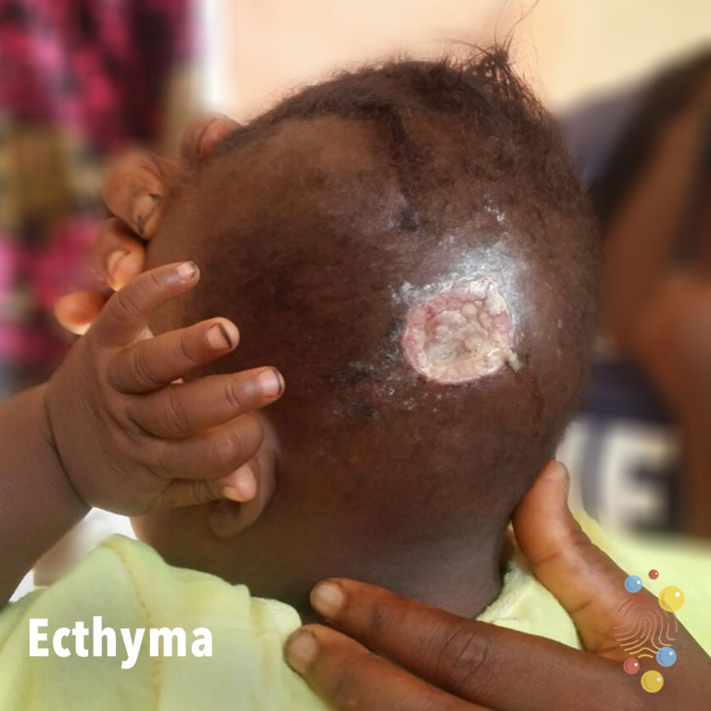
Ecthyma
Learn more about ecthymas
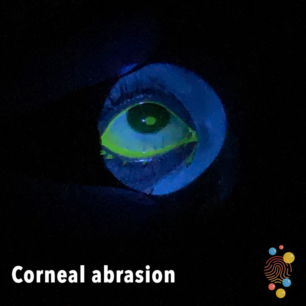
Corneal Abrasion
Learn more about corneal abrasions

Strawberry Tongue
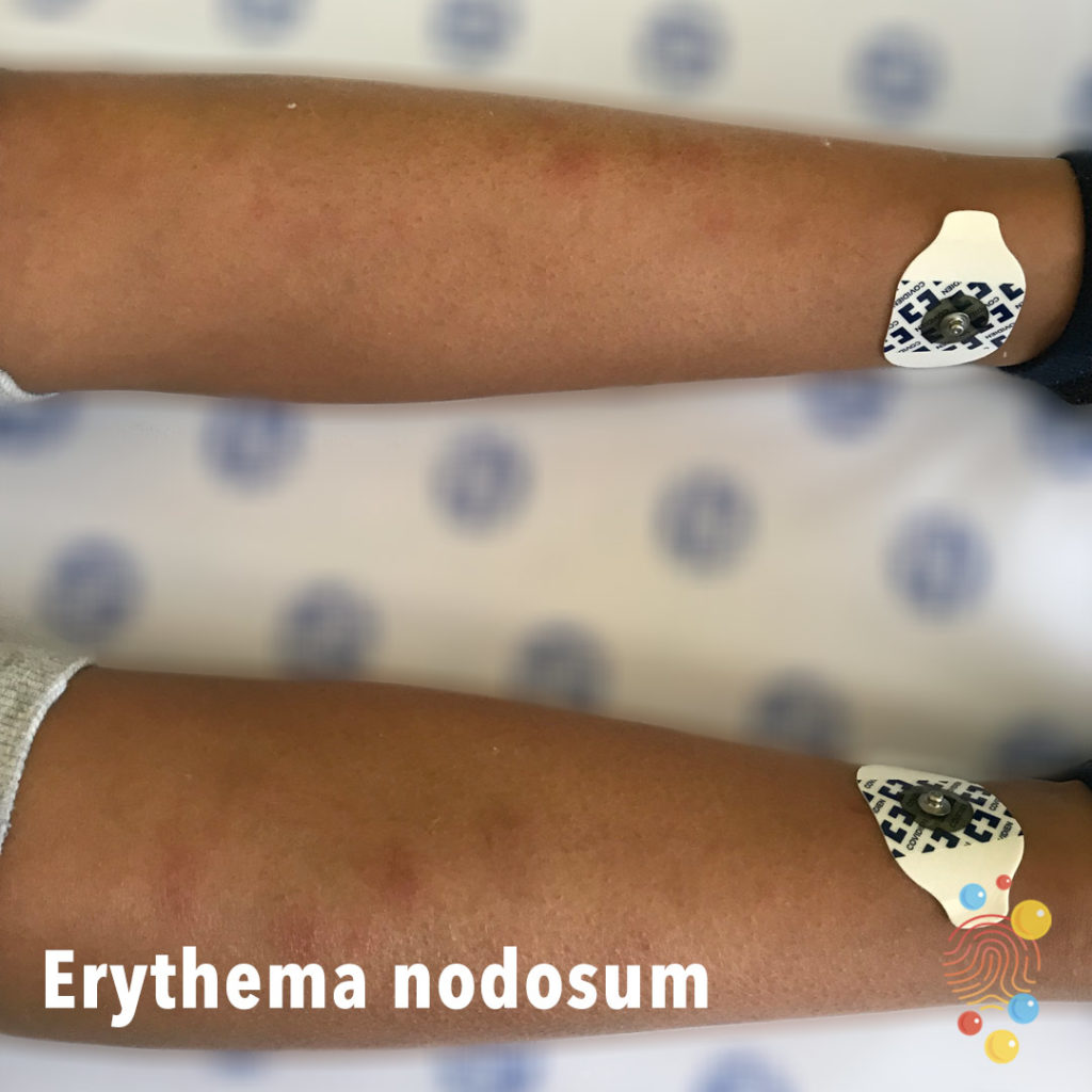
Erythema Nodosum
Learn more about erythema nodosum
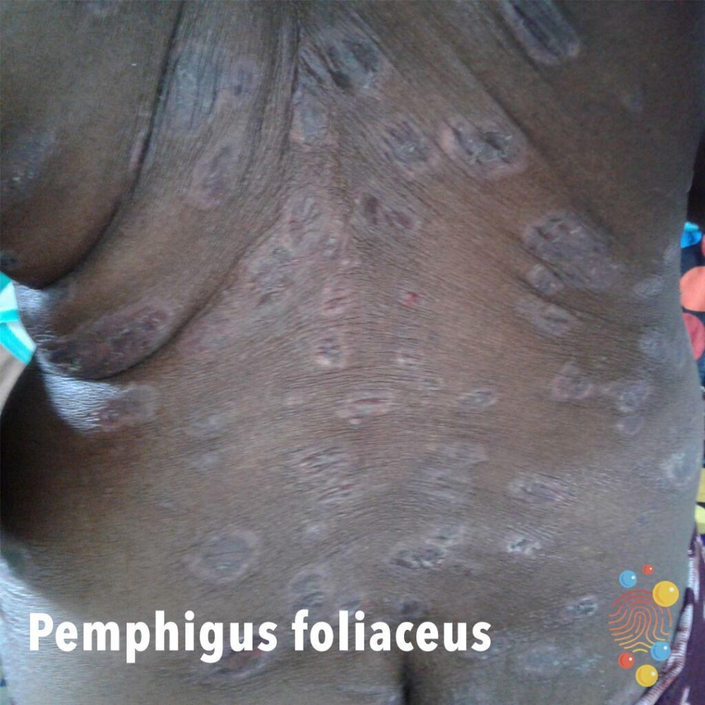
Pemphigus foliaceus
Learn more about pemphigus
Eczema
Learn more about eczema
Psoriasis
Learn more about psoriasis
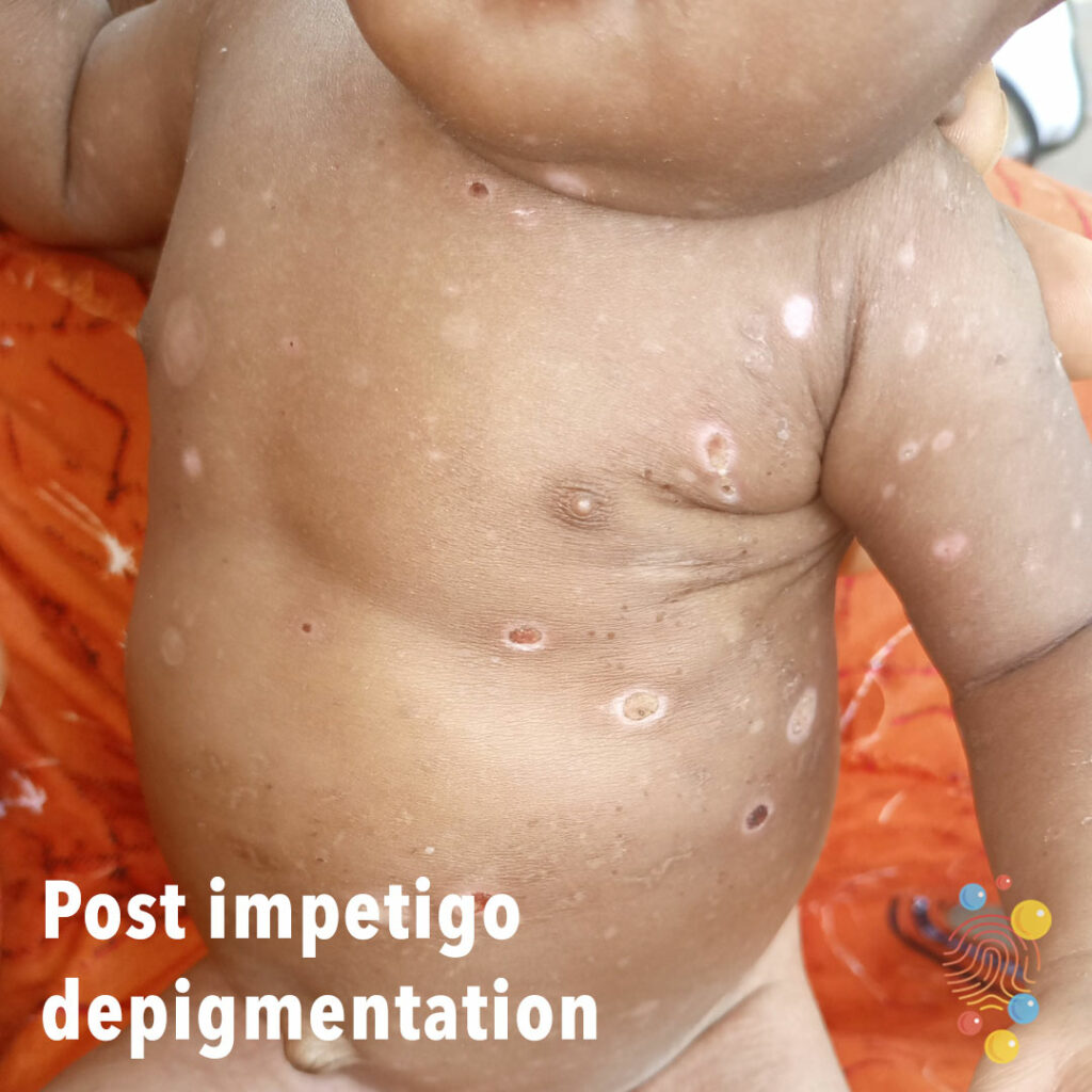
Post Impetigo Depigmentation
Learn more about impetigo
Eczema
Learn more about eczema
Eczema
Learn more about eczema

Strawberry tongue
Strawberry tongue in child with scarlet fever.
Impetigo
Learn more about bullous impetigo
PIMS-TS
Learn more about PIMS-TS
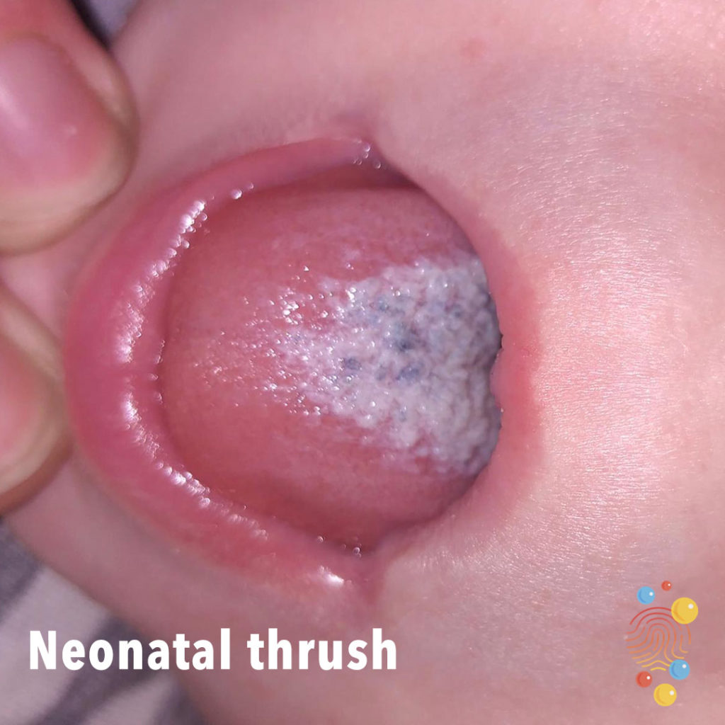
Neonatal Thrush
Learn more about neonatal thrush

Parvovirus
Bright red rash in symmetrical distribution
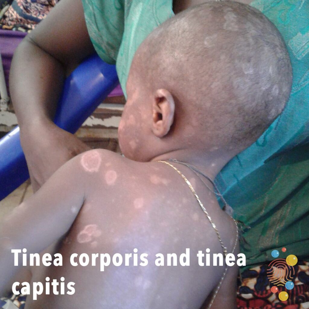
Tinea Corporis And Tinea Capitis
Learn more about tinea corporis
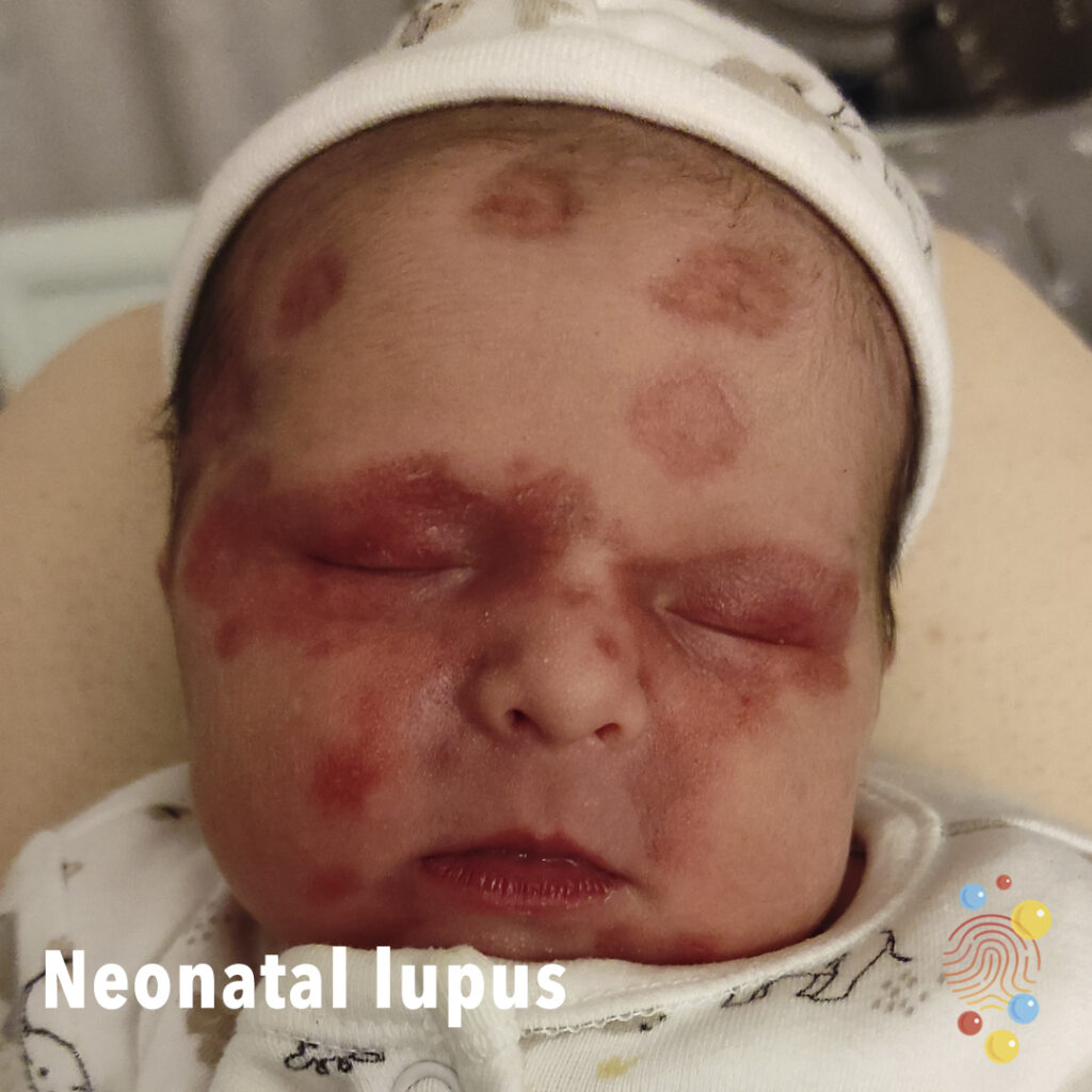
Neonatal Lupus
Discoid erythematous plaques affecting forehead and eyes, with a ‘raccoon-eye’ appearance, in a neonate with a mother with anti-SSA (Ro) antibodies.
Haemangioma
Learn more about haemangiomas.

Post Scarlet Fever
Extensive desquamation on back post scarlet fever.
Scarlet Fever

Psoriasis
Learn more about psoriasis
Scarlet Fever
Learn more about scarlet fever
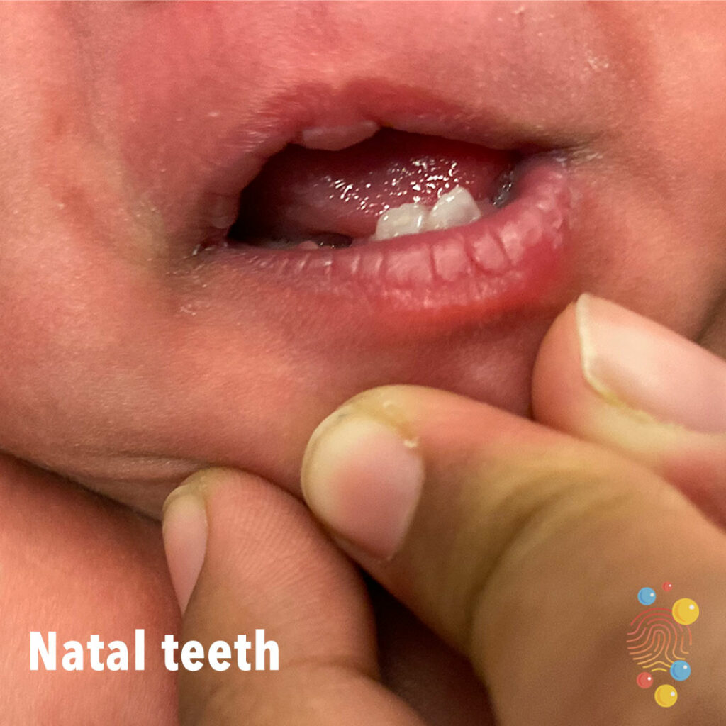
Natal Teeth
Learn more about natal teeth
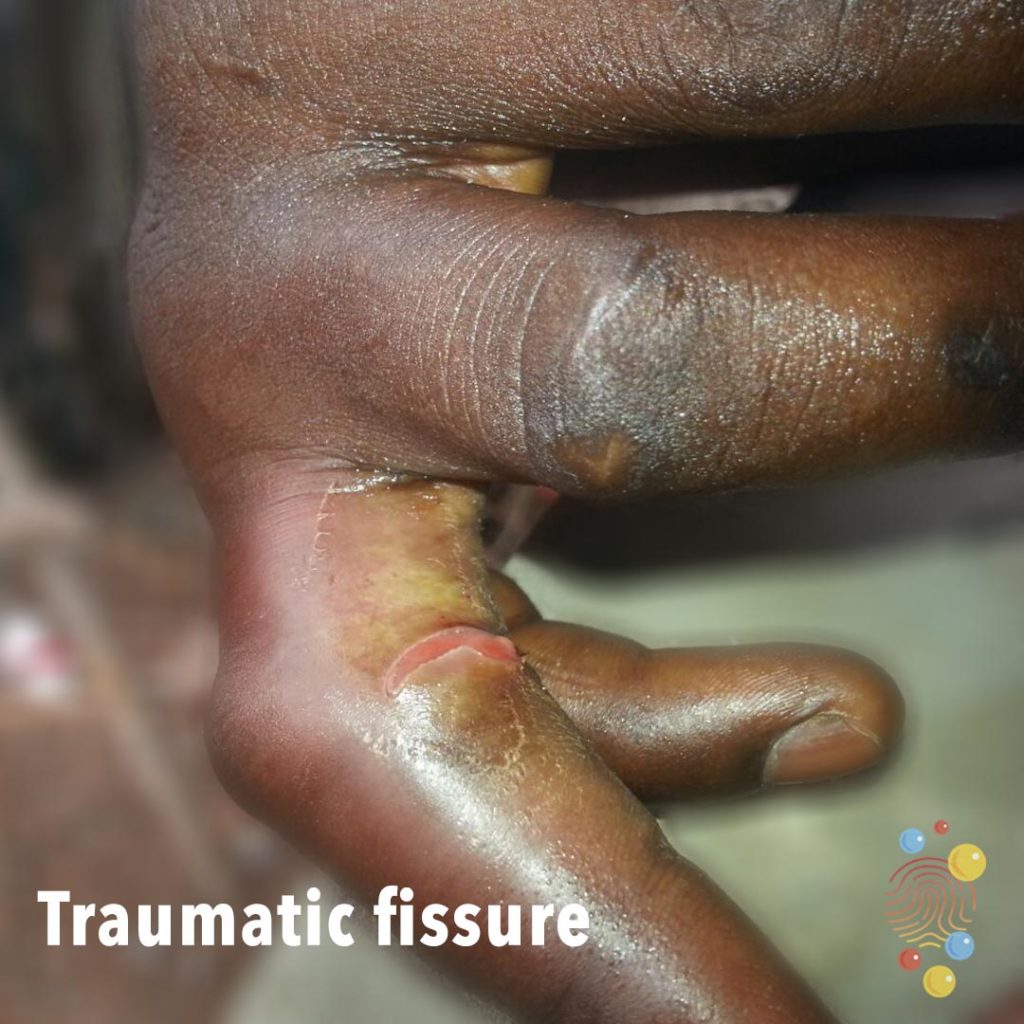
Traumatic Fissure
Learn more about traumatic fissures
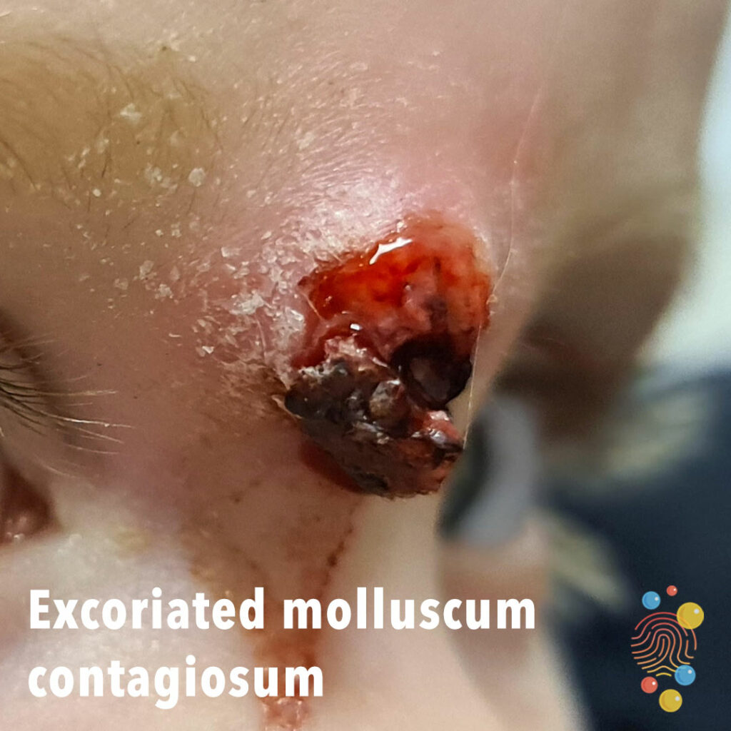
Excoriated molluscum contagiosum
Learn more about molluscum contagiosum
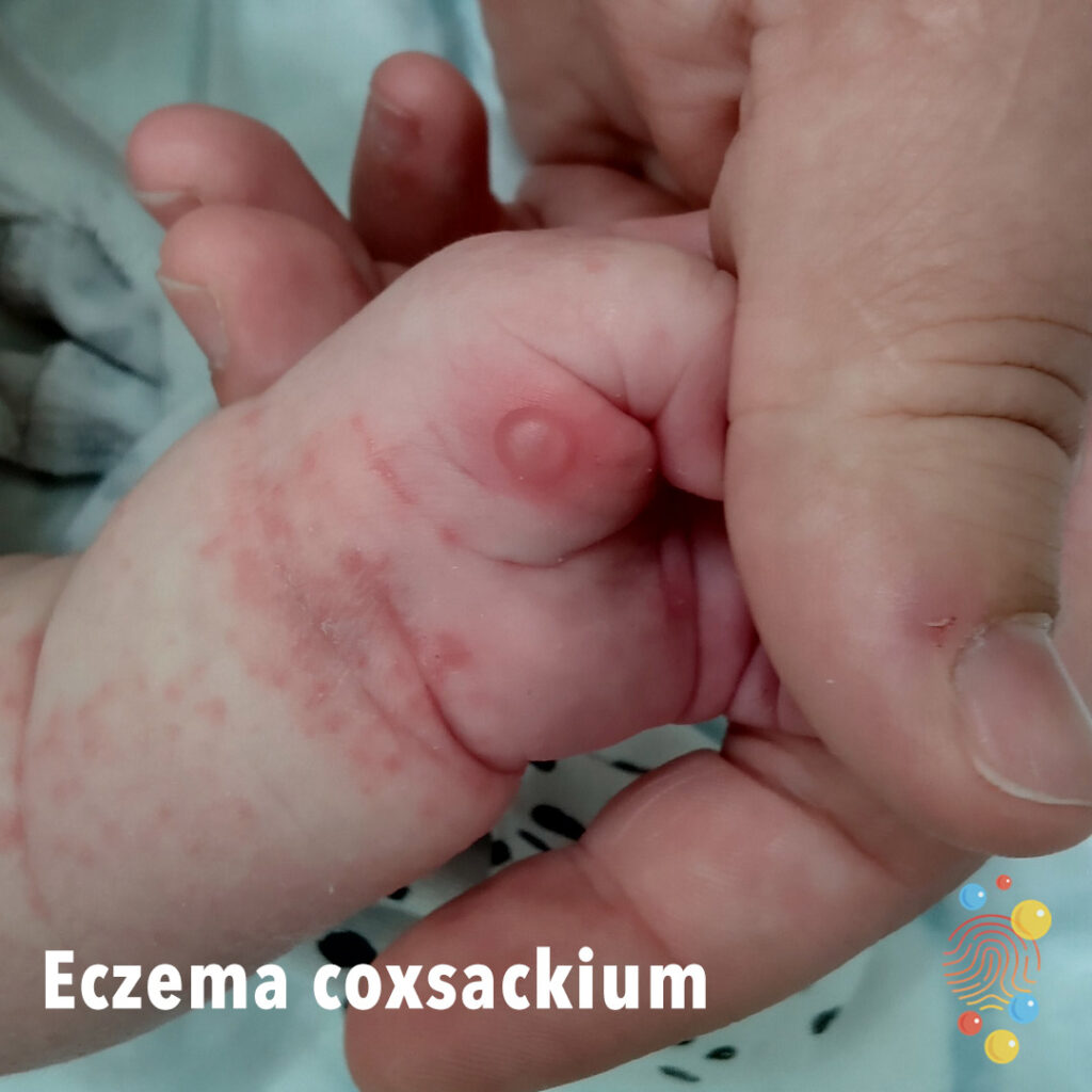
Eczema Coxsackium

Contact Dermatitis
Learn more about eczema
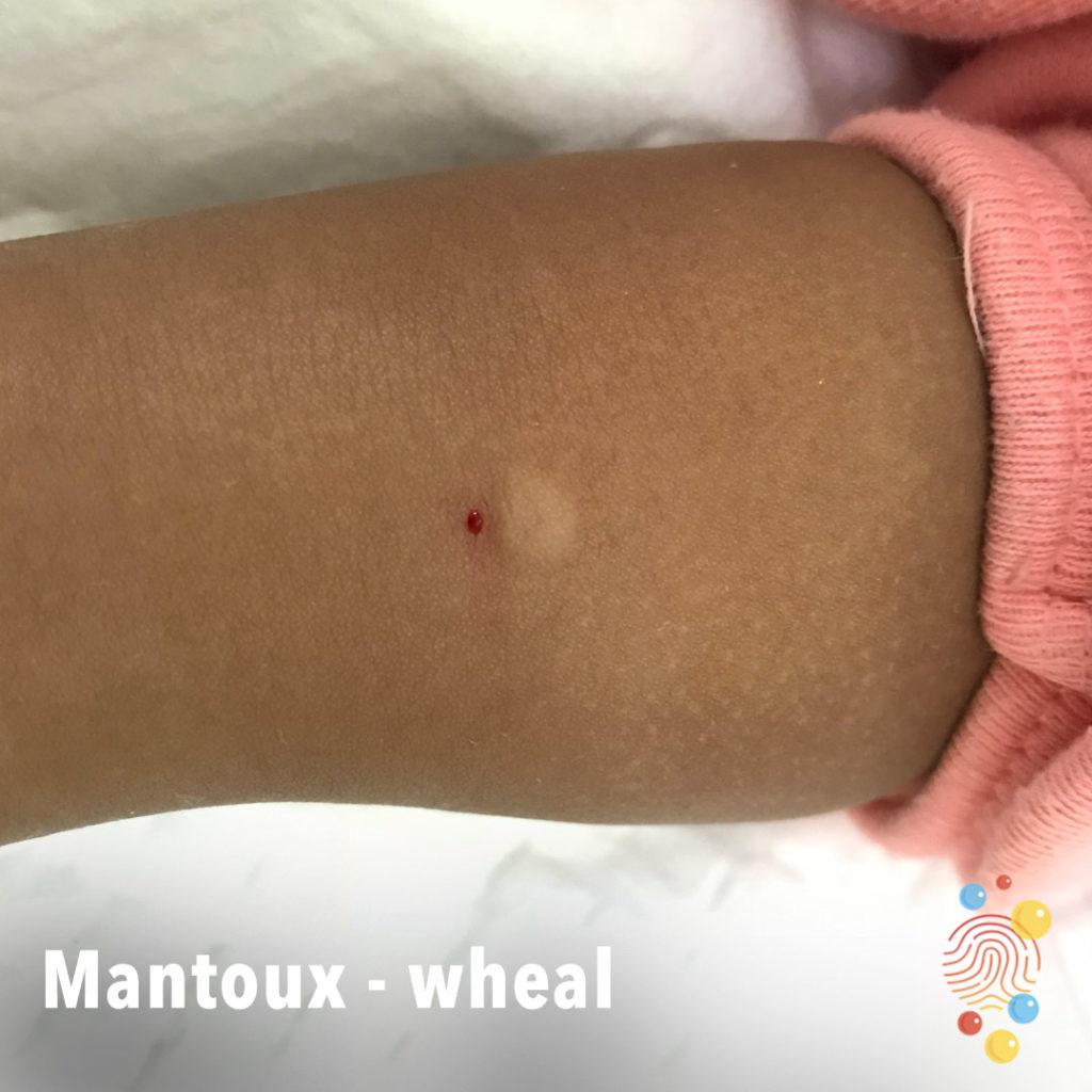
Mantoux Wheal
Learn more about the Mantoux test

Eczema Coxsackium
Eruption of dark red macules, vesicles, and erosions distributed across areas previously affected by atopic dermatitis, with relative sparing of the trunk

Flexor sheath infection (ring finger)
Suspected flexor sheath infection of right ring finger with insect bites on her hand.
Keloid Scar
Learn more about keloid scars.
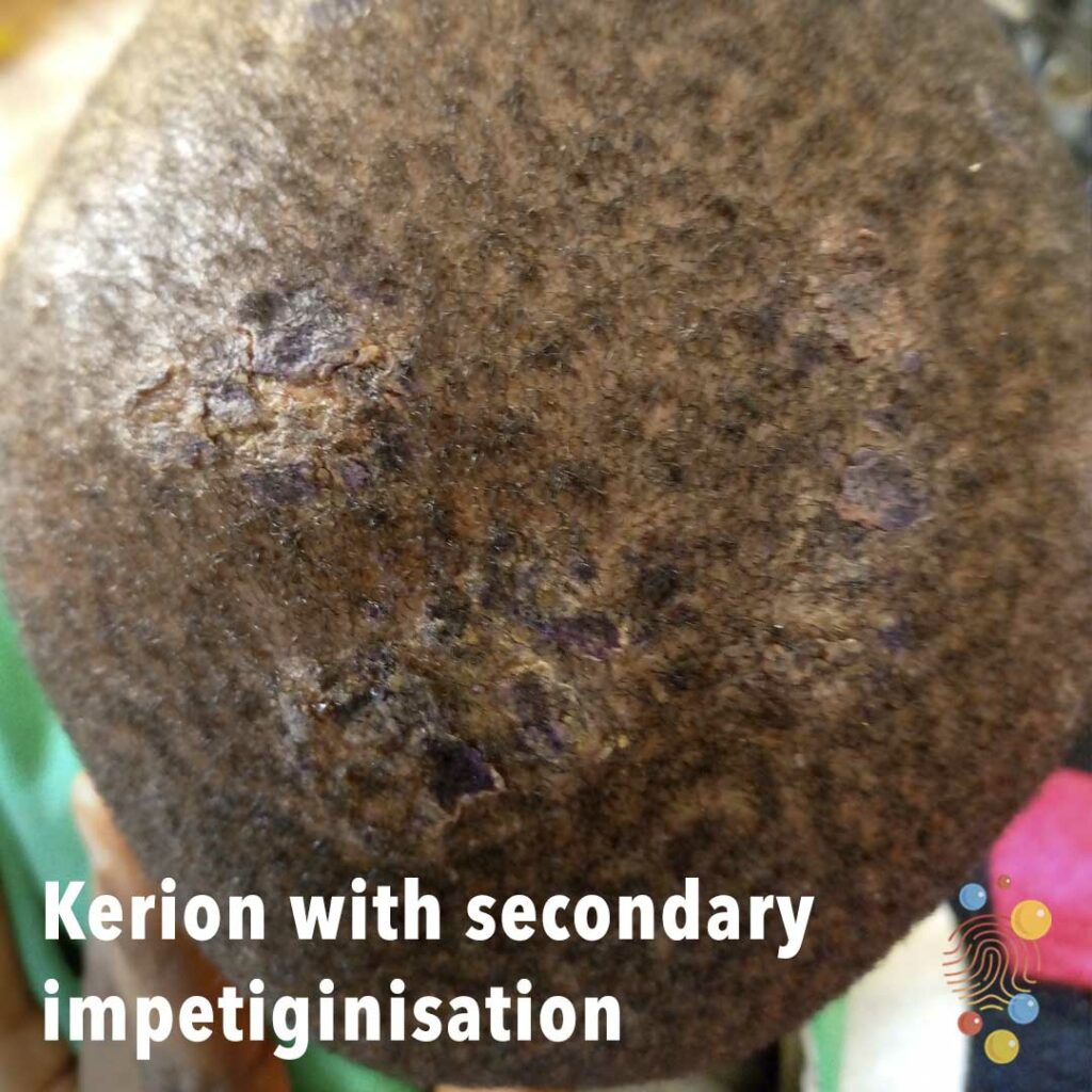
Kerion With Secondary Impetiginisation
Learn more about kerions

Pre- And Post-Deroofing Of A Bulla (With A Wart)
Learn more about warts
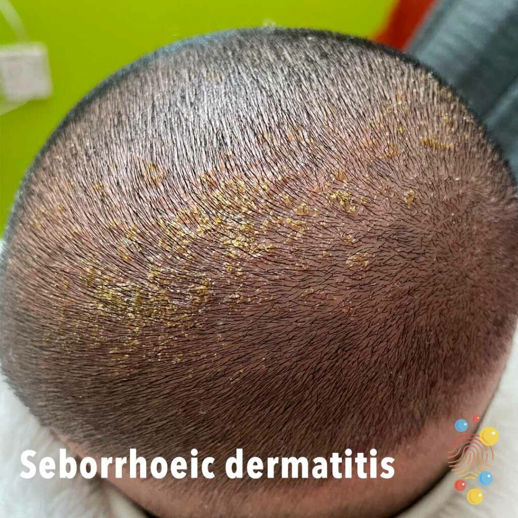
Seborrhoeic dermatitis
Learn more about seborrhoeic dermatitis
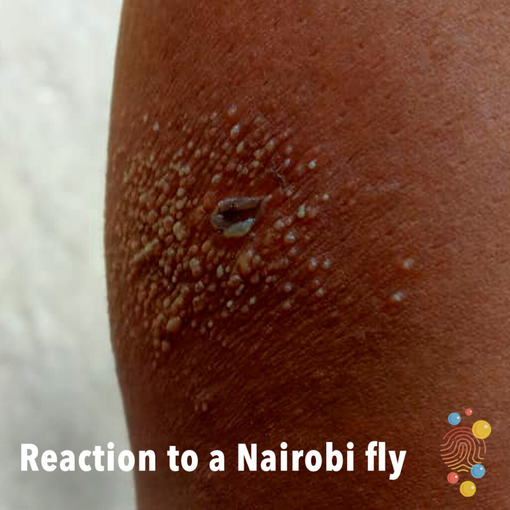
Reaction To A Nairobi Fly
Learn more about bites
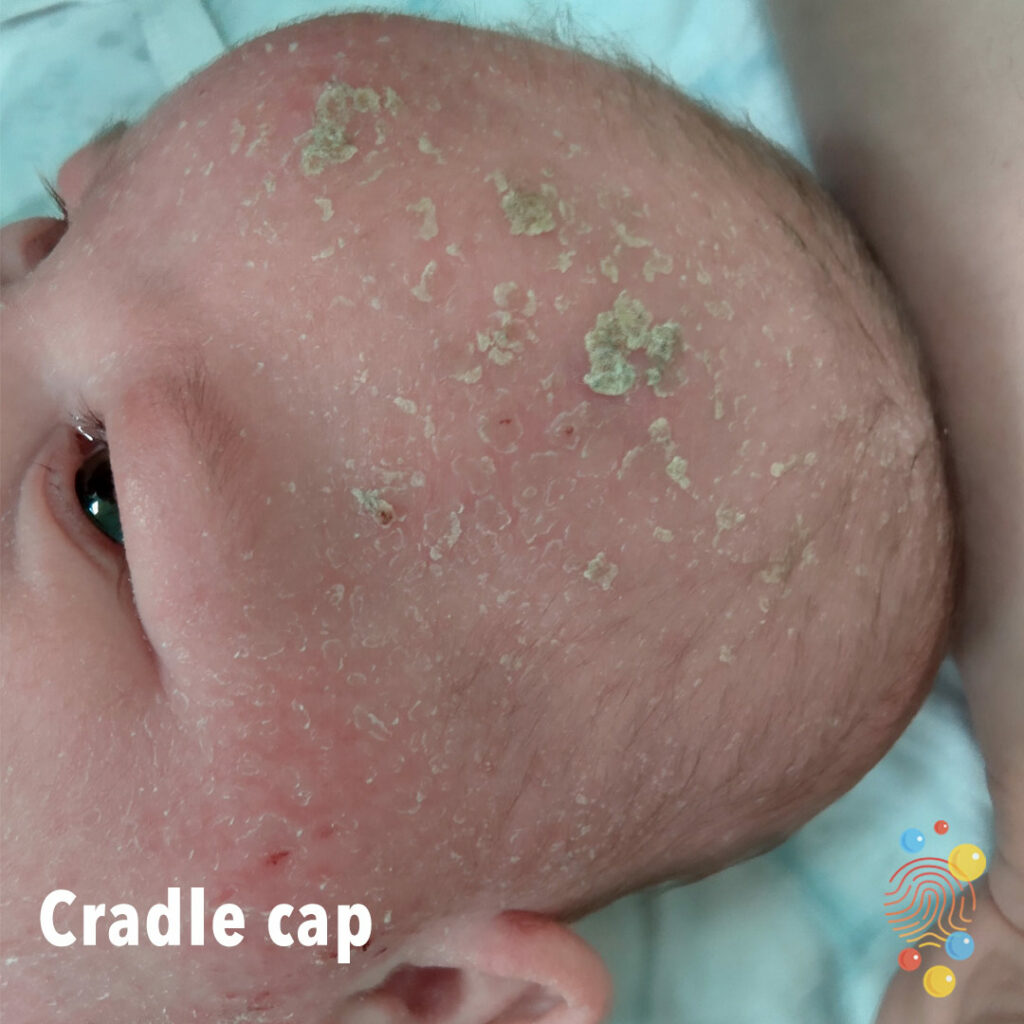
Cradle Cap

Chillblains
Oedema and erythema of the toes circumferentially.
Chalazion
Learn more about chalazion
Erythema Toxicum
Erythematous rash forehead interspersed with pinpoint papules in a young infant
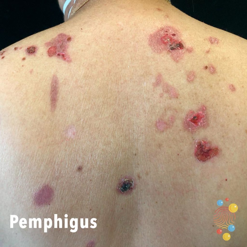
Pemphigus
Learn more about pemphigus
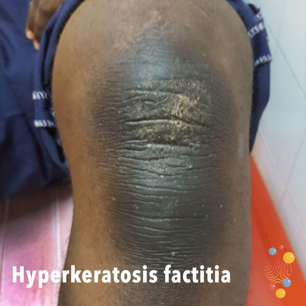
Hyperkeratosis Factitia
Learn more about hyperkeratosis factitia
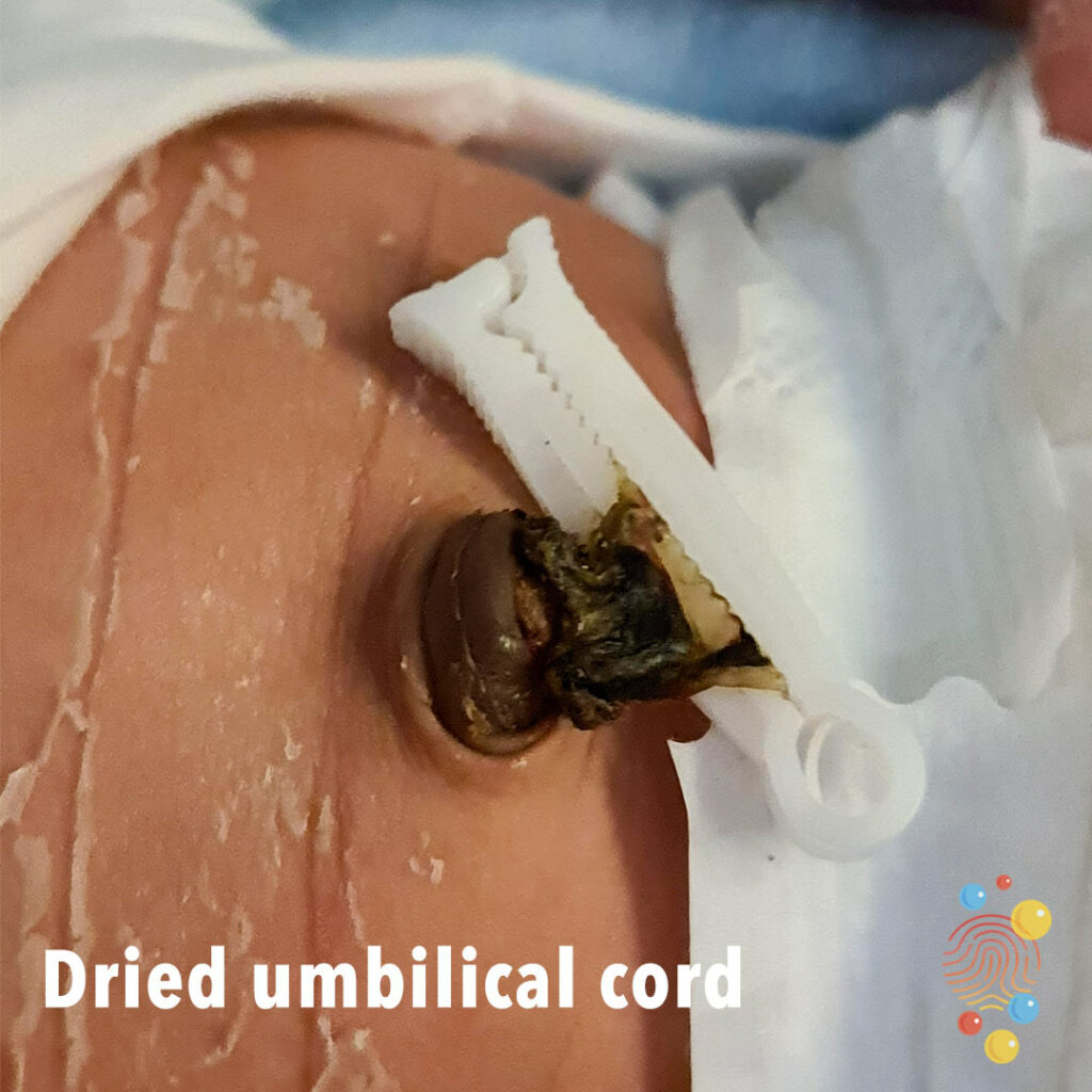
Dried umbilical cord
Learn about umbilical hernias
Eczema
Learn more about eczema
Eczema
Erythema and lichenification of the dorsal hands, with excoriations and bleeding.

Leukaemia Cutis
Learn more about leukaemia cutis
Scarlet Fever
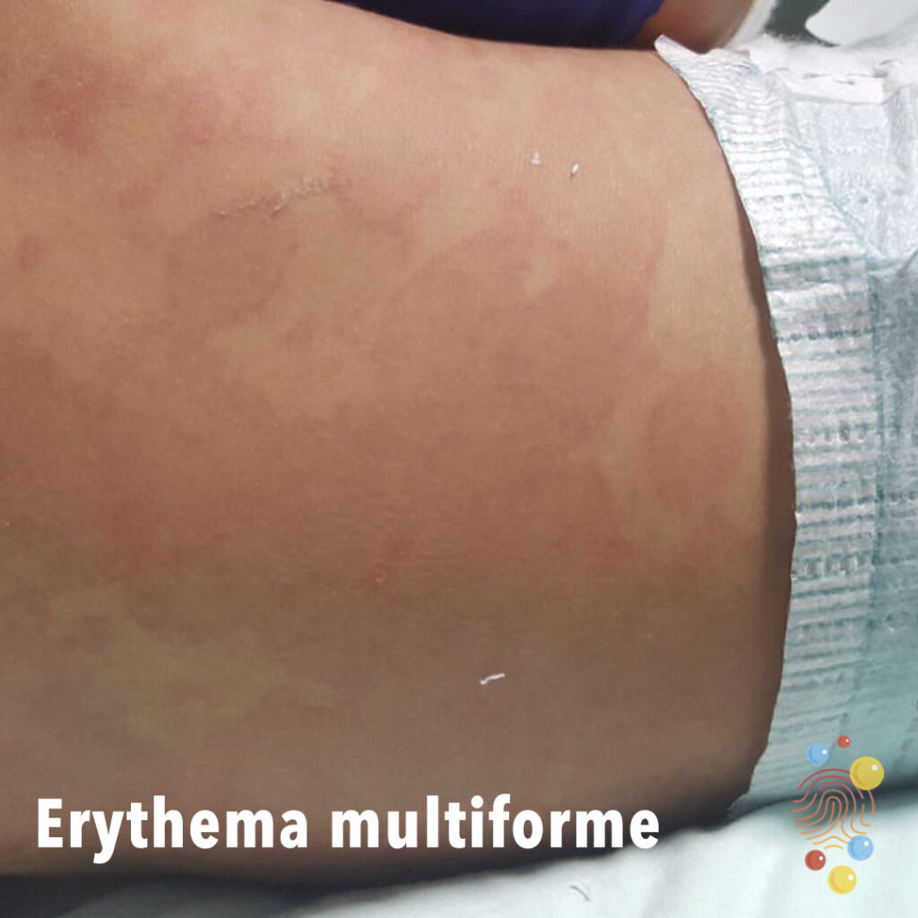
Erythema Multiforme
Learn more about erythema multiforme

Lip laceration

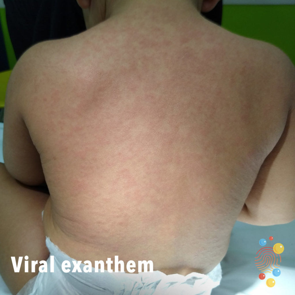
Viral Exanthem
Learn more about viral exanthem
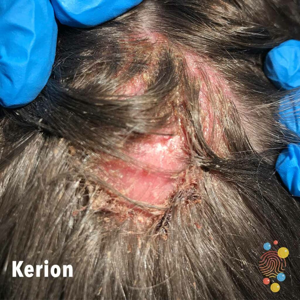
Kerion
Learn more about kerions

Mouth Injury Impacted Tooth
Mouth injury with impacted tooth.
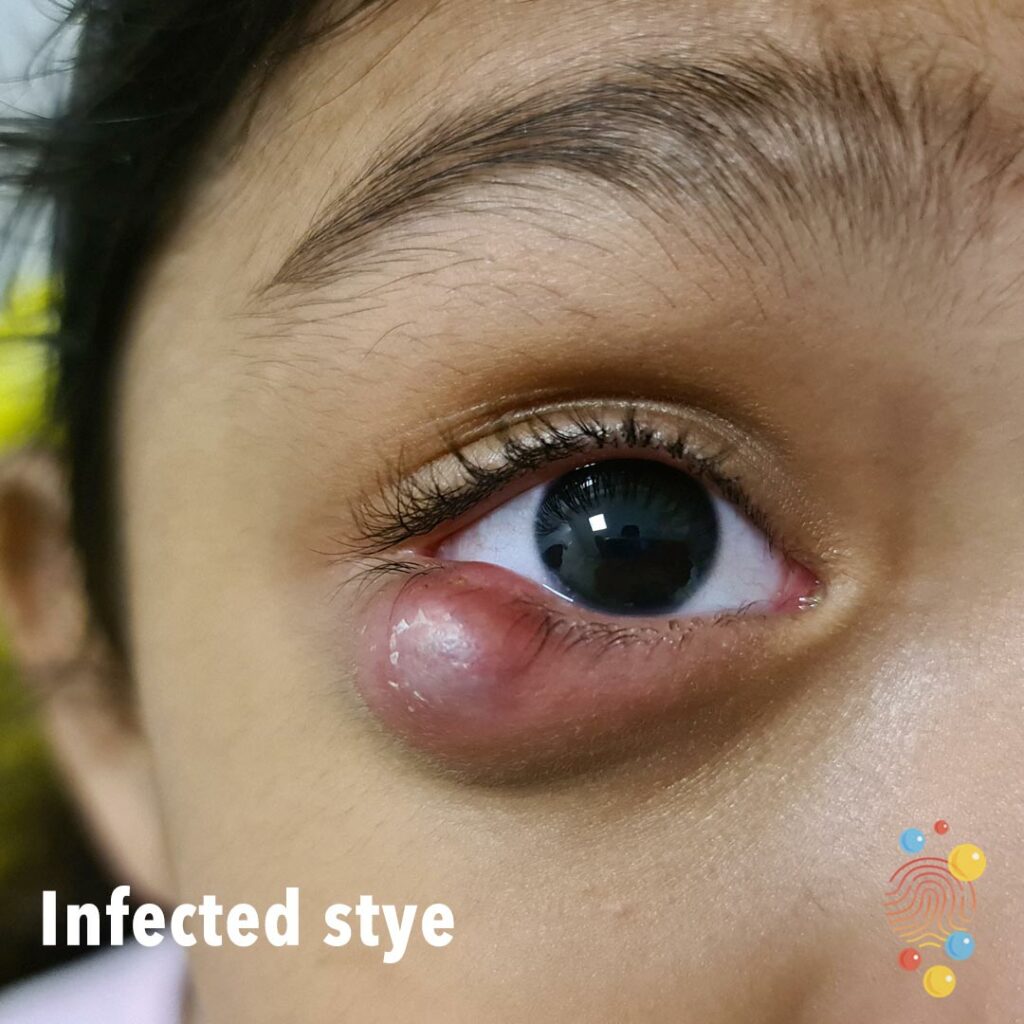
Infected Stye
Infected stye
Infected Eczema
Learn more about eczema

Acute haemorrhagic oedema of infancy
Multiple urticated bruises, some of which have a targetoid appearance

Perioral Dermatitis
Learn more about eczema
Chalazion
Learn more about chalazion

Gianotti-Crosti Syndrome
Learn more about Gianotti-Crosti syndrome
Dermal Melanocytosis
Learn more about dermal melanocytosis
Scarlet Fever
Scarlet fever is a bacterial illness that develops in some people who have strep throat. Also known as scarlatina, scarlet fever features a bright red rash
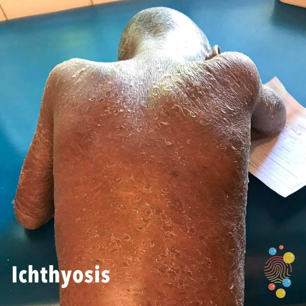
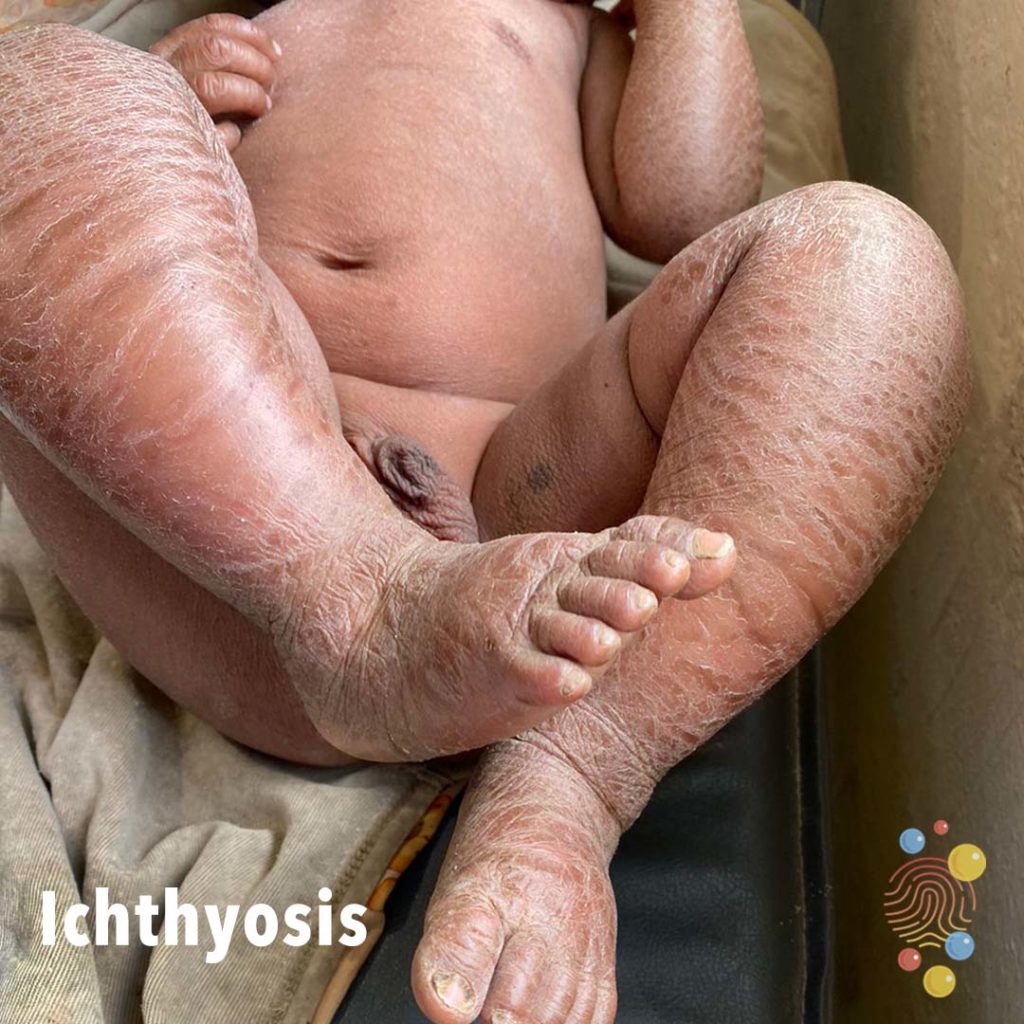
Ichthyosis
Learn more about ichthyosis

Eczema herpeticum
Learn more about eczema herpeticum

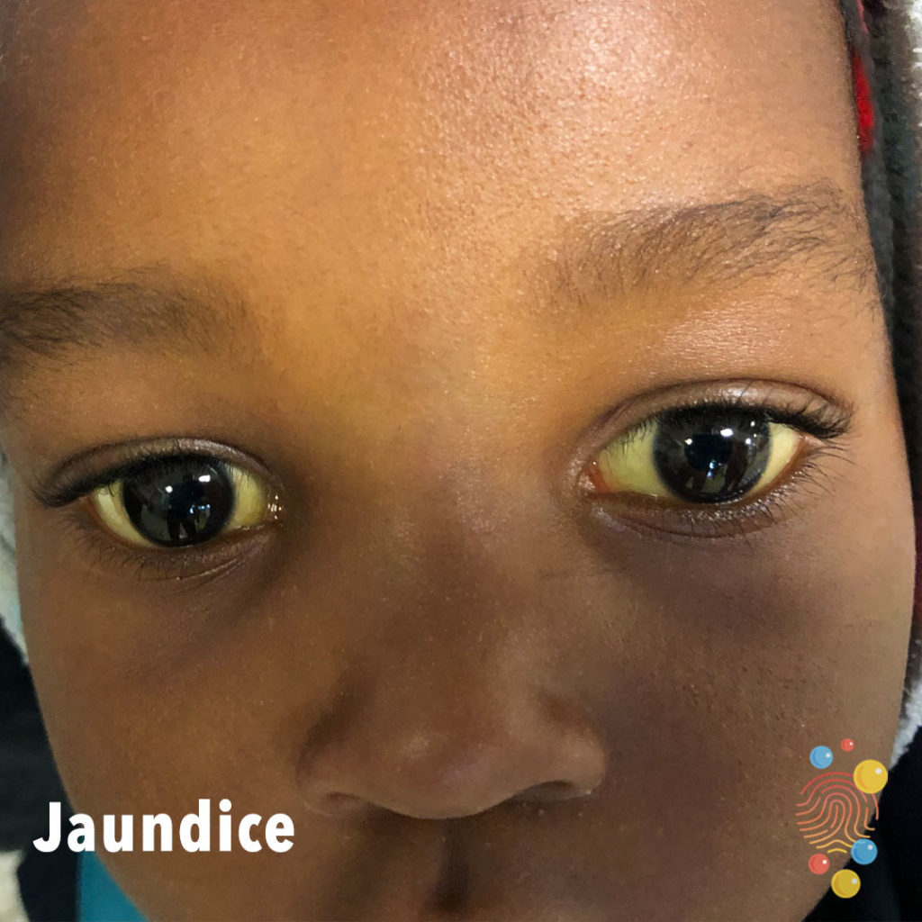
Jaundice
Learn more about jaundice

Stye
Learn more about styes

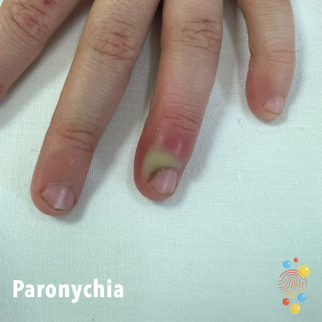
Paronychia
2 week old with paronychia

Eczema Coxsackium
Nummular Eczema
Learn more about eczema

Steven-Johnson-syndrome
Widespread dusky erythema of the posterior trunk with no blistering
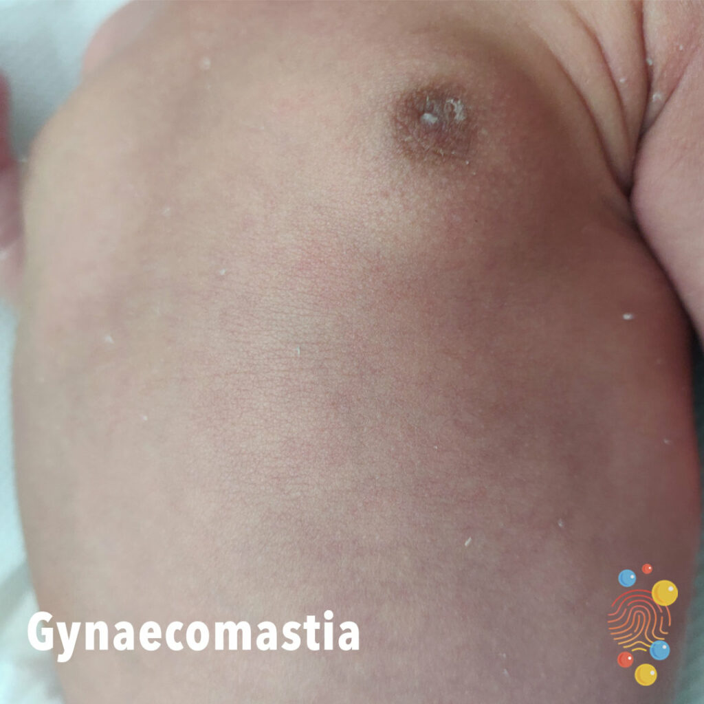
Gynaecomastia
Dermal Melanocytosis
Learn more about dermal melanocytosis
Tinea Capitis
Learn more about tinea capitis

Lymphoedema and hyperkeratosis
Symmetric swelling of lower limbs associated with hyperkeratosis, plantar keratoderma, and dystrophic toenails
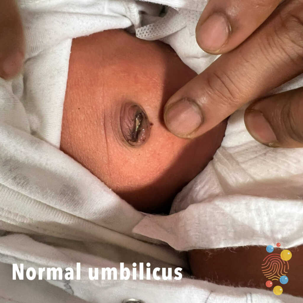
Normal Umbilicus
Psoriasis
Learn more about psoriasis

Neonatal Varicella
Baby is 2 weeks old, born with these papular lesions all over body, which are progressive.
Scarlet Fever
Strawberry tongue (due to reduced filiform papillae with retained fungiform papillae), crusted nodule on left cheek, and desquamation on trunk.
Impetigo
Learn more about bullous impetigo

Nailbed Injury
Toxic Epidermal Necrolysis
Learn more about toxic epidermal necrolysis
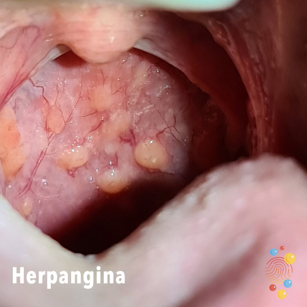
Herpangina
Learn more about herpangina

Erythema Nodosum
Learn more about erythema nodosum
Intertrigo

Stomatitis
Stomatitis in child with bilateral pneumonia, urticaria rash and cardiovascular instability requiring >40ml/kg fluid + inotropes.

Omphalitis
Infection of the cord stump and surrounding skin.
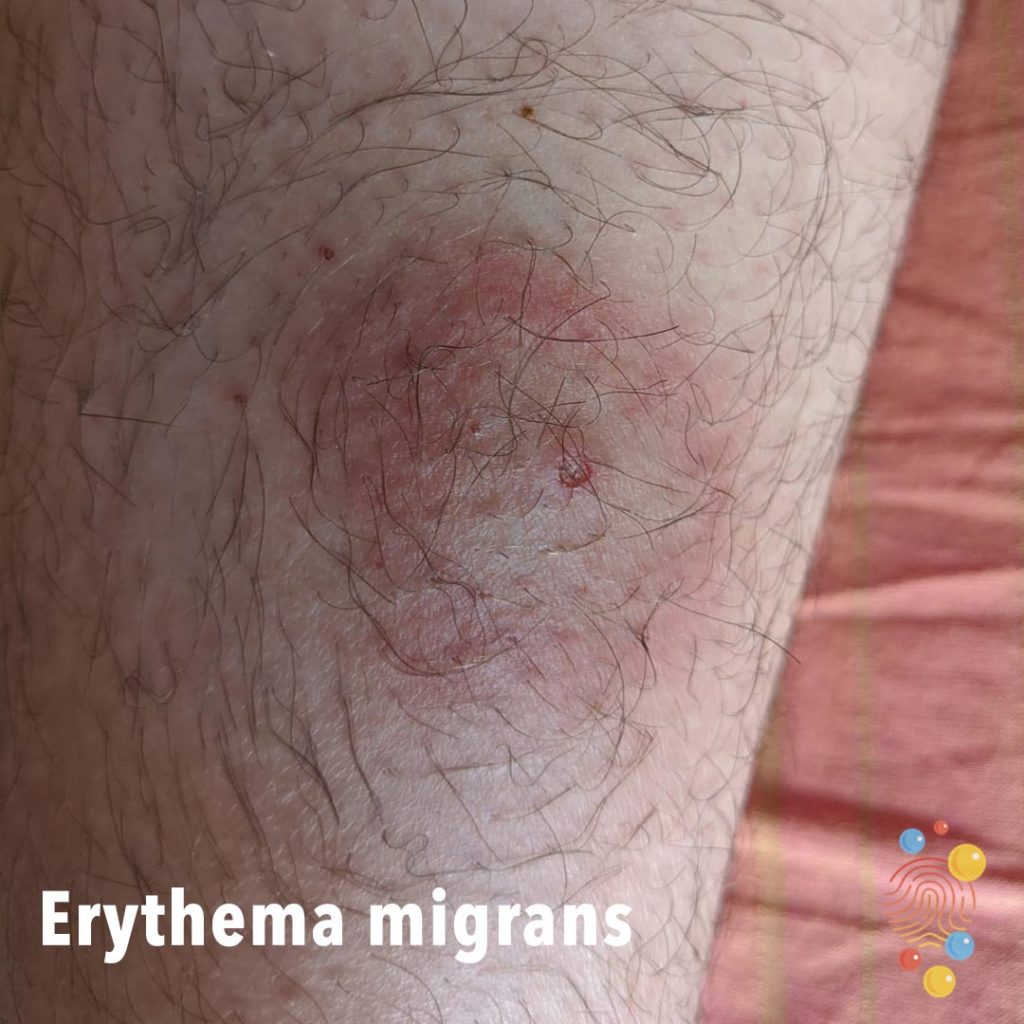
Erythema Migrans
Annular erythematous eruption with central crusting and erosion.
Pityriasis Versicolor
Learn more about pityriasis versicolor
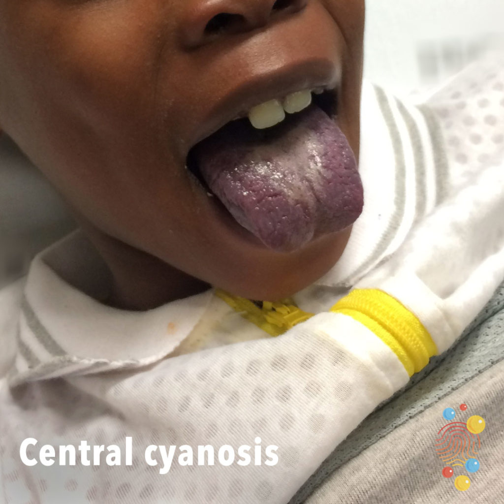
Central Cyanosis
Learn more about central cyanosis
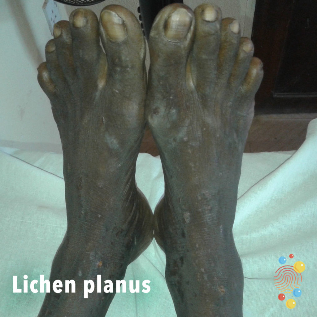
Lichen Planus
Learn more about lichen planus
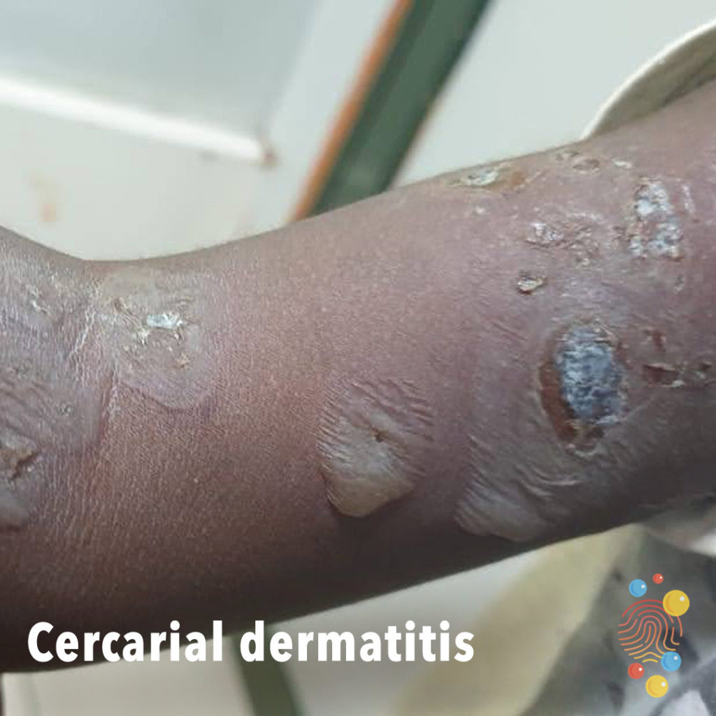
Cercarial Dermatitis
Multiple flaccid bullae with erosions on upper limb.
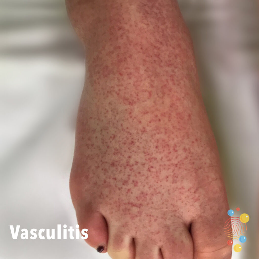
Vasculitis
Learn more about vasculitis
Petechial Rash
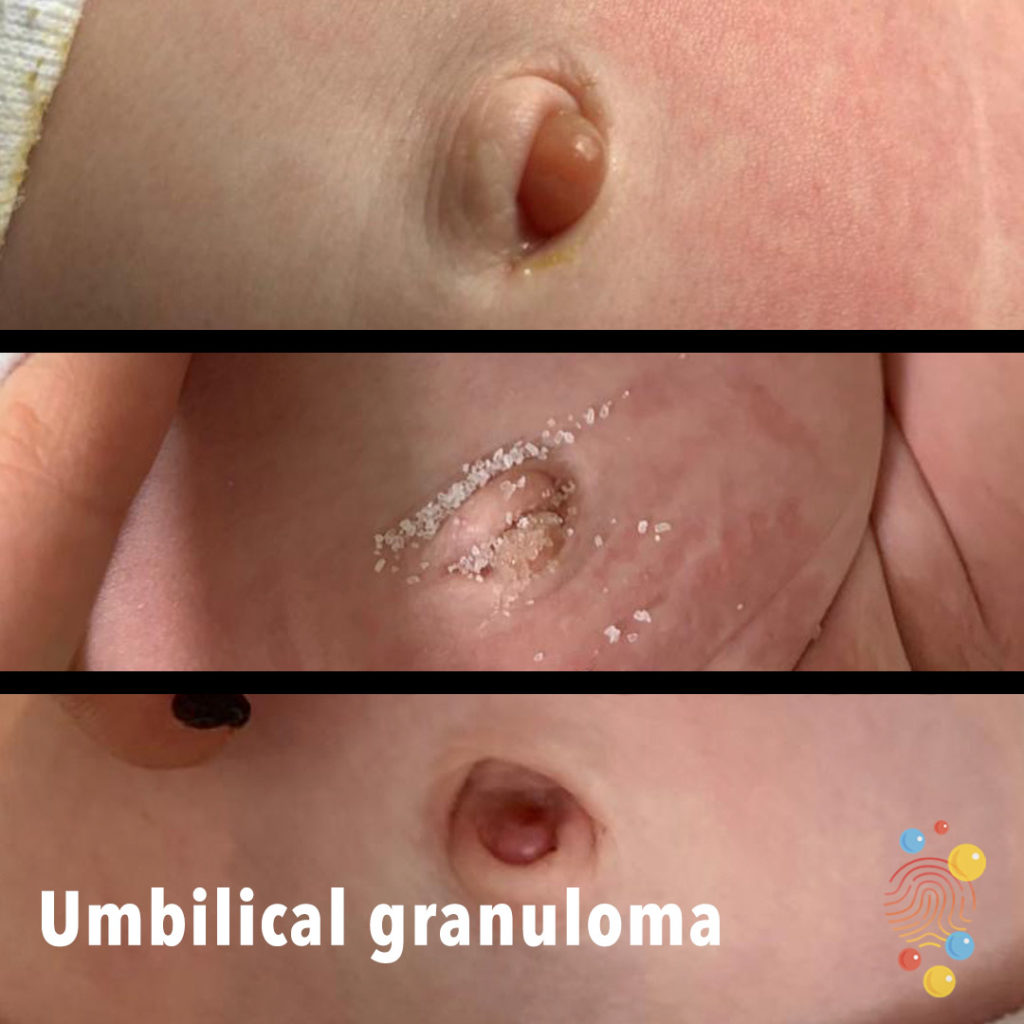
Umbilical Granuloma
Learn more about umbilical granulomata

Kawasaki Disease
Learn more about Kawasaki disease

Post Chickenpox Abscess
A post-chickenpox abscess can be a complication of chickenpox, which is caused by the varicella-zoster virus (VZV).
Eczema
Learn more about eczema
Roseola
Learn more about roseola
Scarlet Fever
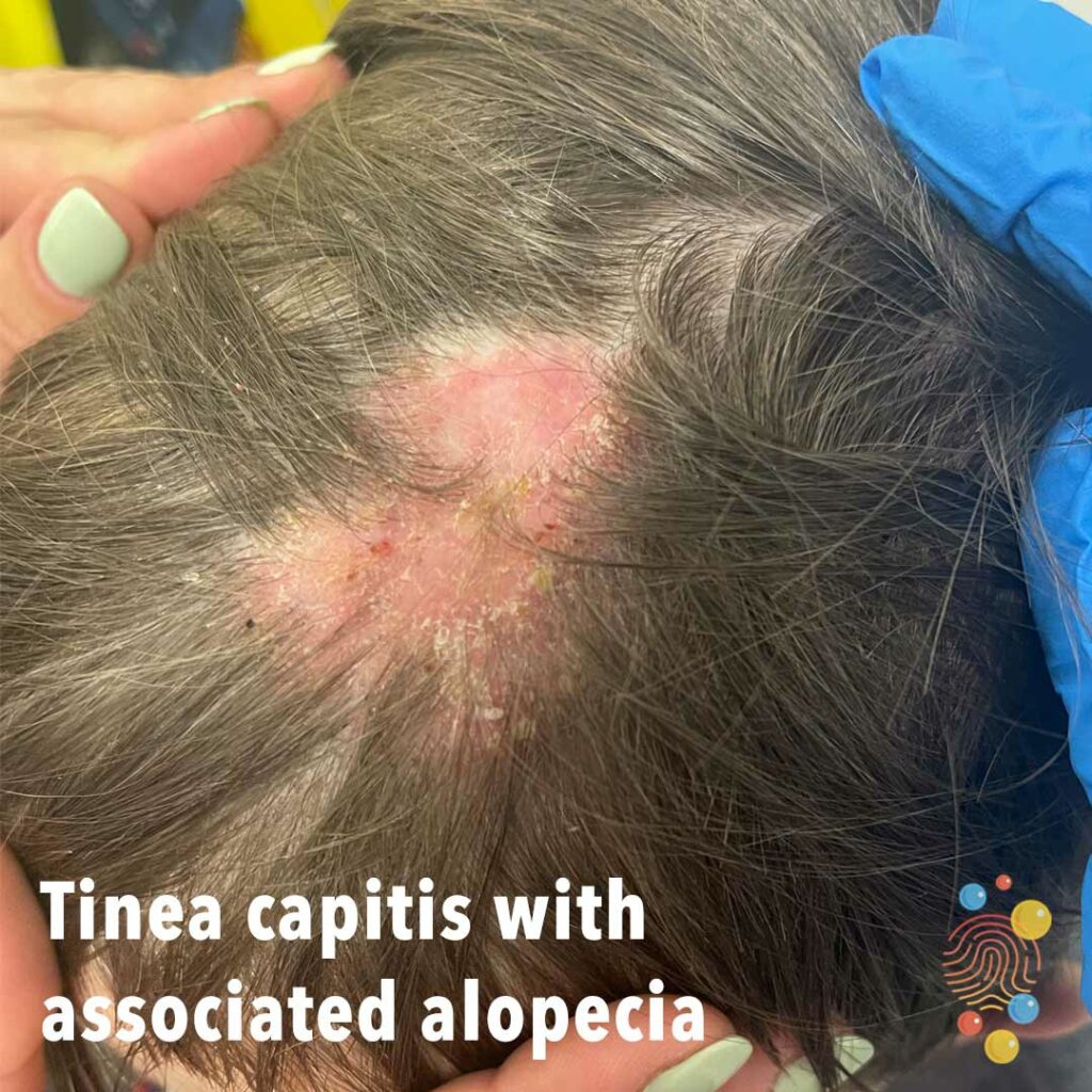
Tinea capitis with associated alopecia

Corneal Abrasion
Learn more about corneal abrasions
Impetigo
Folliculitis
Learn more about folliculitis

Bullous impetigo
Learn more about bullous impetigo
Eczema
Learn more about eczema
Chicken Pox
Learn more about chicken pox
Dermal Melanocytosis
Learn more about dermal melanocytosis

Tinea Corporis
Learn more about tinea corporis

Hand, foot & mouth
Learn more about hand, foot and mouth

Henoch-Schonlein Purpura
Learn more about Henoch-Schonlein purpura
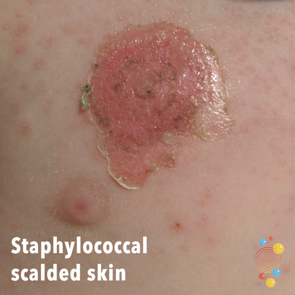
Staphylococcal Scalded Skin
Learn more about staphylococcal scalded skin
Warts
Learn more about warts
Folliculitis
Learn more about folliculitis
Henoch-Schonlein Purpura
Learn more about Henoch-Schonlein purpura

Eczema Herpeticum
Chicken Pox
Learn more about chicken pox
Haemangioma to scalp
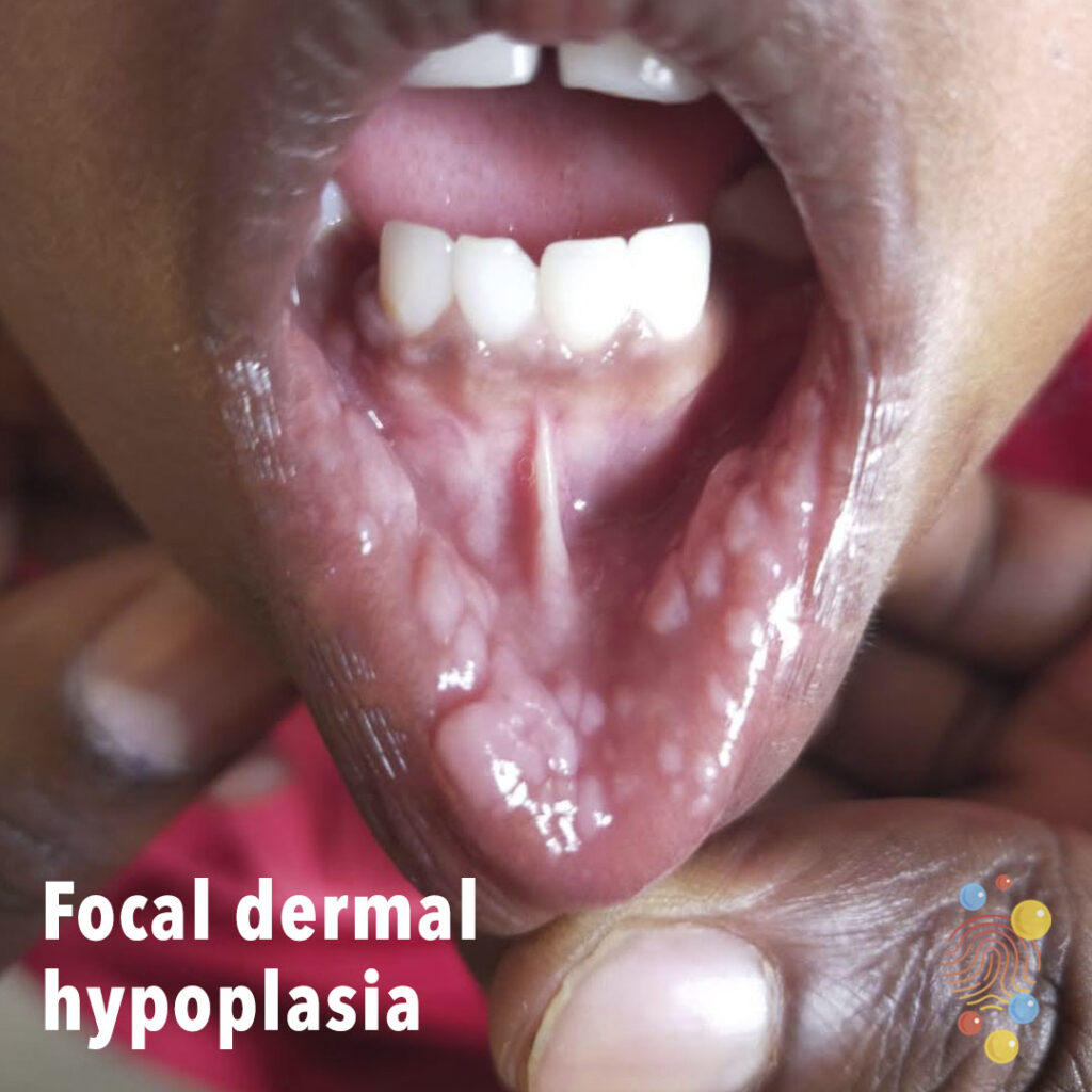
Focal Dermal Hypoplasia
Reaction To A Bite
Learn more about bites.

Gastrostomy
Learn more about gastrostomies

Warts
Learn more about warts
Scarlet Fever

Syphilis
Learn more about syphilis
Eczema
Learn more about eczema
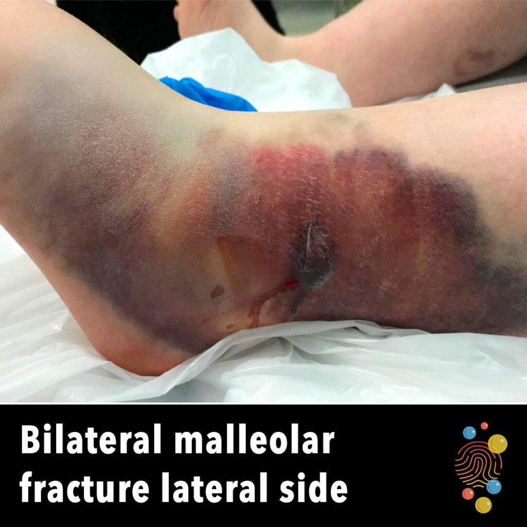
Bilateral Malleolar Fracture Lateral Side
Learn more about ecchymosis
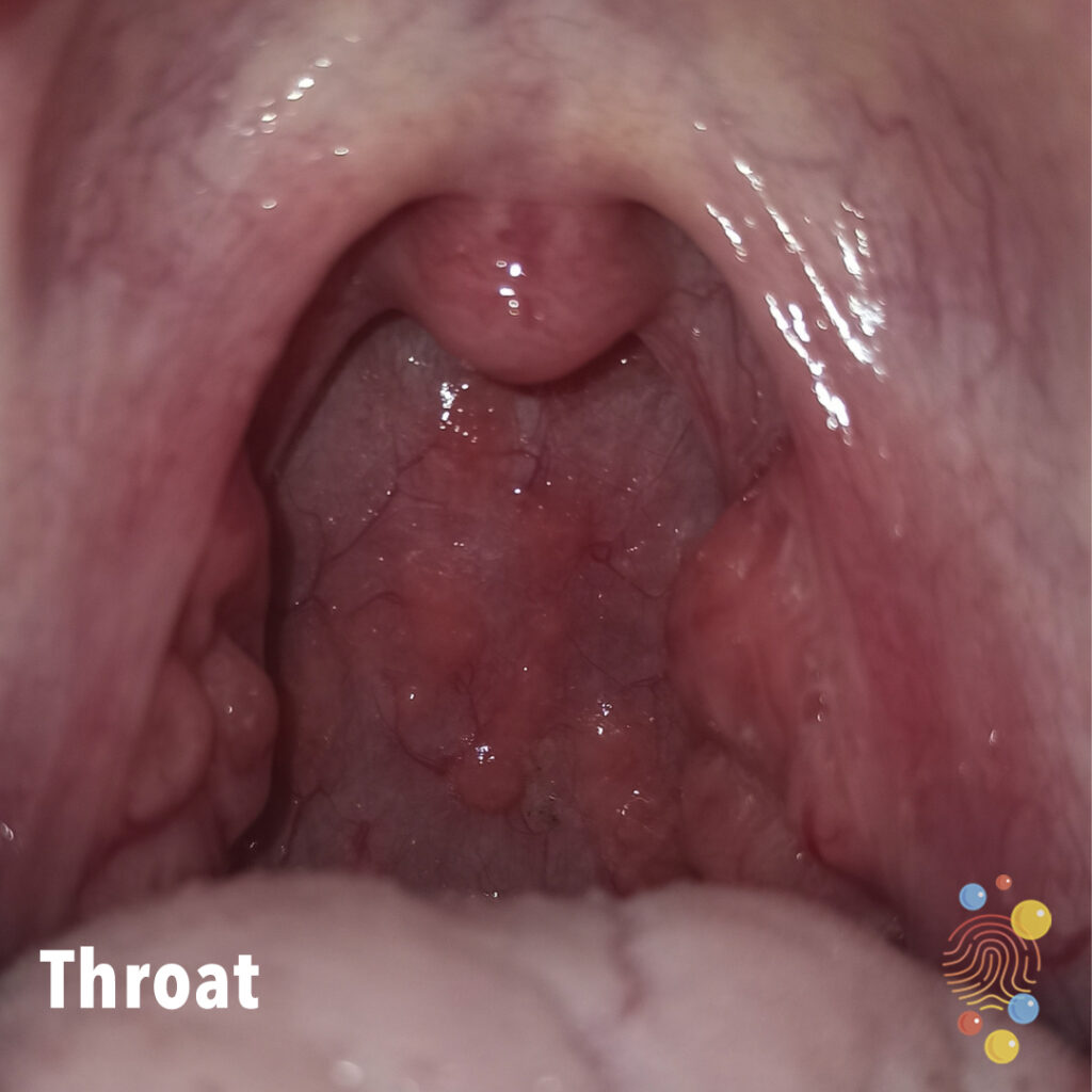
Throat
Throat burning with bubbles at the back of the mouth.

Umbilical hernia and umbilical granuloma
Learn more about umbilical hernias

Proximal phalanx fracture
left little finger proximal phalanx fracture
Pityriasis Versicolor
Learn more about pityriasis versicolor
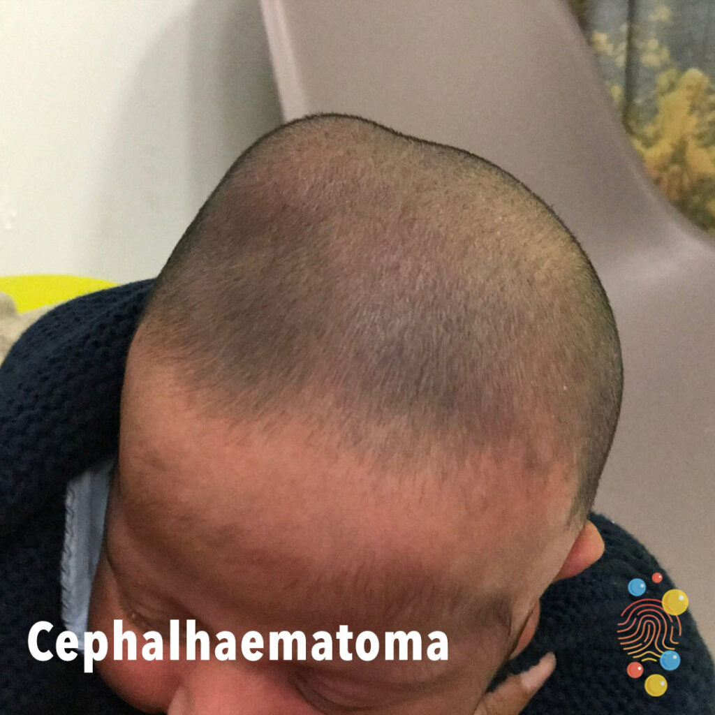
Cephalhaematoma
Learn more about cephalhaematoma
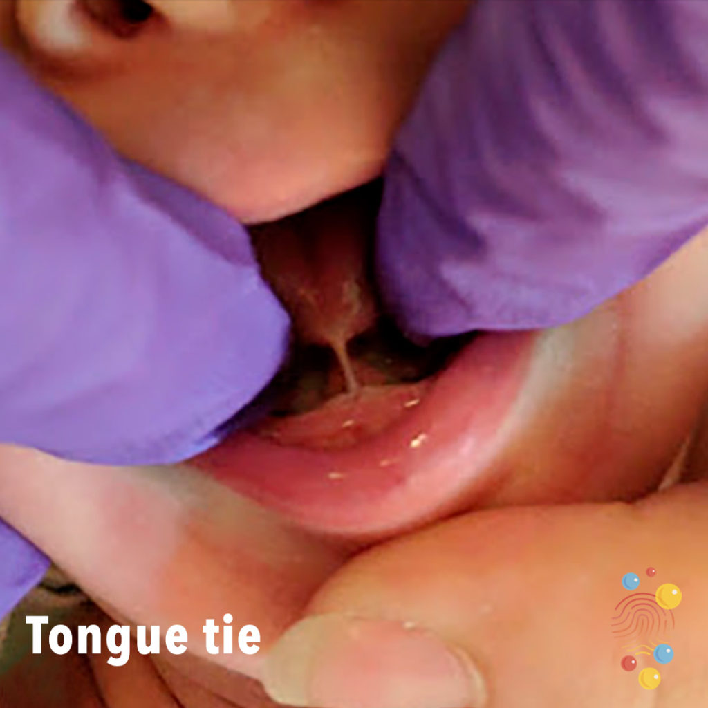
Tongue Tie
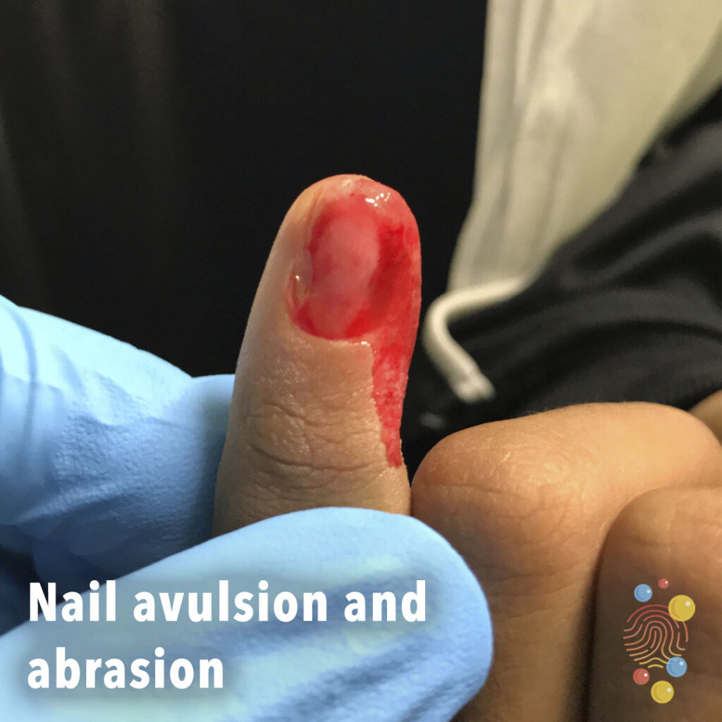
Nail Avulsion And Abrasion
Nail avulsion and abrasion

Steven’s Johnson syndrome
Stevens–Johnson syndrome is a type of severe skin reaction. Together with toxic epidermal necrolysis and Stevens–Johnson/toxic epidermal necrolysis overlap, they are considered febrile mucocutaneous drug reactions and probably part of the same spectrum of disease, with SJS being less severe.
Staphylococcal Abscess
Learn more about staphylococcal abscesses
Folliculitis
Follicular based erythematous papules.
Learn more about folliculitis
Jaundice
Learn more about jaundice
Tinea Capitis
Learn more about tinea capitits
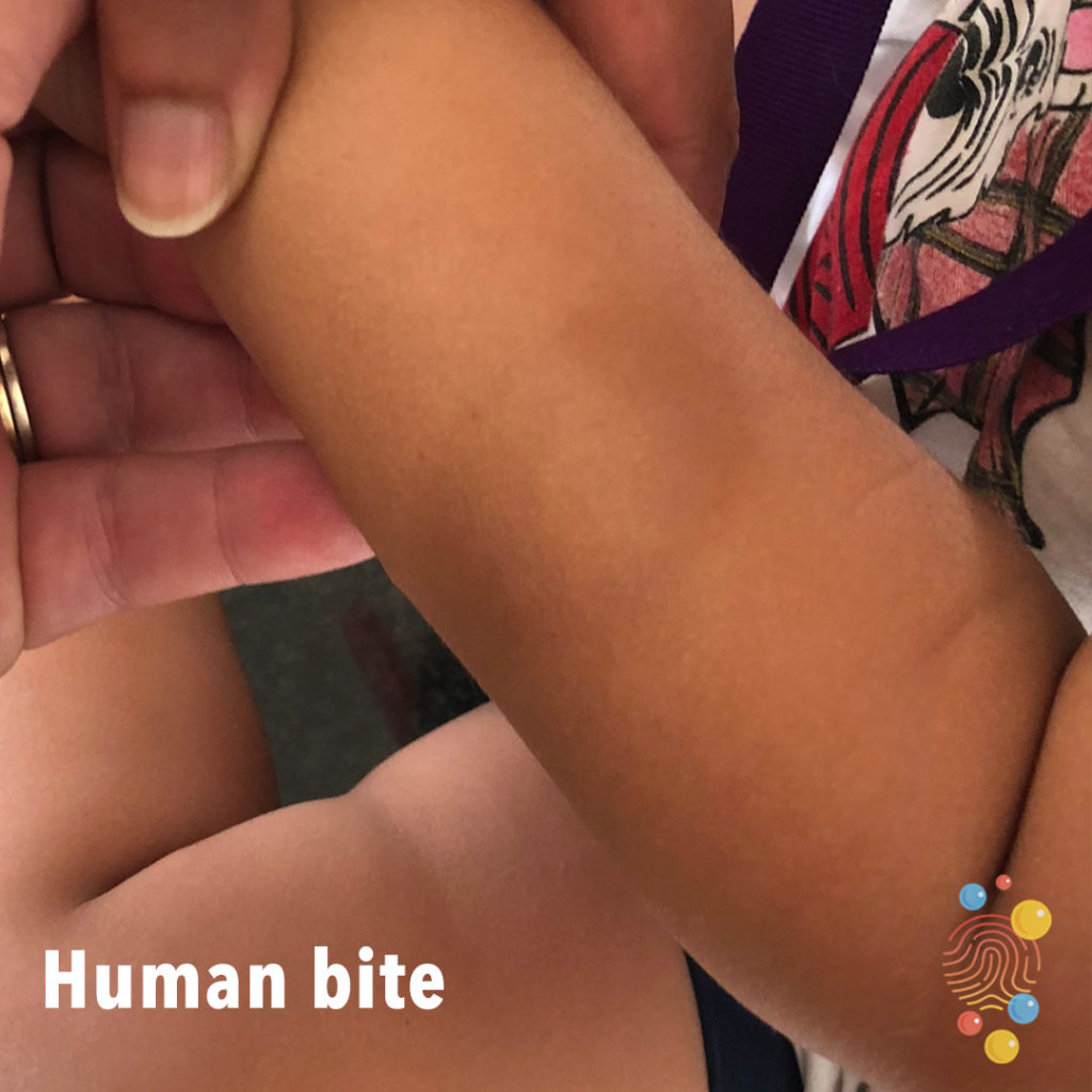
Human Bite
Learn more about bites
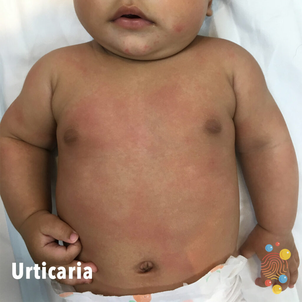
Urticaria
Learn more about urticaria
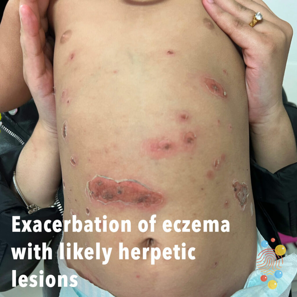
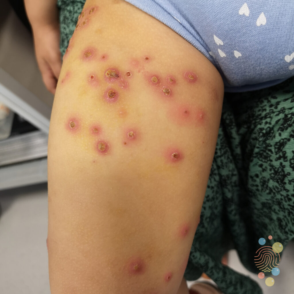
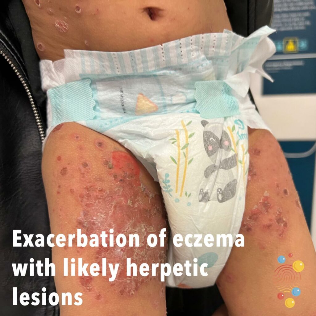
Exacerbation of eczema with likely herpetic lesions
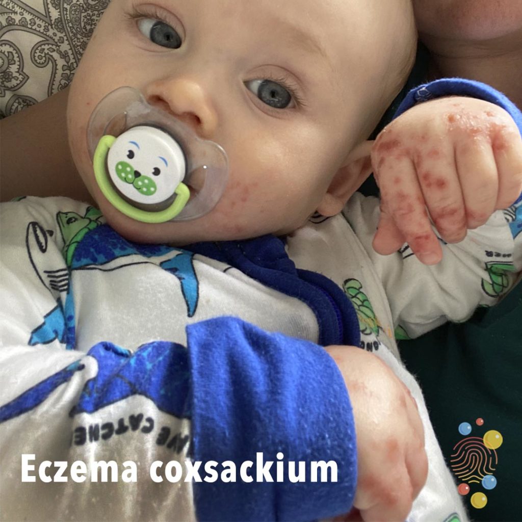
Eczema Coxsackium
Learn more about eczema coxsackium

Discoid Lupus
Learn more about discoid lupus
Psoriasis
Learn more about psoriasis
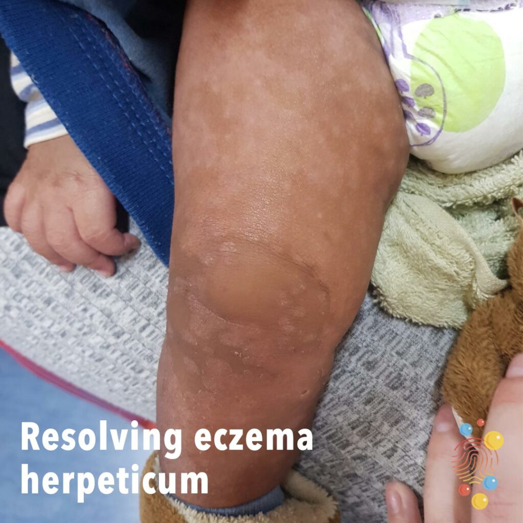
Resolving eczema herpeticum
Learn more about eczema herpeticum
Impetigo
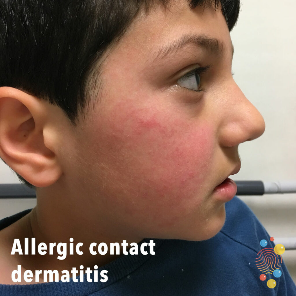
Allergic contact dermatitis
Learn more about eczema
Ichthyosis
Learn more about ichthyosis

Impetigo
Learn more about bullous impetigo

Eczema With Secondary Impetiginisation
Learn more about eczema
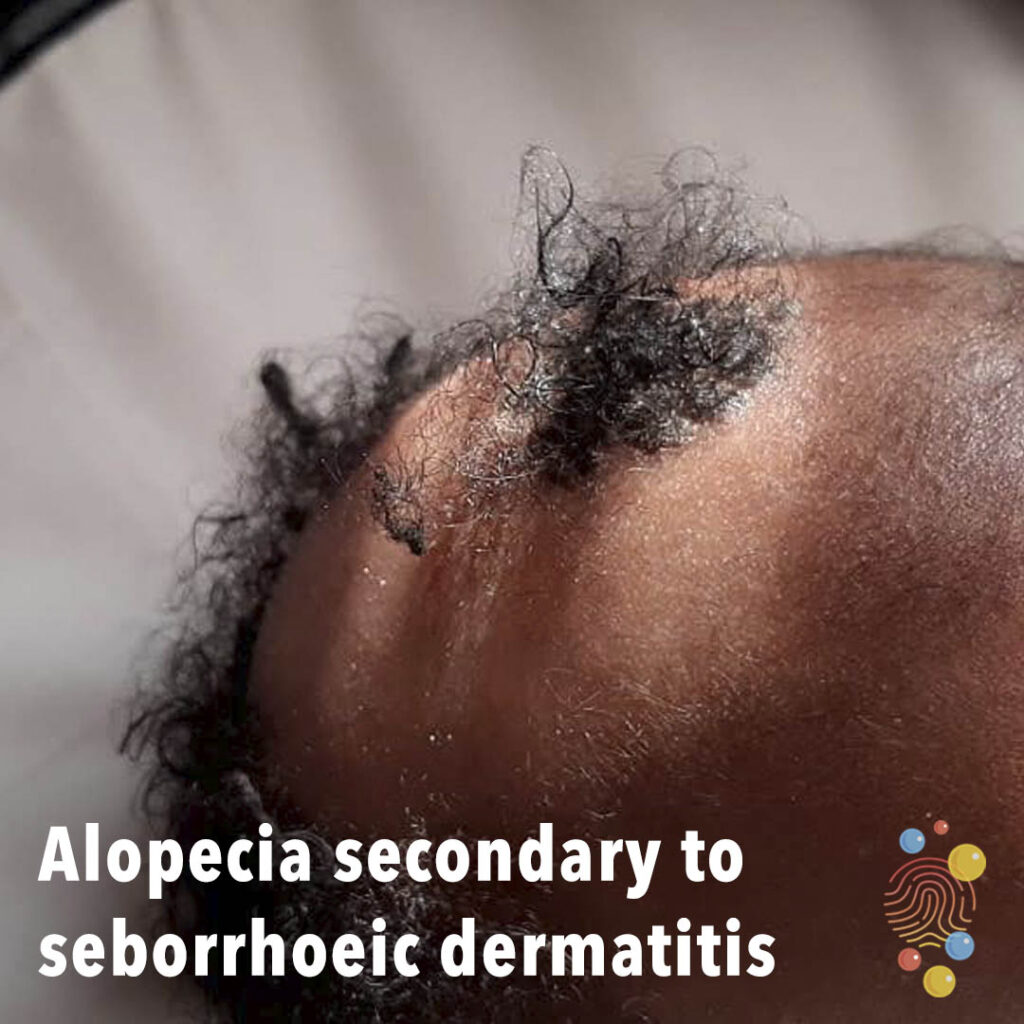
Alopecia Secondary To Seborrhoeic Dermatitis
Multi-focal non-scarring alopecia with preservation of follicular ostia. Scaly, adherent plaque on the scalp.
Learn more about seborrhoeic dermatitis
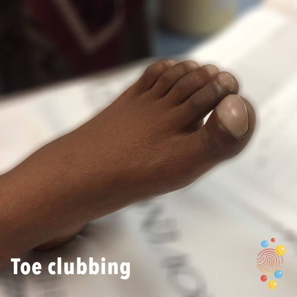
Toe Clubbing
Learn more about clubbing

Streptococcal Pharyngitis
Learn more about streptococcal pharyngitis
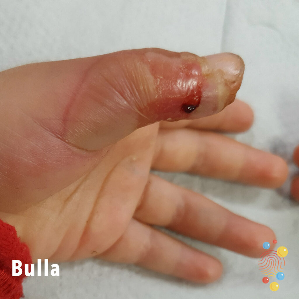
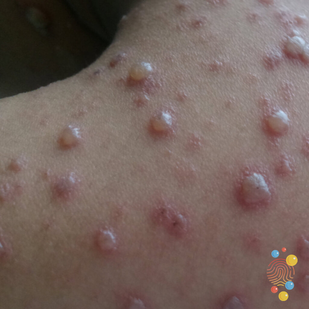
Bulla
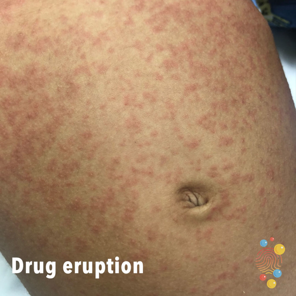
Drug Eruption
Learn more about drug eruptions
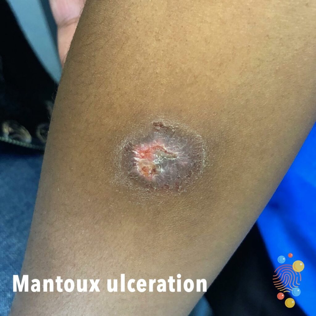
Mantoux Ulceration
Learn more about Mantoux ulceration

Herpes Simplex Virus
Learn more about herpes simplex virus
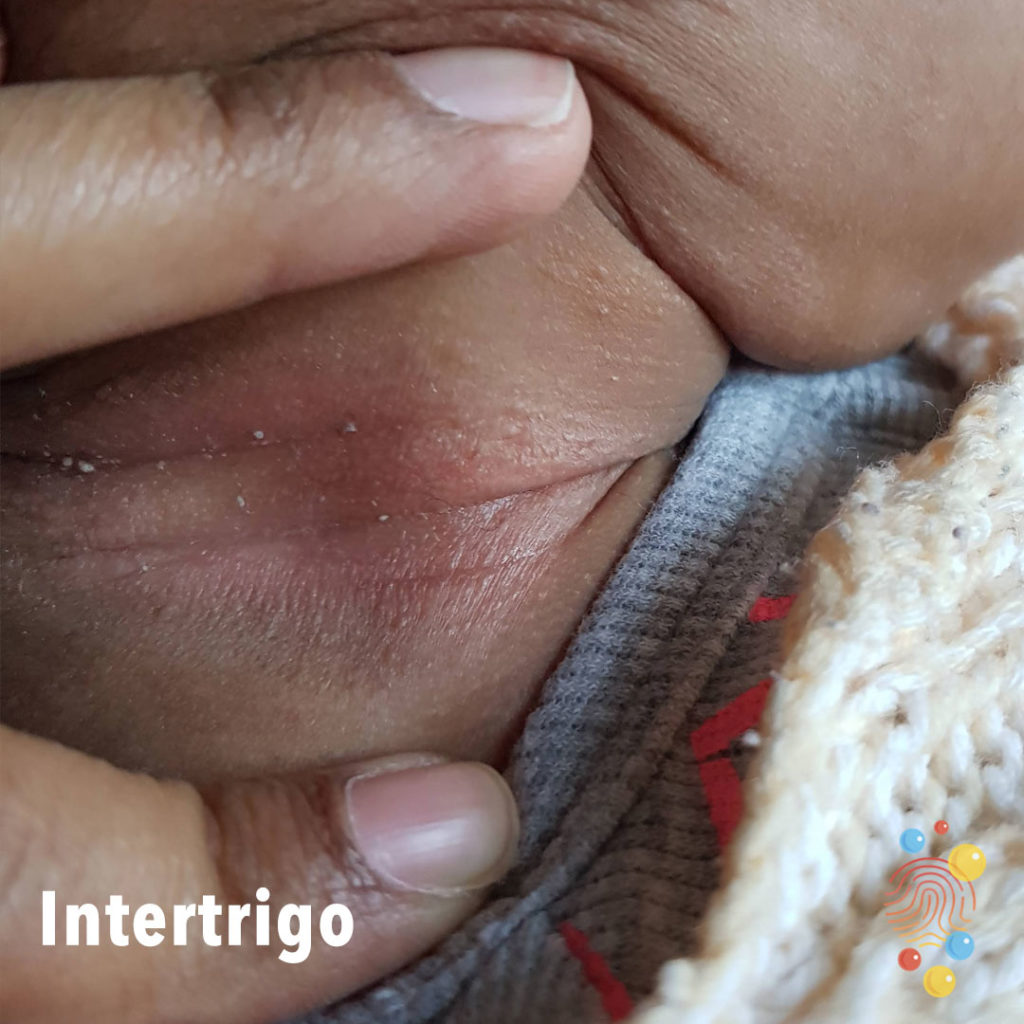
Intertrigo
Learn more about intertrigo

Urticaria
Learn more about urticaria

Viral Exanthem
Learn more about viral exanthem

Flexor sheath infection (ring finger)
Suspected flexor sheath infection of right ring finger with insect bites on her hand.

Dermal Melanocytosis
Learn more about dermal melanocytosis
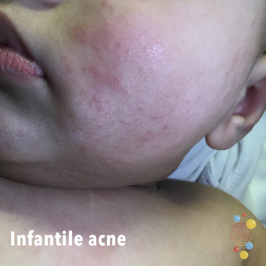
Infantile Acne
Learn more about infantile acne
Eczema
Learn more about eczema
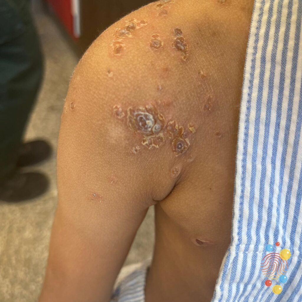
Eczema Herpeticum
Eczema herpeticum (EH) is a rare but serious and contagious skin infection that occurs when the human herpes simplex virus (HSV) infects damaged skin

Periorbital Oedema
Learn more about periorbital oedemas

Balloon Gastrojejunostomy
Learn more about gastrostomies

Steven’s Johnson syndrome
Bruised Toe

Scrofuloderma
Learn more about scrofulderma
Impetigo
Learn more about bullous impetigo
Urticaria
Learn more about urticaria
Hand Foot And Mouth Disease
Learn more about hand, foot and mouth
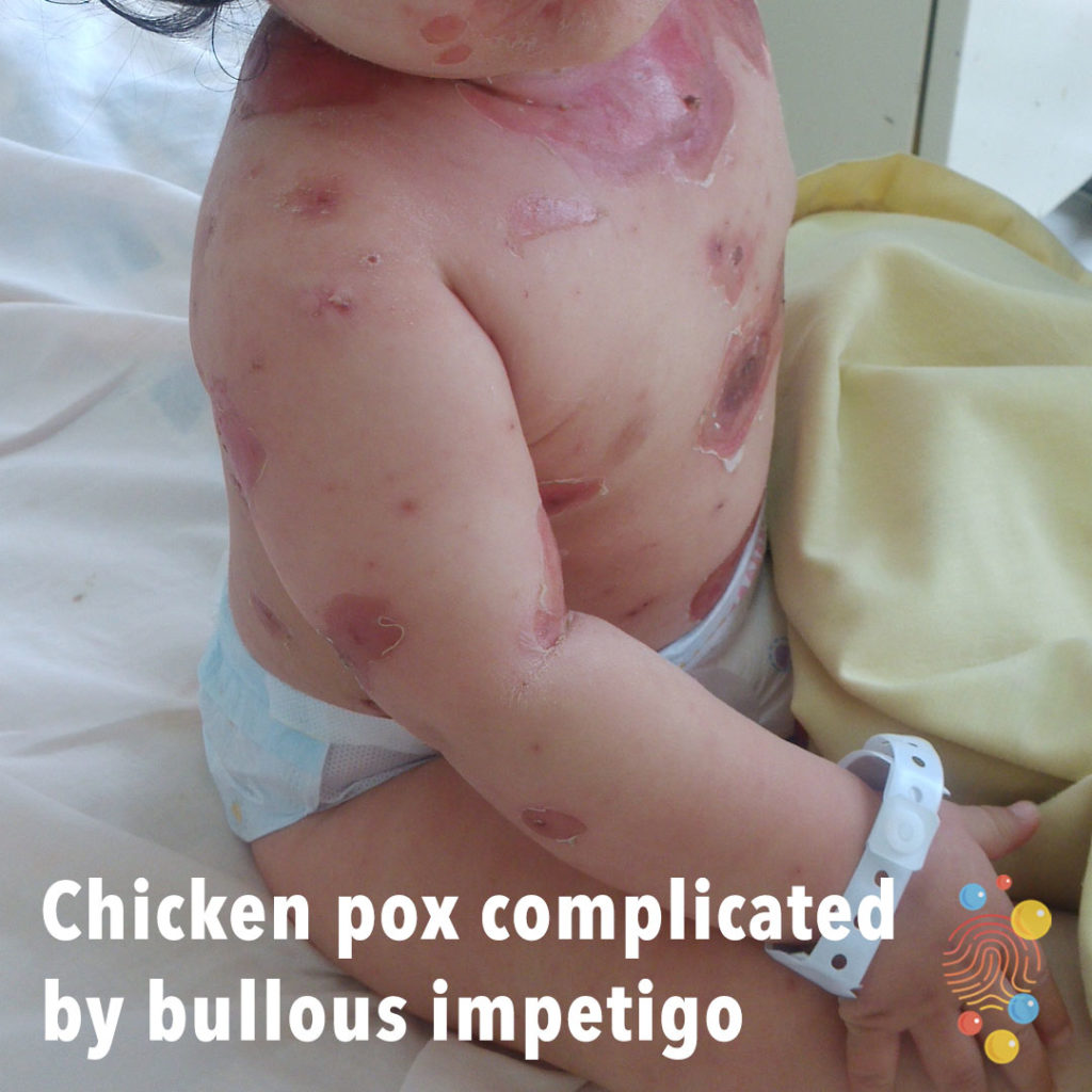
Chicken Pox Complicated By Bullous Impetigo
Learn more about chicken pox |
Learn more about bullous impetigo

Dermal melanocytosis
Learn more about dermal melanocytosis
Viral Exanthem
Learn more about viral exanthem

Eczema
Learn more about eczema
Erythema Toxicum
Learn more about erythema toxicum
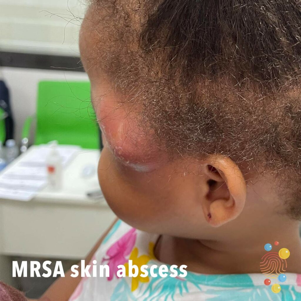
MRSA Skin Abscess
Red tender fluctuant swelling consistent with abscess in this case caused by MRSA.
Eczema
Learn more about eczema
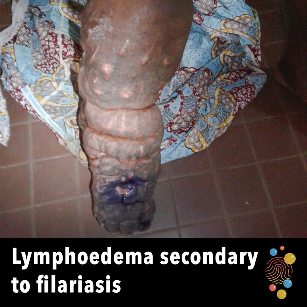
Lymphoedema secondary to filariasis
Learn more about lymphoedema
Henoch-Schonlein Purpura
Learn more about Henoch-Schonlein purpura
Granuloma Annulare
Learn more about granuloma annulare
Folliculitis
Learn more about folliculitis
Roseola
Roseola is a common infection that usually affects children by age 2.
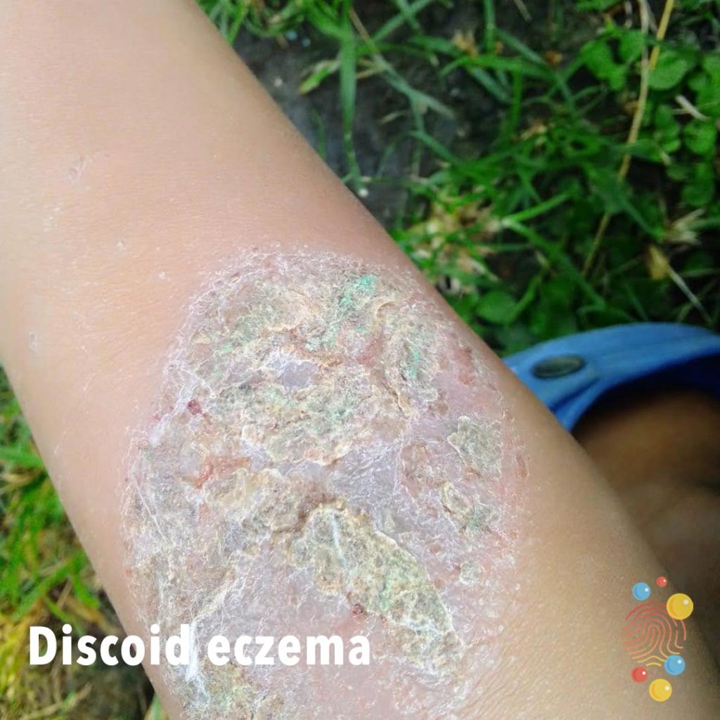
Discoid Eczema
Learn more about eczema
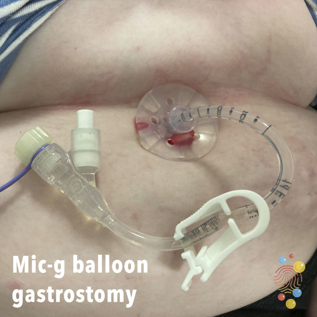
Mic-G Balloon Gastrostomy
Learn more about gastrostomies
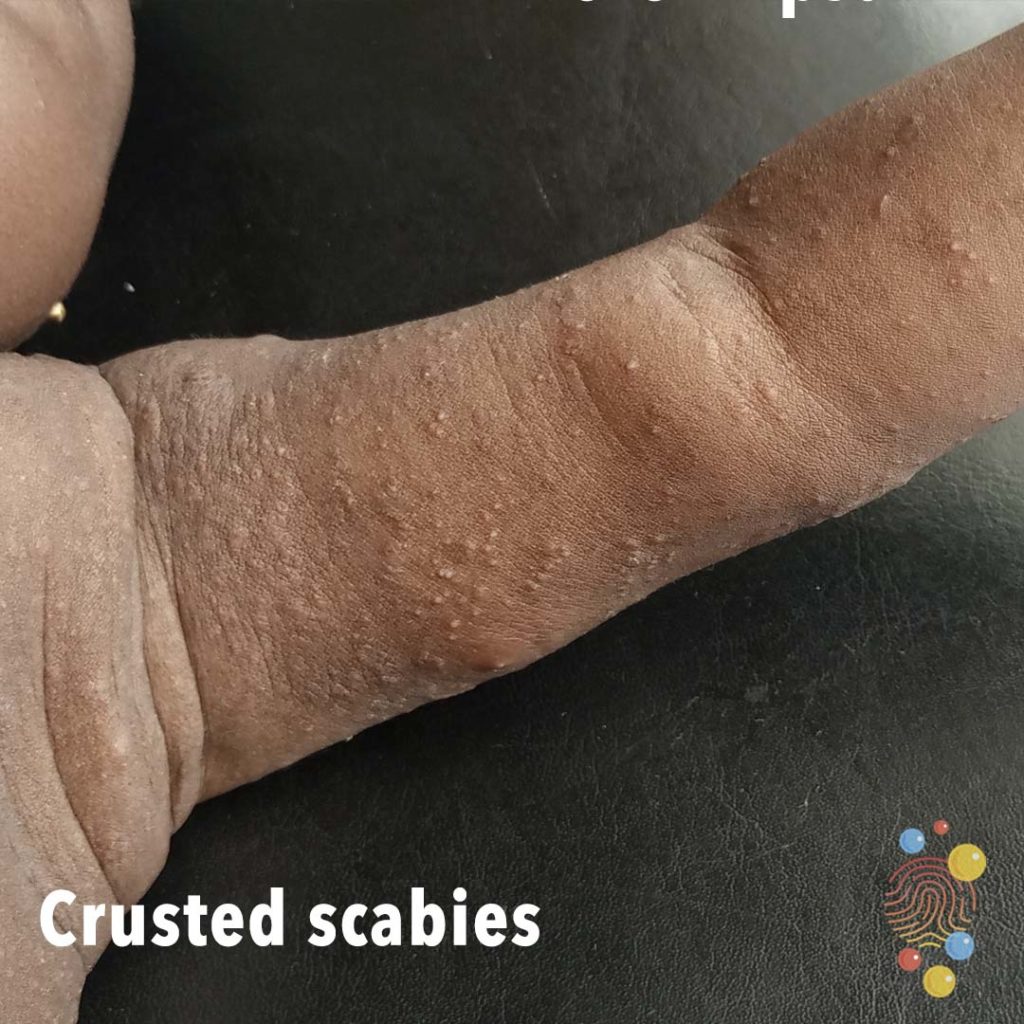
Crusted Scabies
Learn more about scabies
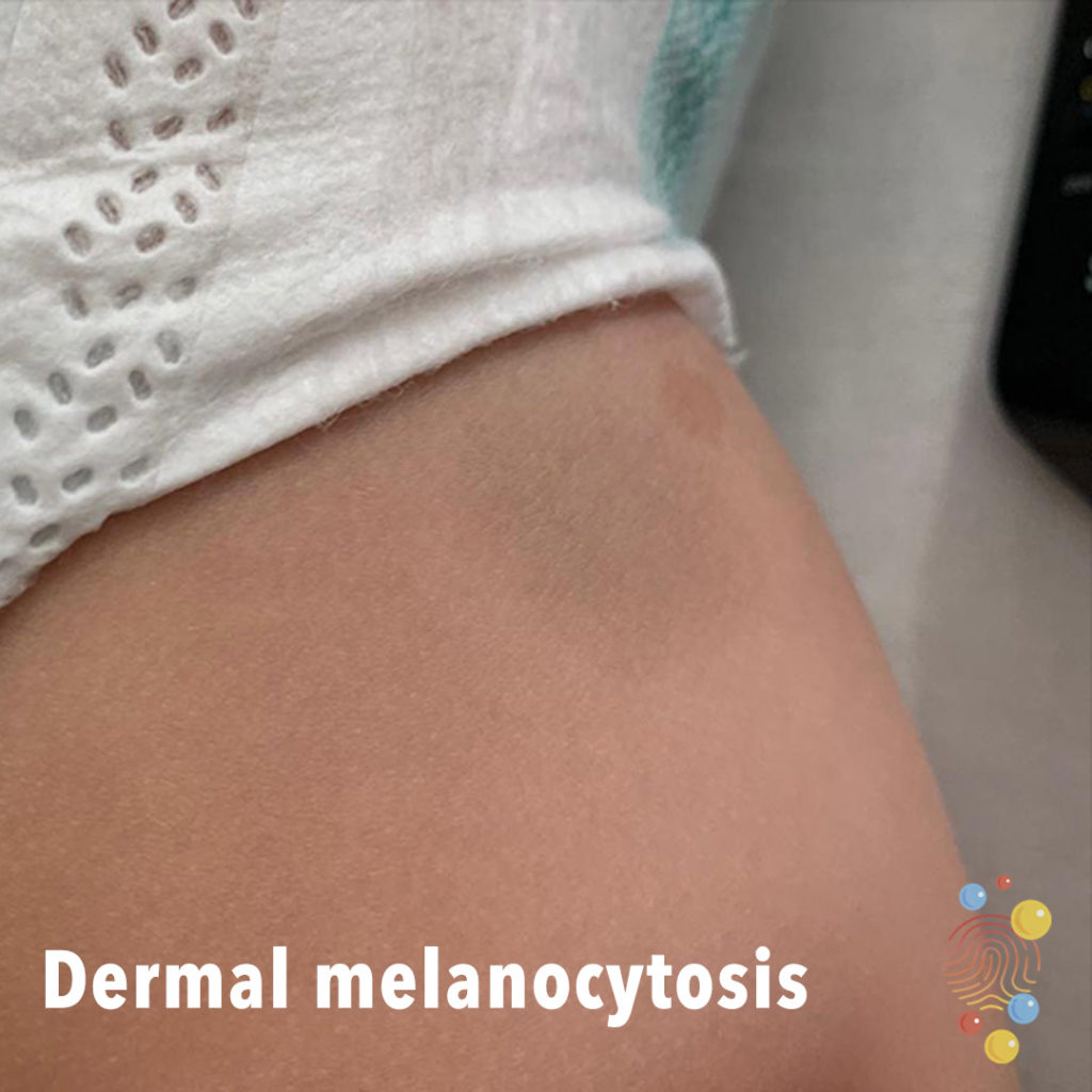
Dermal Melanocytosis
Learn more about dermal melanocytosis

Mouth Injury
Eczema
Learn more about eczema
Follicular Eczema
Learn more about eczema
Abrasion
Scabies
Learn more about scabies
Eczema
Learn more about eczema
Molluscum contagiosum
Learn more about molluscum contagiosum

Steven’s Johnson syndrome
Stevens–Johnson syndrome is a type of severe skin reaction. Together with toxic epidermal necrolysis and Stevens–Johnson/toxic epidermal necrolysis overlap, they are considered febrile mucocutaneous drug reactions and probably part of the same spectrum of disease, with SJS being less severe.

Eczema
Learn more about eczema
PIMS-TS
Scattering of erythematous papules.

Petechial rash
Petechiae are tiny spots of bleeding under the skin. They can be caused by a simple injury, straining or more serious conditions. If you have pinpoint-sized red dots under your skin that spread quickly, or petechiae plus other symptoms, seek medical attention.
Dermal melanocytosis
Learn more about dermal melanocytosis

Gianotti Crosti
Gianotti-Crosti syndrome (GCS) is a skin condition that usually affects children, but can also occur in adolescents and adults
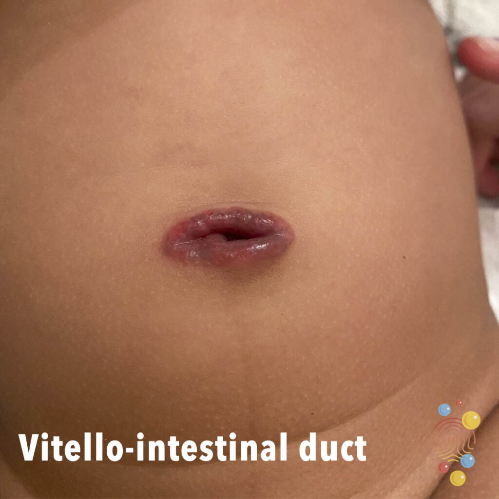
Vitello Intestinal Duct
Well circumscribed violaceous umbilical plaque.
Exacerbation of eczema with likely herpetic lesions

Acute haemorrhagic oedema of infancy
Multiple urticated bruises, some of which have a targetoid appearance

Bullous insect bite reaction
Learn more about bites
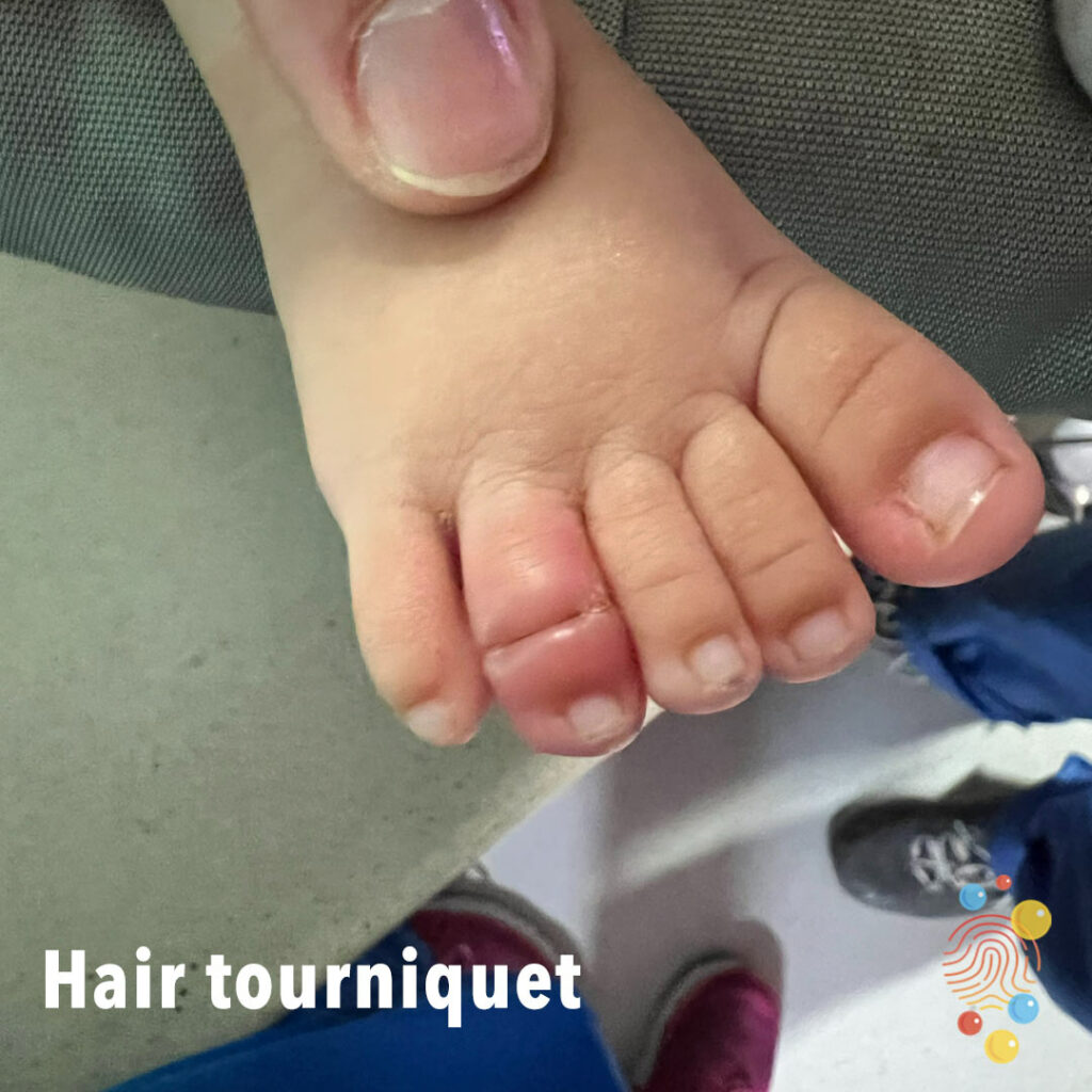
Hair Tourniquet