Melanocytic nevi are a rapid increase in the number of melanocytes, the pigment producing cells in our bodies. These lesions, which are sometimes called moles, are benign, meaning that they are not malignant. When present at birth, or developing within the first 2 years of life, they are called congenital melanocytic nevi (CMN).
Cause
All melanocytic nevi are caused by an overproliferation, or a rapid increase, in the number of melanocytes within the epidermal layer of the skin. The reason for this overproliferation is not fully understood but localized genetic abnormalities are thought to play an immense role.
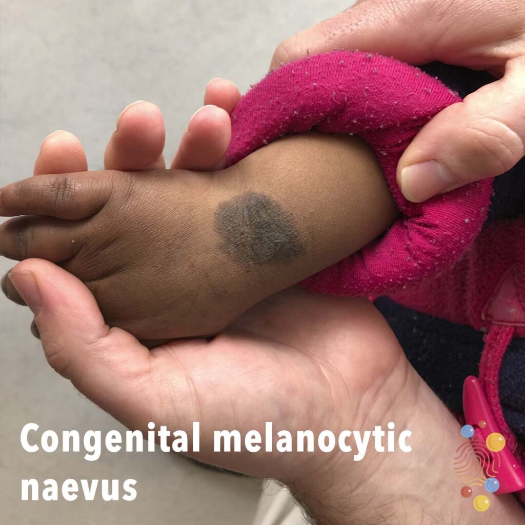
Appearance
CMN can vary in color, size, and texture. In the majority of cases, CMN lesions appear brown to black, but in some individuals, they can look pink or red. It is normal for the CMN lesions to darken over time but in some individuals, they may lighten. CMN lesions can appear as small spots or as very large patches covering entire regions of skin, such as the face, back, or neck. They can be smooth, bumpy, or even covered in hair.
Symptoms
Aside from the appearance, CMN is usually asymptomatic. Xerosis, otherwise known as dry skin, can commonly occur as a result of decreased function of the oil (sebaceous) and sweat (eccrine) producing glands in the body. These lesions can ulcerate. Smaller lesions can also form close to the site of the main lesion. These smaller lesions are called satellite lesions. In some cases, CMN can be accompanied by neurological abnormalities.
Complications
Most melanocytic nevi are without complications; however, individuals with CMN covering large areas of skin are at higher risks for melanomas (a type of skin cancer) and neurocutaneous melanosis, a condition where melanocytes overproliferate in the brain and spinal cord leading to neurological manifestations like seizures and delay in development. There may also be slight abnormalities in the homeostasis of hormones; more specifically, signs of puberty, such as secondary breast development and pubic hair development, may occur earlier than normally in some patients. Disturbances in the levels of LH and FSH hormones usually accompany these early signs of puberty. Quality of life and self confidence can also be affected when the lesions are very large.
Treatment
If the lesion is small and asymptomatic, no treatment is needed. Larger lesions or symptomatic lesions are often first biopsied and then treated by surgical removal. Surgery is indicated when there is risk that the lesion can transform from benign to malignant, when melanoma develops, or if the patient is unsatisfied with the appearance of the lesion. Surgery is also indicated if the lesion is in a region that is difficult for the patient to monitor, such as on the back, or if there are changes to the lesion. Surgical treatment can result in permanent scarring; therefore, patients need to consider this before undergoing surgical treatment.
References
Congenital melanocytic naevus. Congenital melanocytic naevus | DermNet NZ. (n.d.). https://dermnetnz.org/topics/congenital-melanocytic-naevi/
Primary Care Dermatology Society. (n.d.). Congenital melanocytic naevus. Primary Care Dermatology Society. https://www.pcds.org.uk/clinical-guidance/congenital-melanocytic-naevus