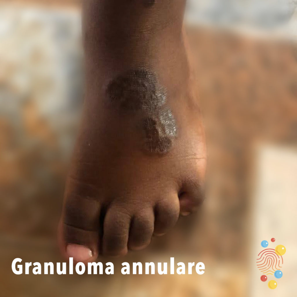
Raised purplish confluent are in a ring pattern on the dorsal area of the foot.
Learn more about granuloma annulare

Erythematous papules + microvesicles.
Learn more about miliaria


Multiple planar warts on the sole of the foot , some in a mosaic appearance.
Learn more about warts

Multiple scaly lesions on the lower legs, some appear papular.
Learn more about Gianotti-Crosti syndrome

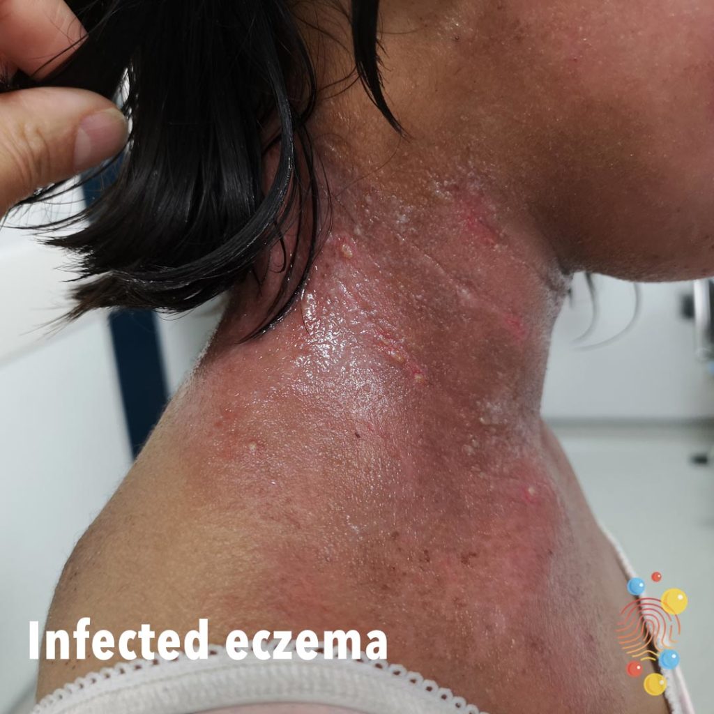
Impetiginized Eczema

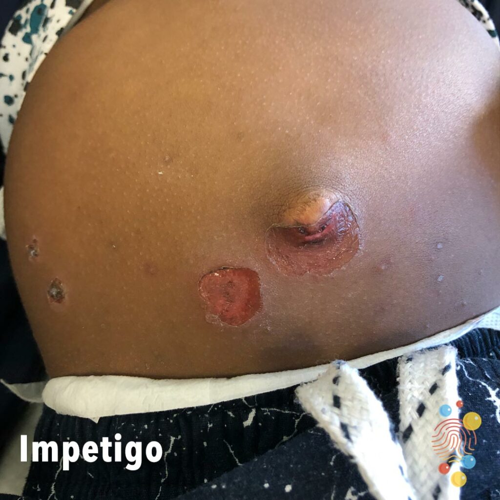
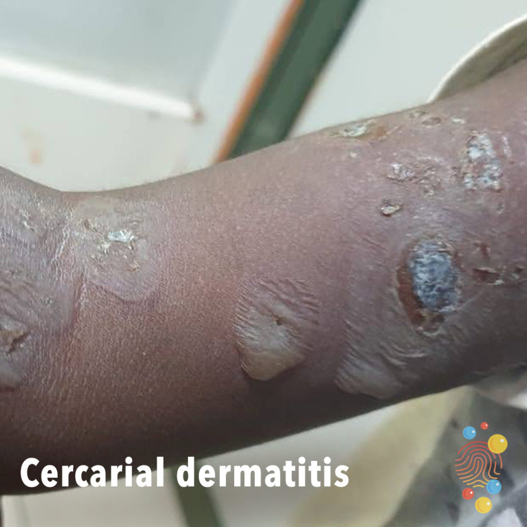
Extensive healing erosions with haemorrhagic crust and a collarette of scale
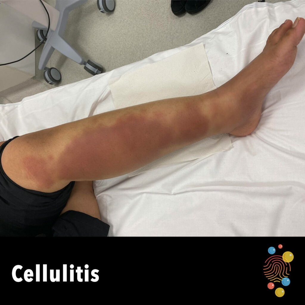
Erythematous swollen leg.
Learn more about cellulitis
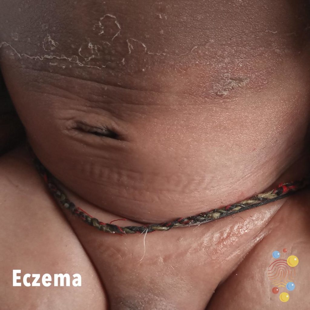
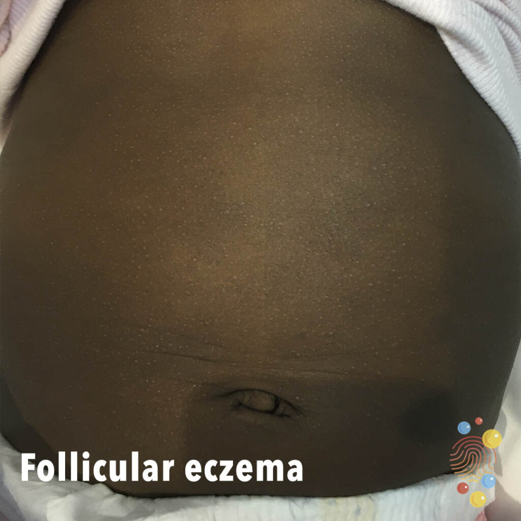
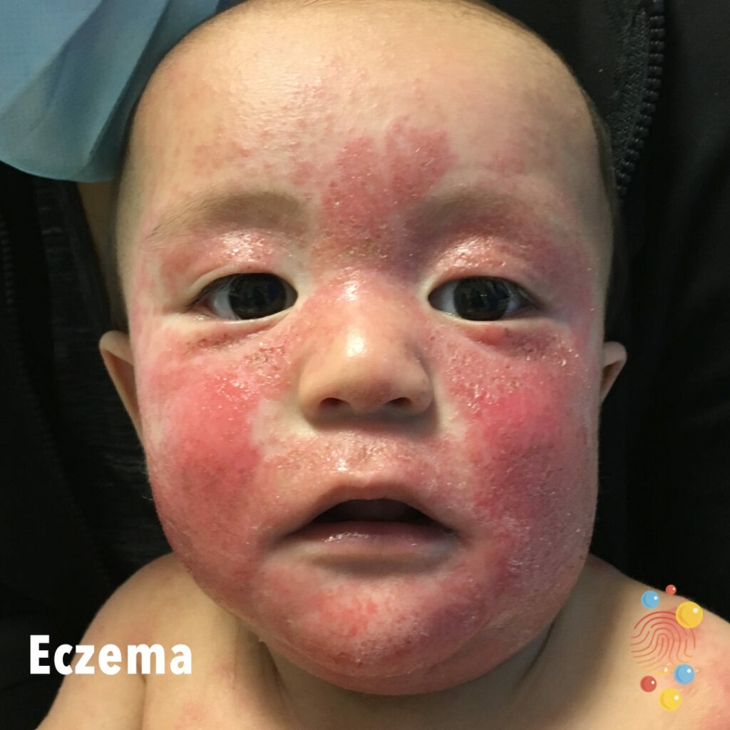
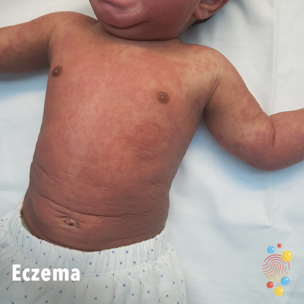
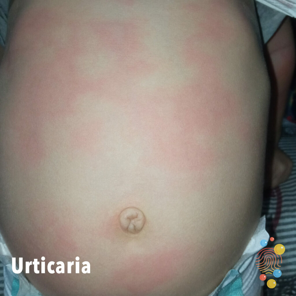
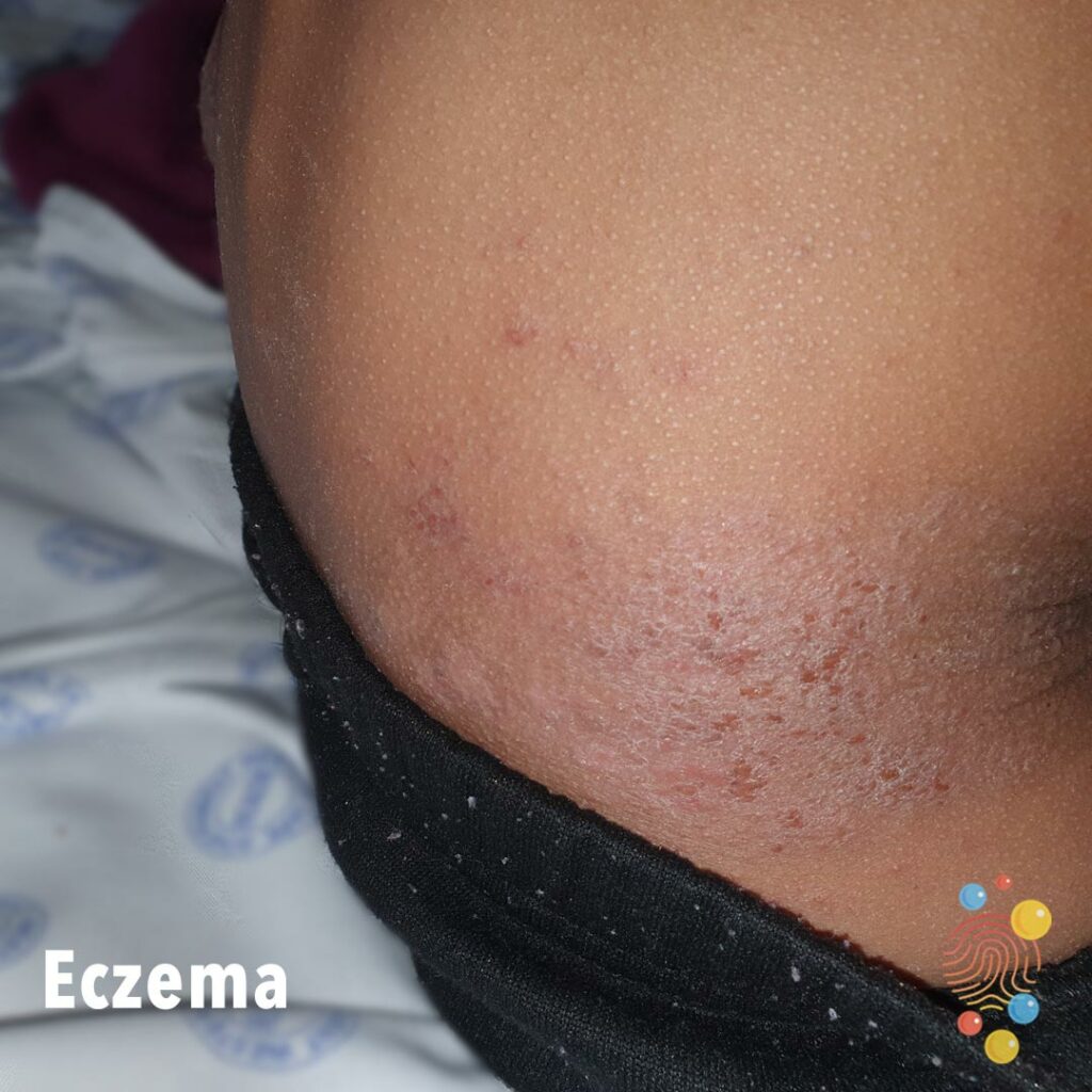
Faint scale on the abdomen with periumbilical wrinkling and an eczematous patch on the upper abdomen.
Learn more about eczema
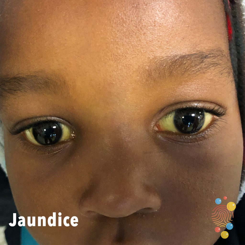
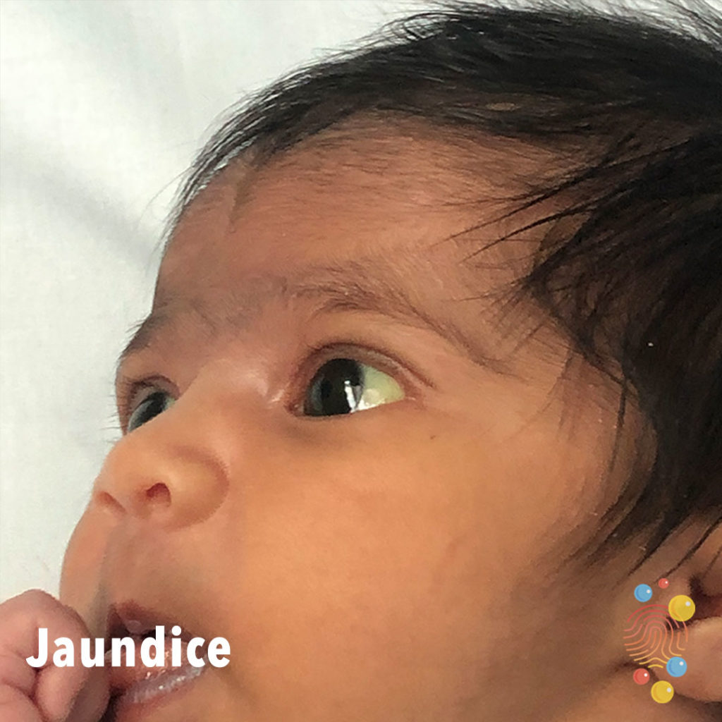
Scleral icterus.
Learn more about jaundice
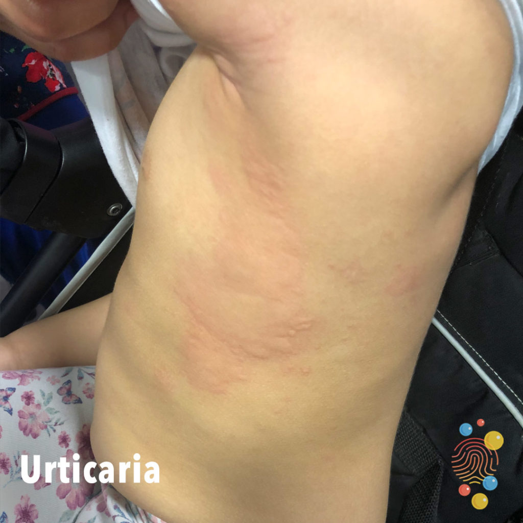
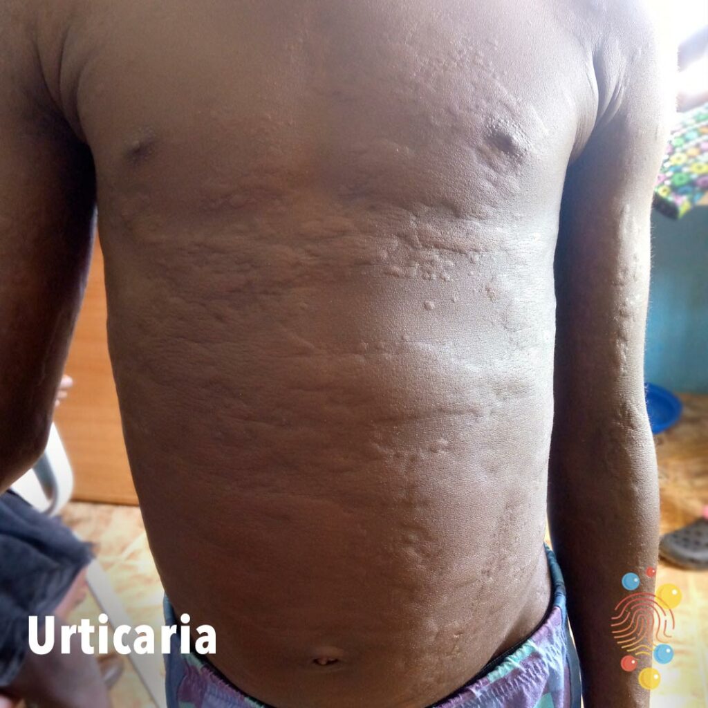
Superficial raised erythematous swelling/wheals affecting the chest.
Learn more about urticaria
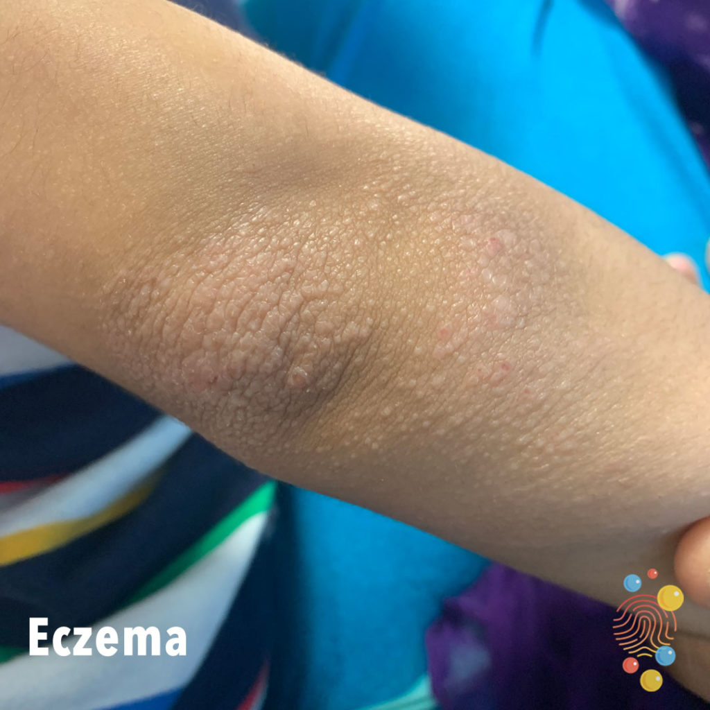
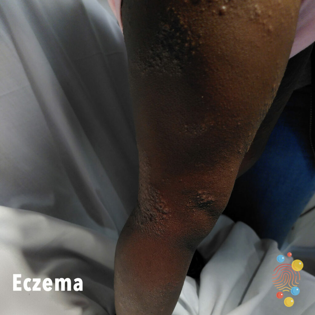
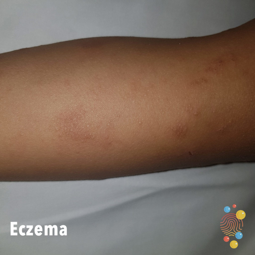
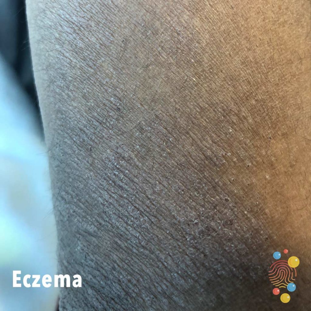
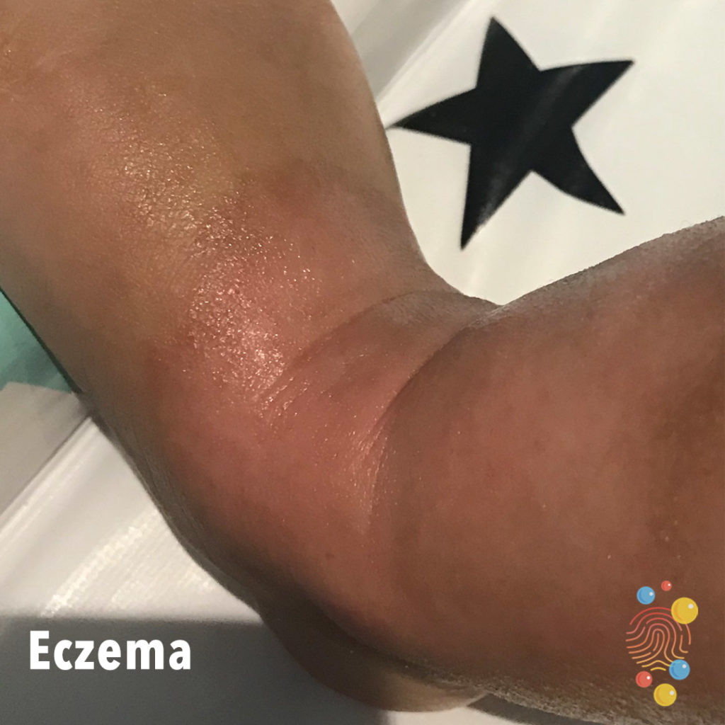
Lichenified, pale, papular eruption on the extensor surface of the arm. Some excoriation. Possibly some xerosis.
Learn more about eczema
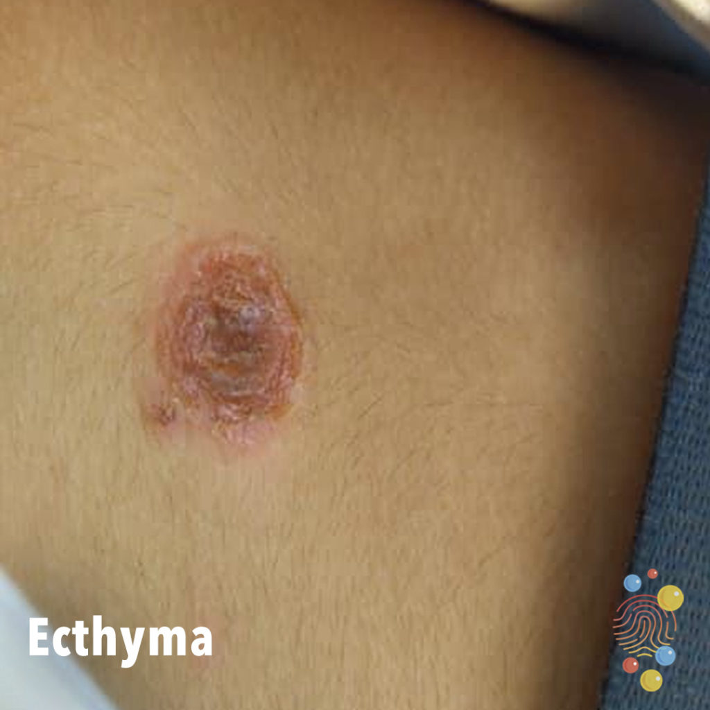
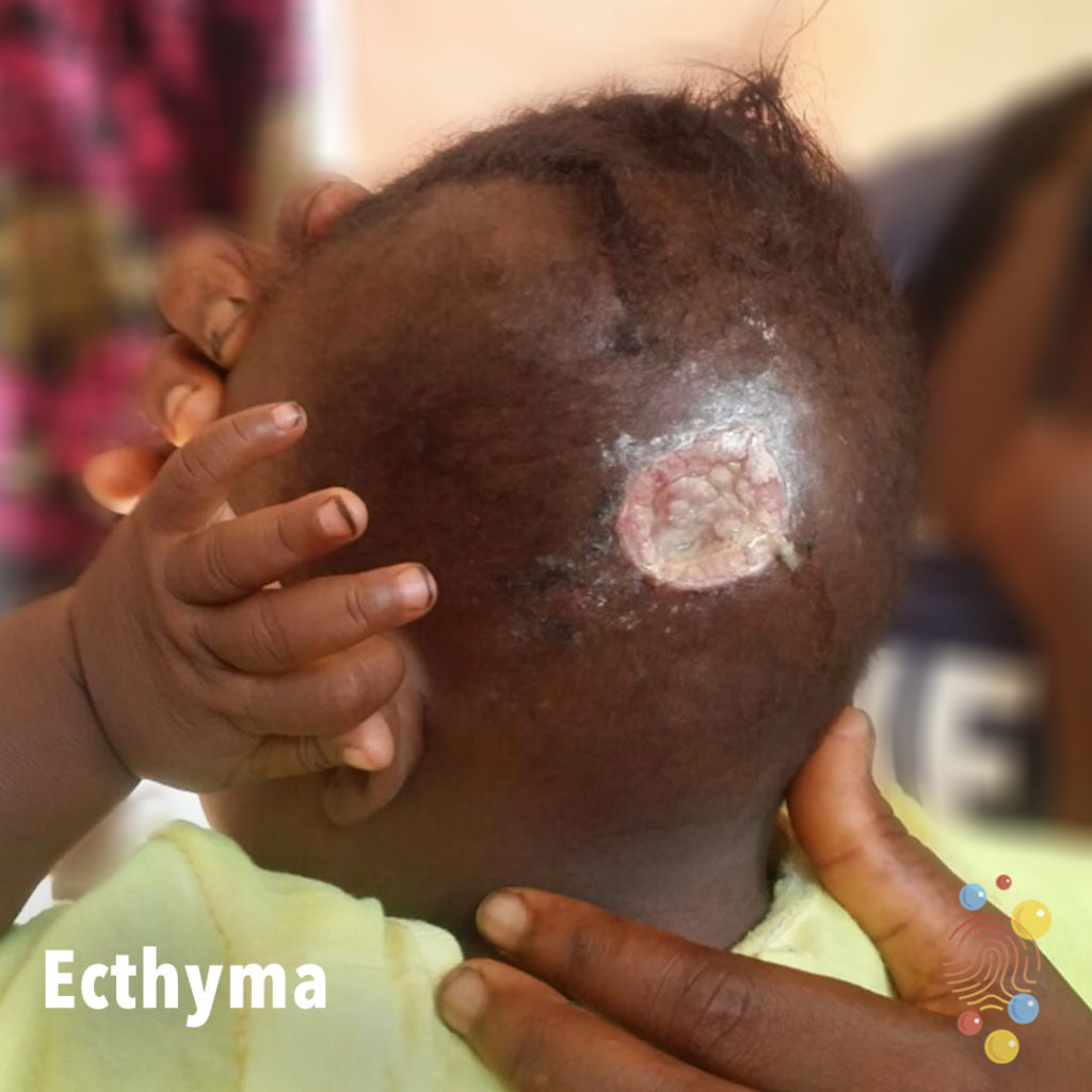
Infected, round ulcer with erythematous borders.
Learn more about ecthymas
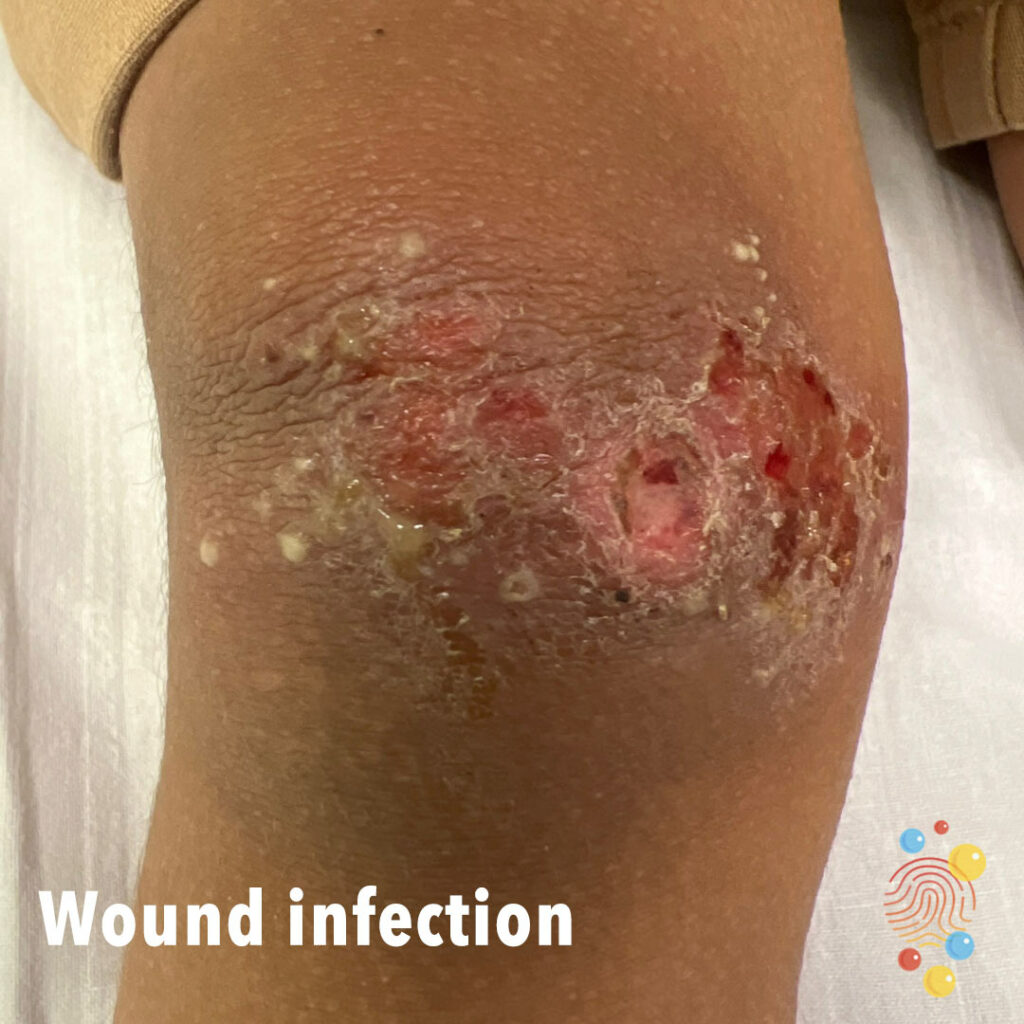
3 year old boy. Tripped and fell twice in a week, a few days later noted to have pus in wound. Skin infection secondary to wound.
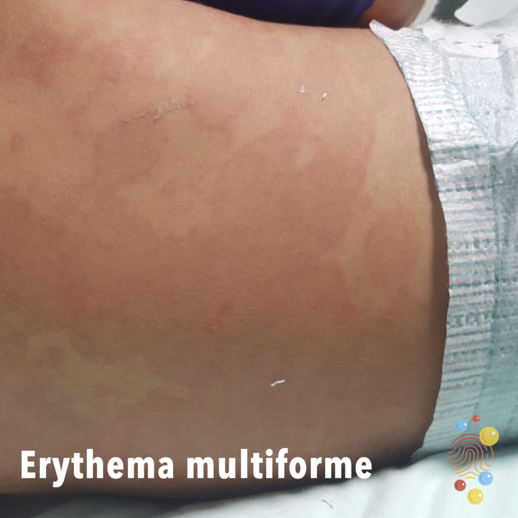
Multiple erythematous targetoid macular lesions on the back.
Learn more about erythema multiforme
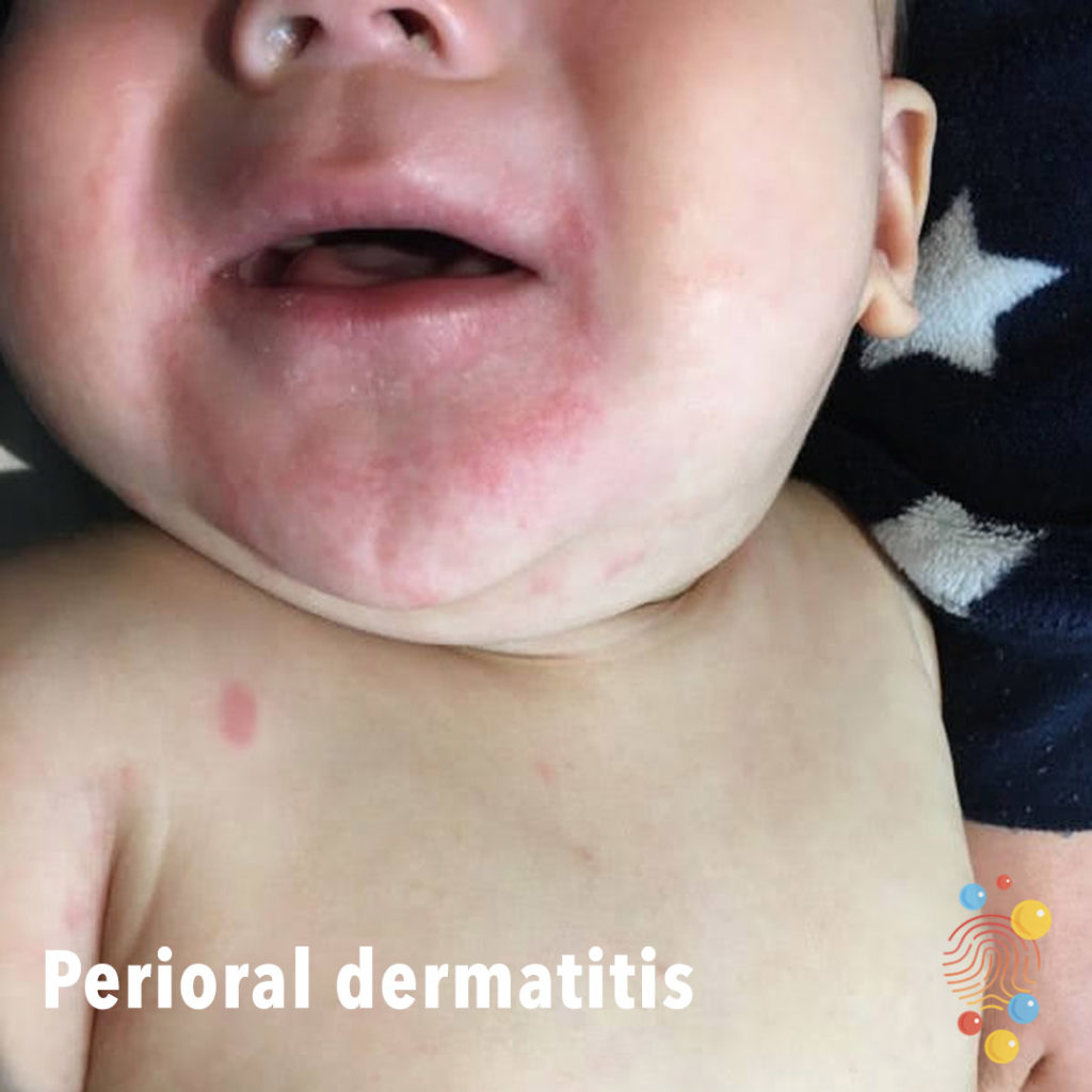
Erythematous, maculopapular eruption with sparing of vermillion.
Learn more about eczema

Multiple, small, superficial, clear blisters.
Learn more about miliaria
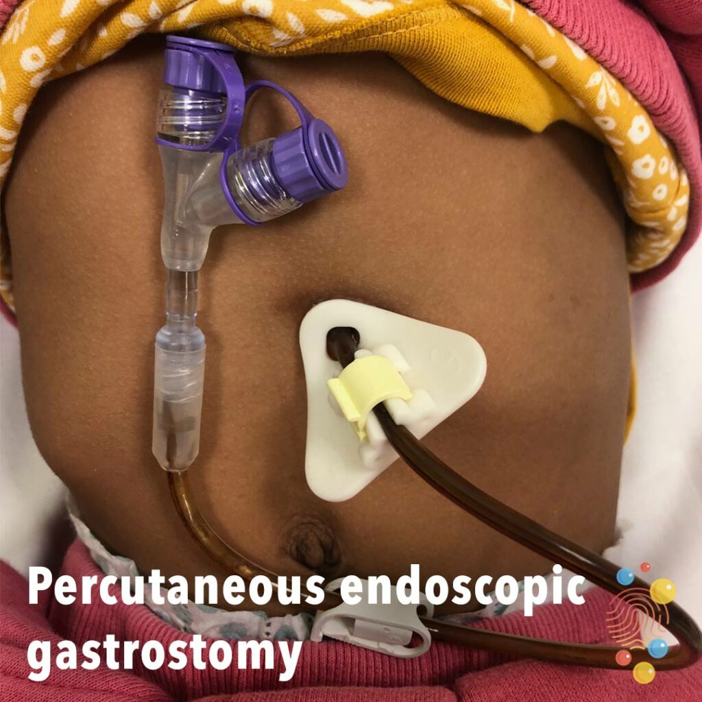
This tube has a triangular Freka PEG fixation plate and Cor-Flo end part. The tube itself is held in place by a plastic disc in the stomach and by the triangular plate on the outside. The tube has been in place for some time and the plastic has darkened with use.
Learn more about gastrostomies
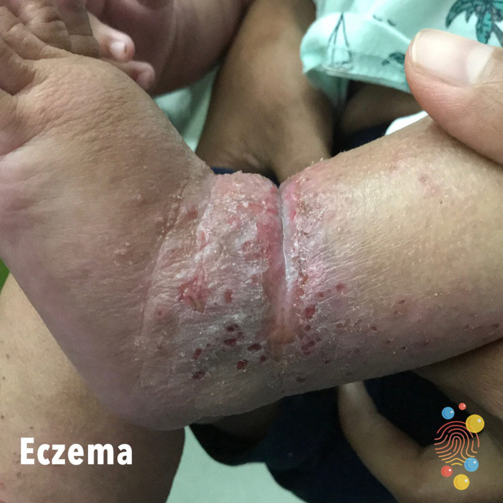
Lichenified excoriated patch with erosions and weeping
Learn more about eczema

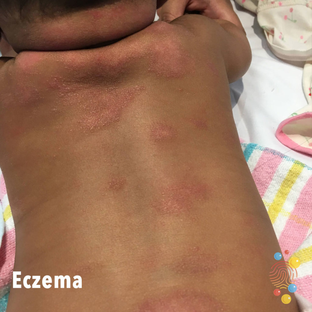
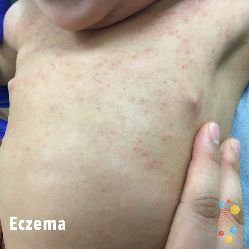
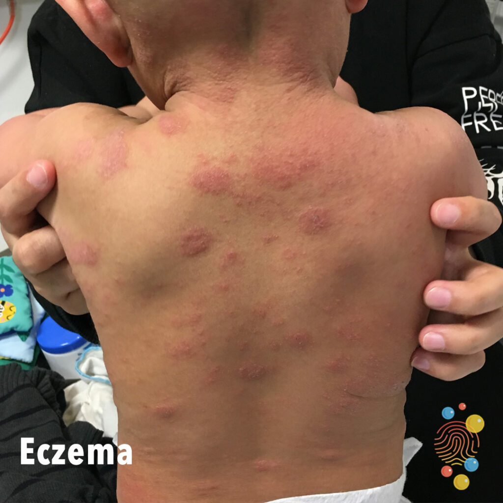
Grouped erythematous slightly urticated papules on trunk with surrounding dry skin.
Learn more about eczema
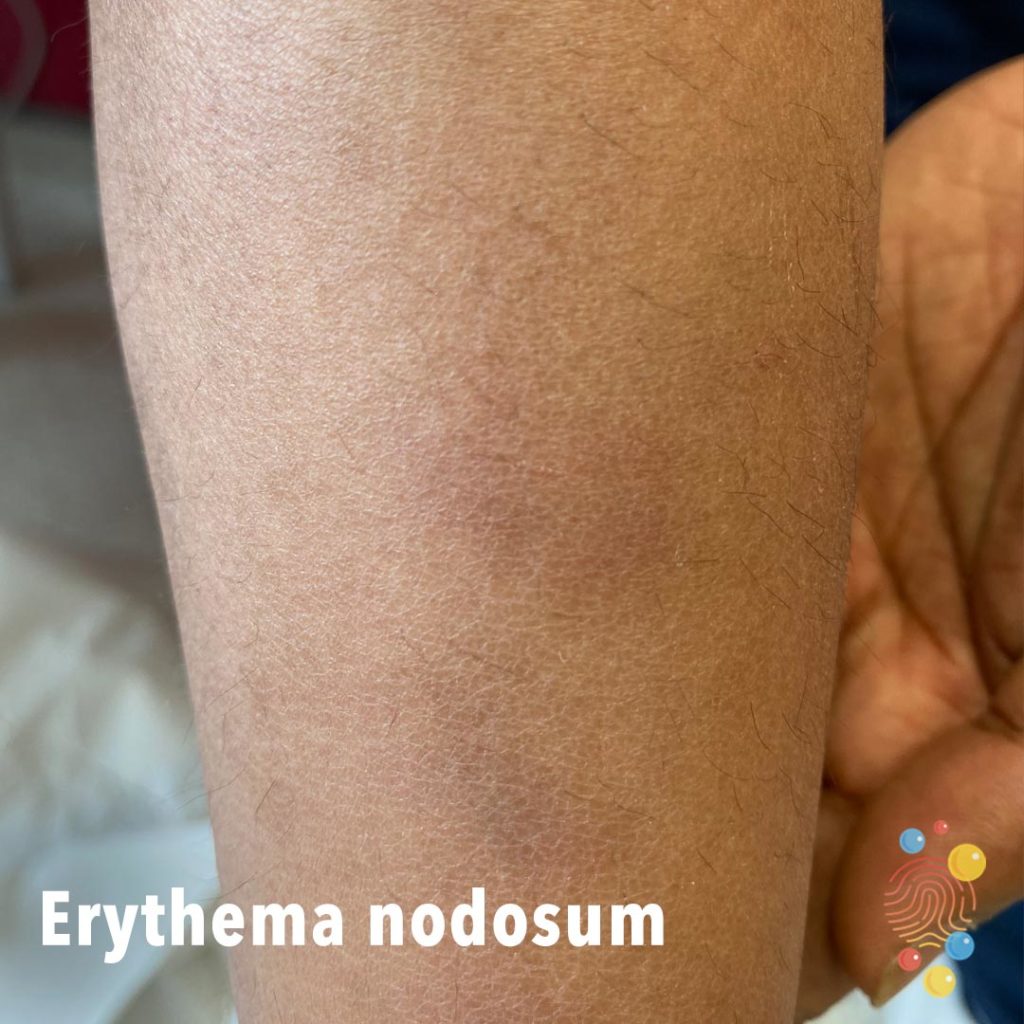
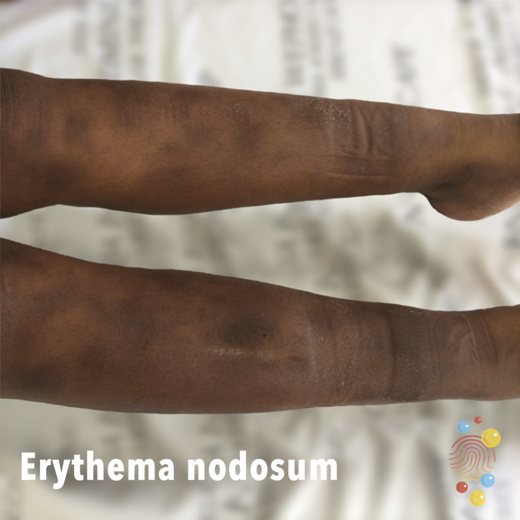
Multiple erythematous to violaceous subcutaneous nodules.
Learn more about erythema nodosum
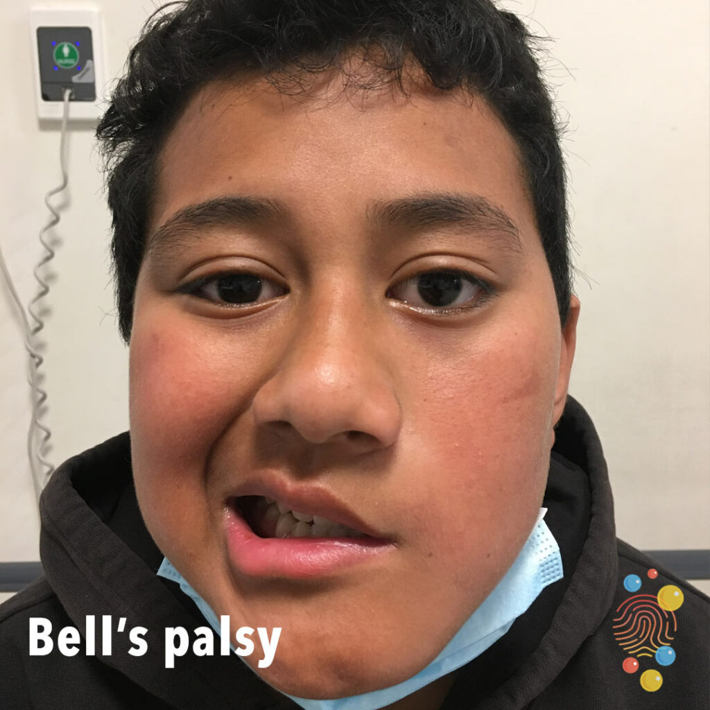
Left facial droop.
Learn more about Bell’s palsy

Petechiae of the hard palate. Common in viral infections.
Learn more about petechiae

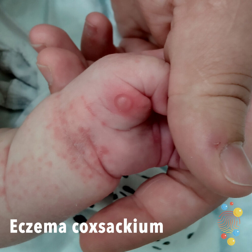
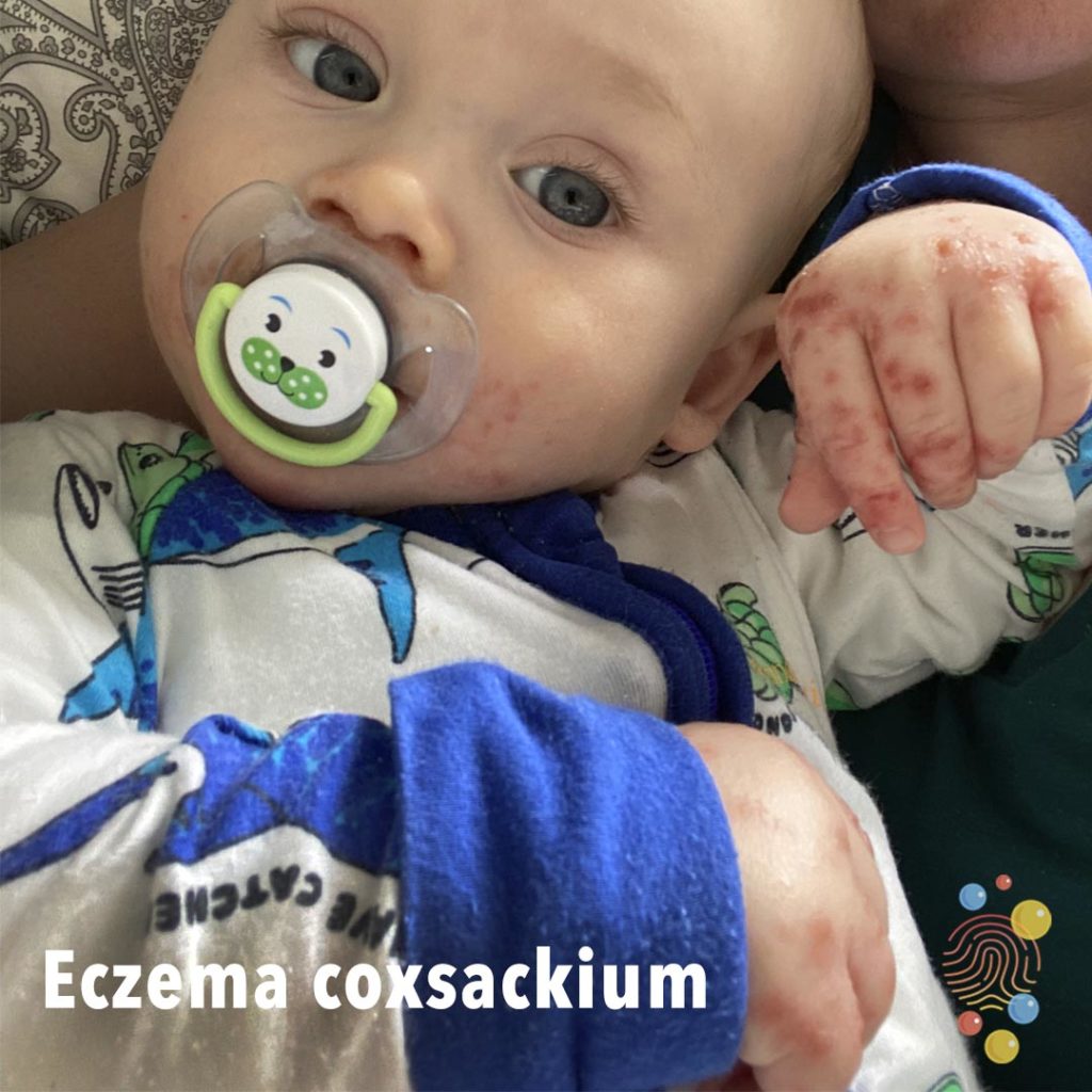
Eczema Coxsackium
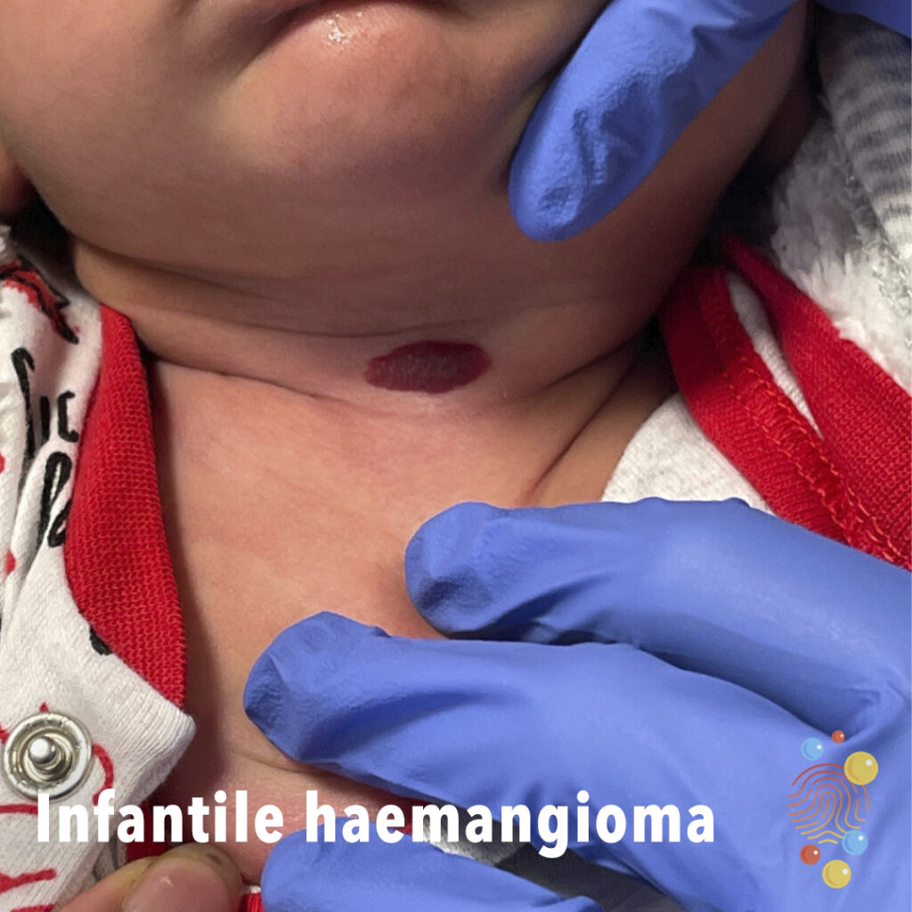
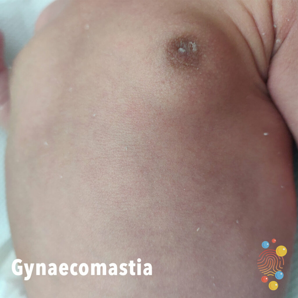
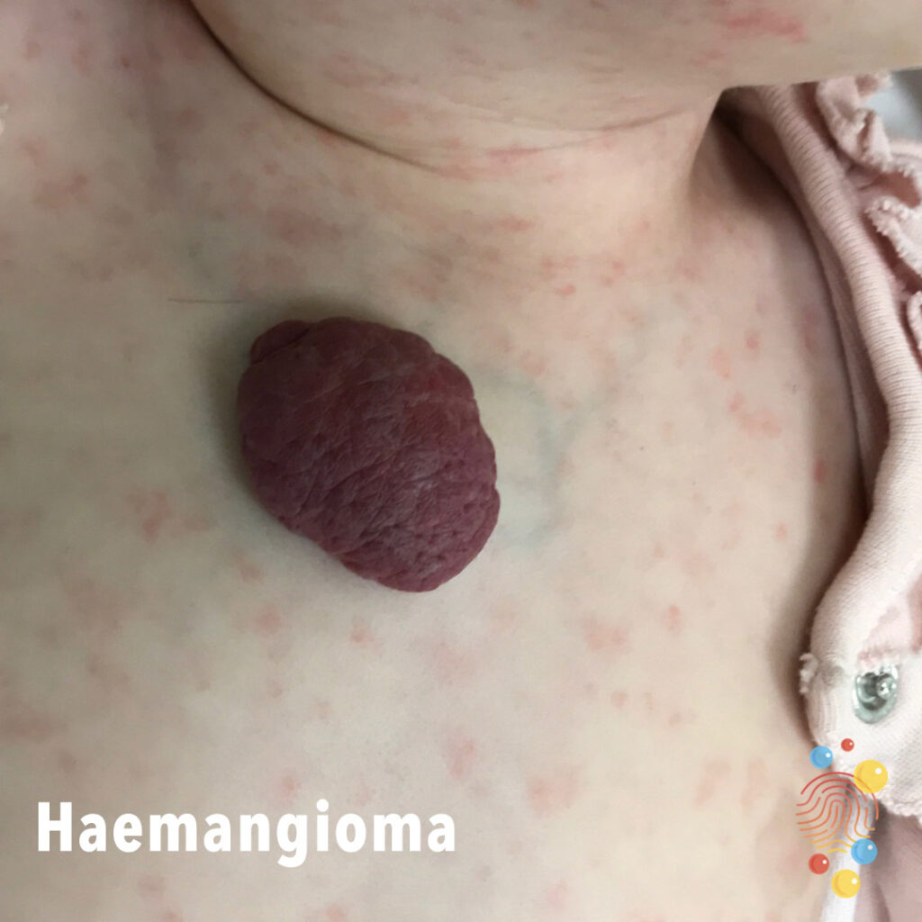
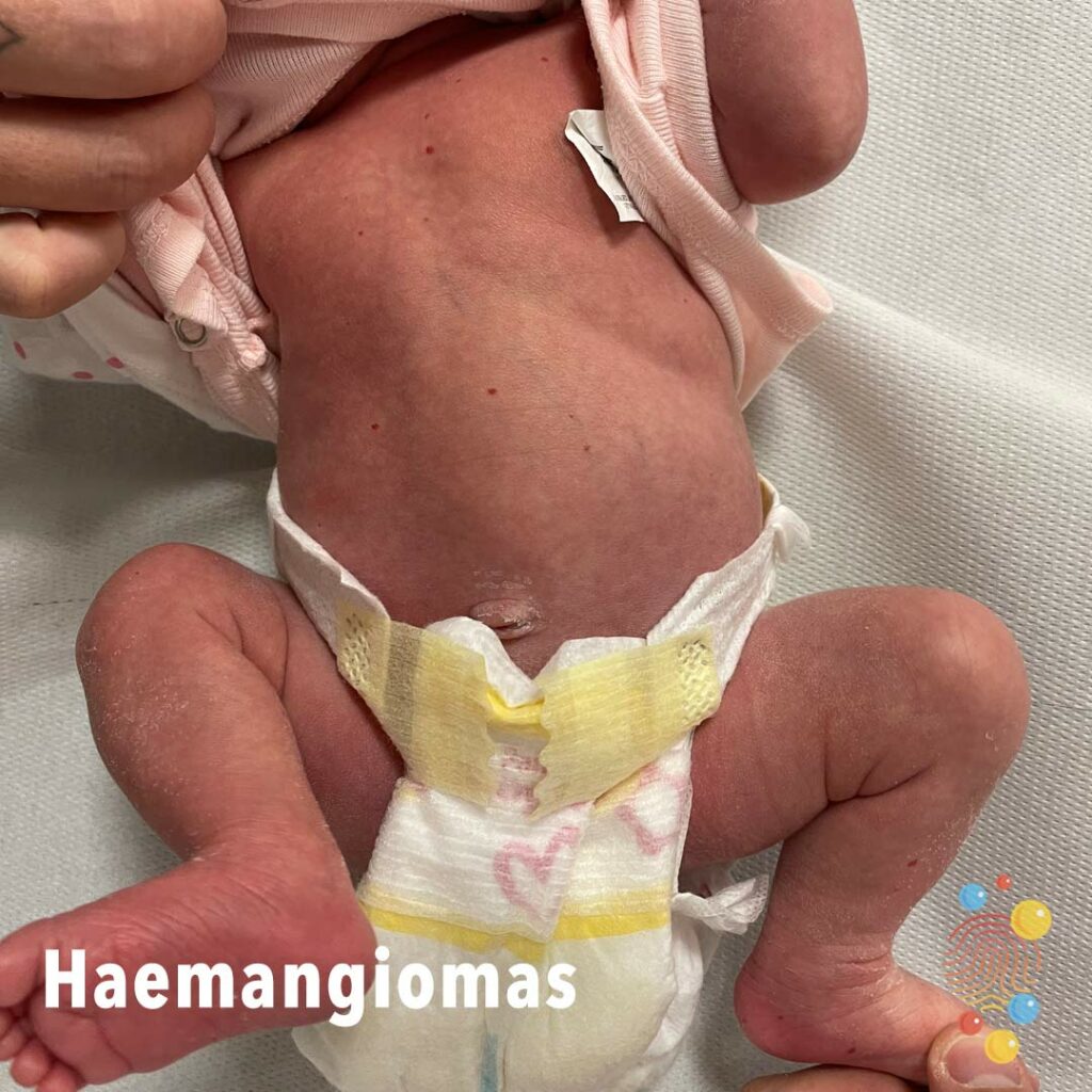
Superficial infantile haemangioma on the anterior neck.
Lichenified hyperpigmented plaque with excoriations.
Learn more about eczema
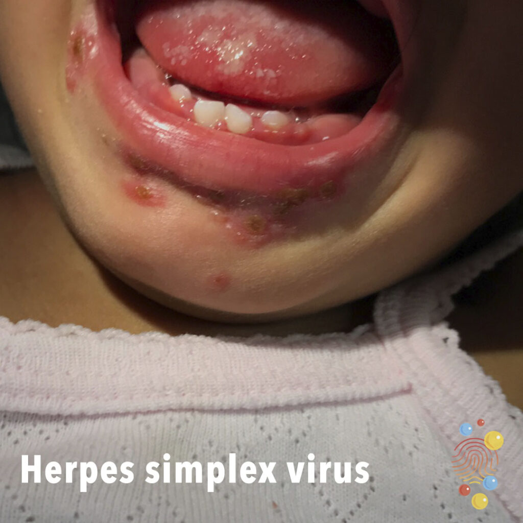
Punctuate grouped ulcers on tongue and crusted vesicles in perioral region.
Learn more about herpes simplex virus
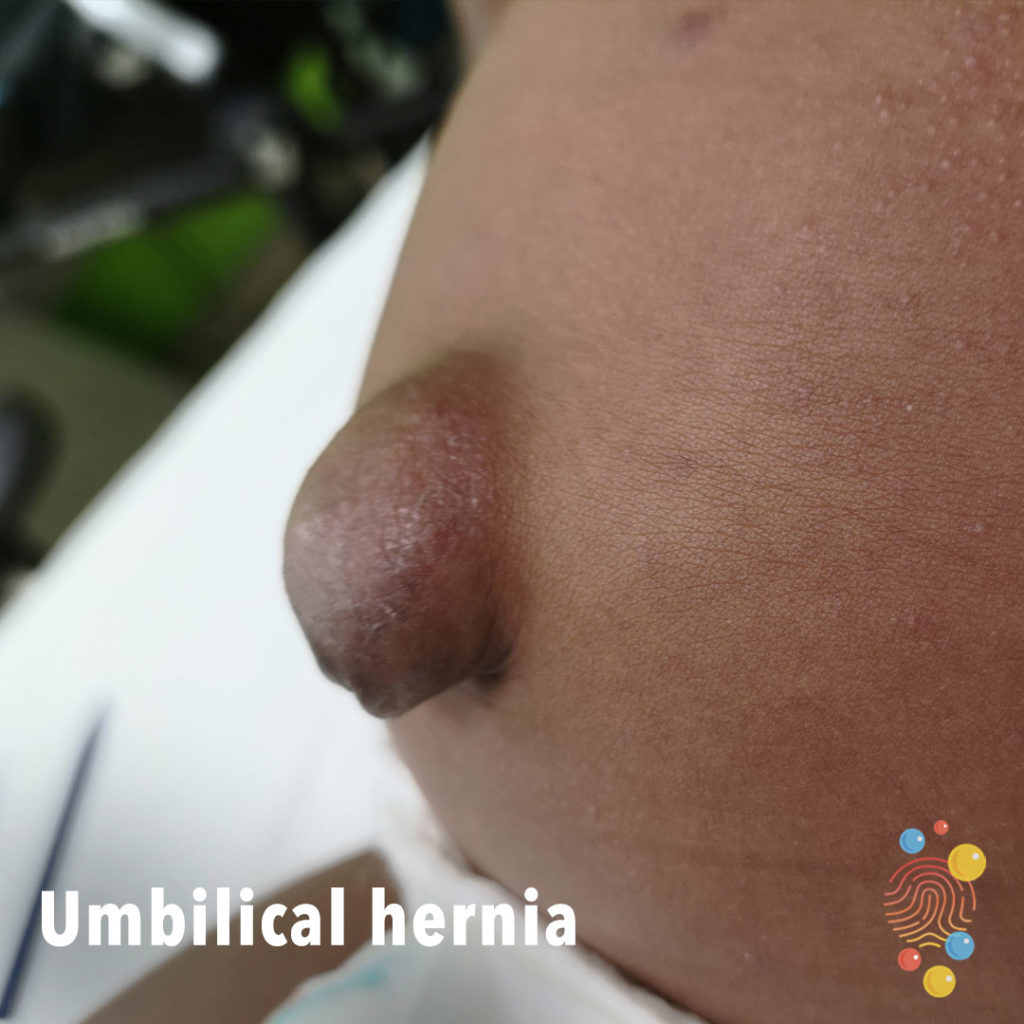
Skin-coloured bulge emanating from the umbilicus with erythema and scale consistent with lichen simplex chronicus.
Learn more about umbilical hernia
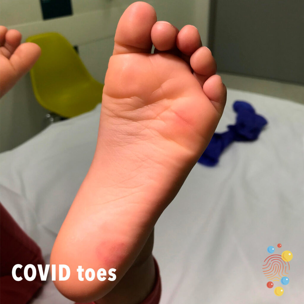
Erythematous macular patch on heel with some surface peeling.
Learn more about COVID

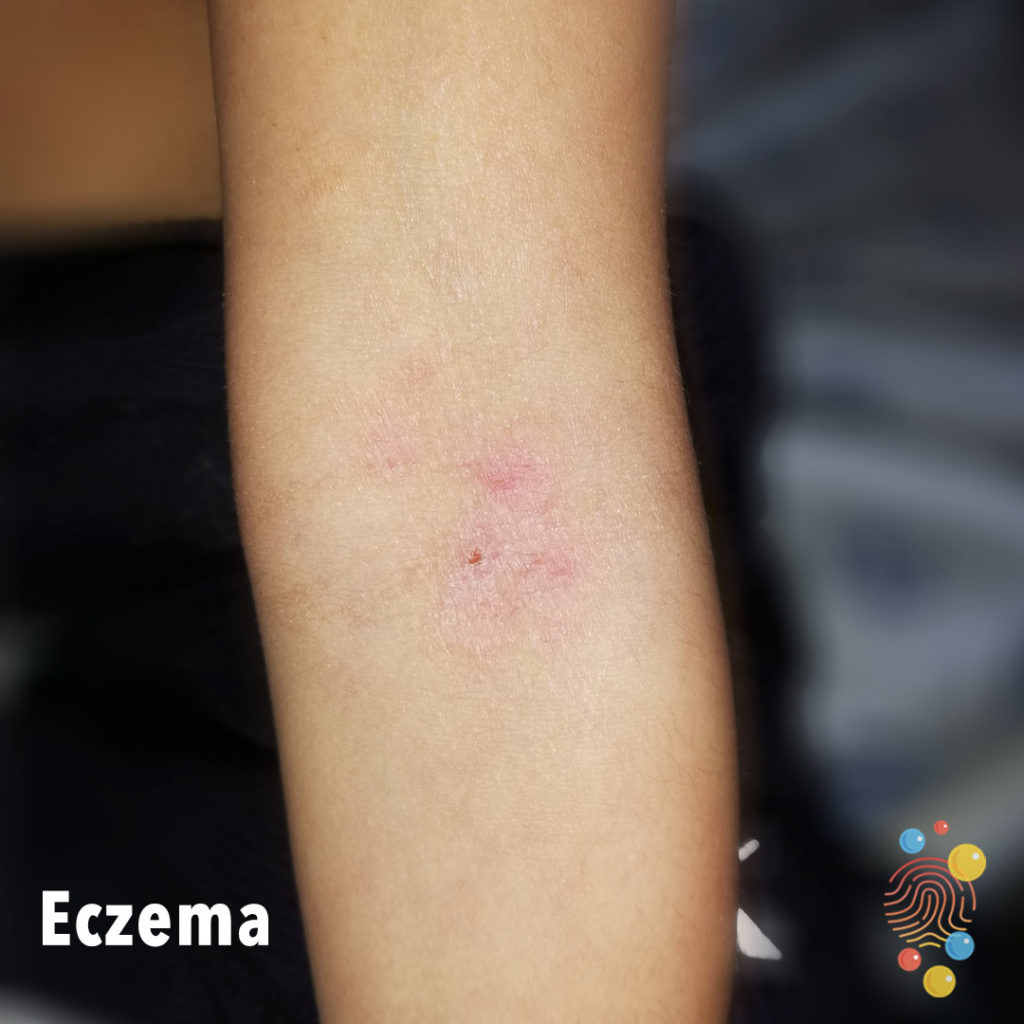
Eczematous patch at antecubital fossa with lichenification, scale, and excoriations.
Learn more about eczema
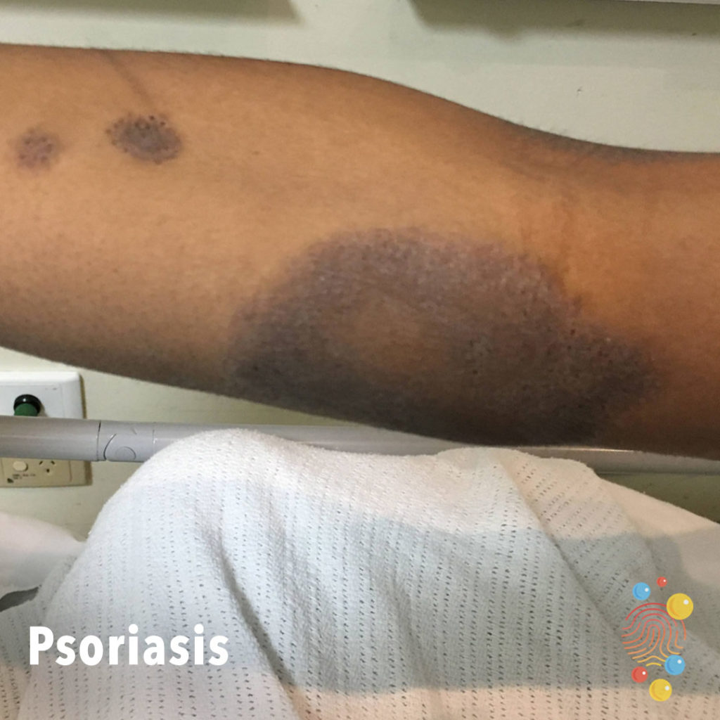
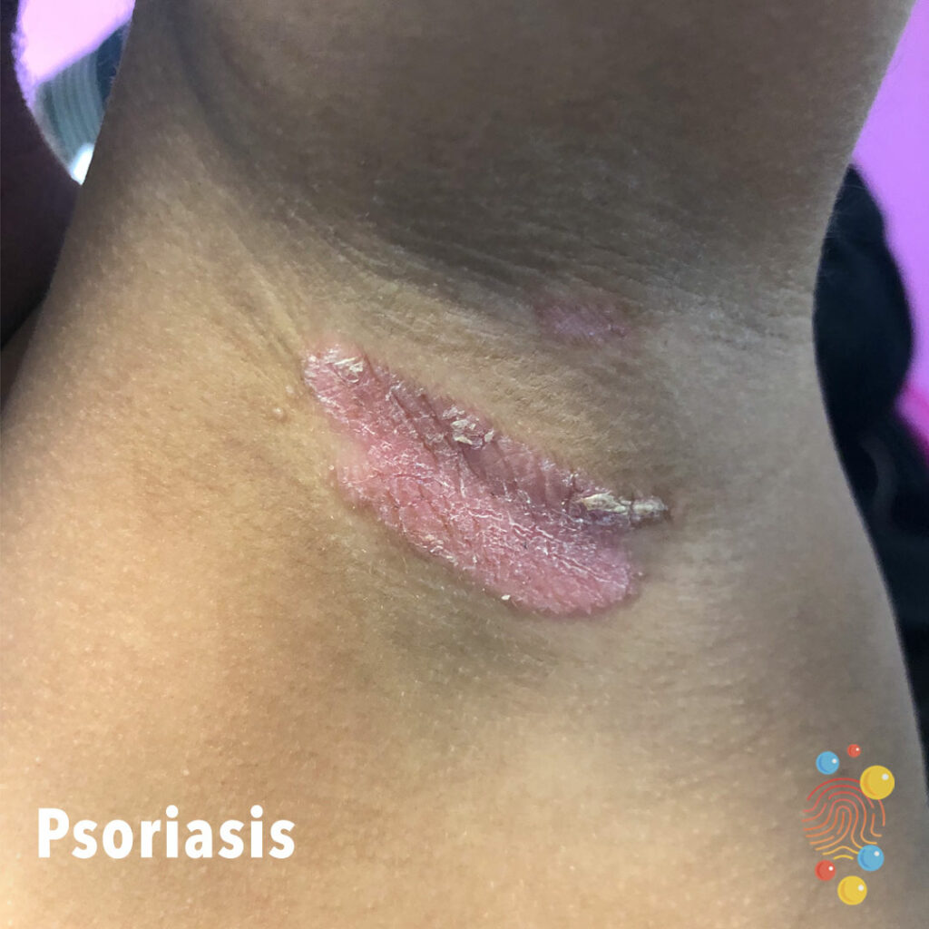
Well circumscribed, hyperpigmented plaques of varying sizes on the flexor surfaces. There is central clearing in areas. There is fine overlying scale.
Learn more about psoriasis
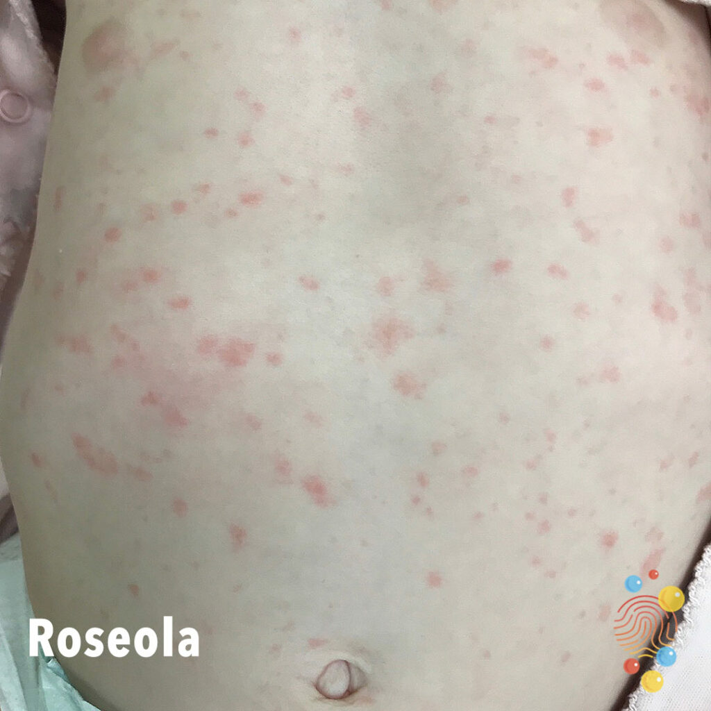
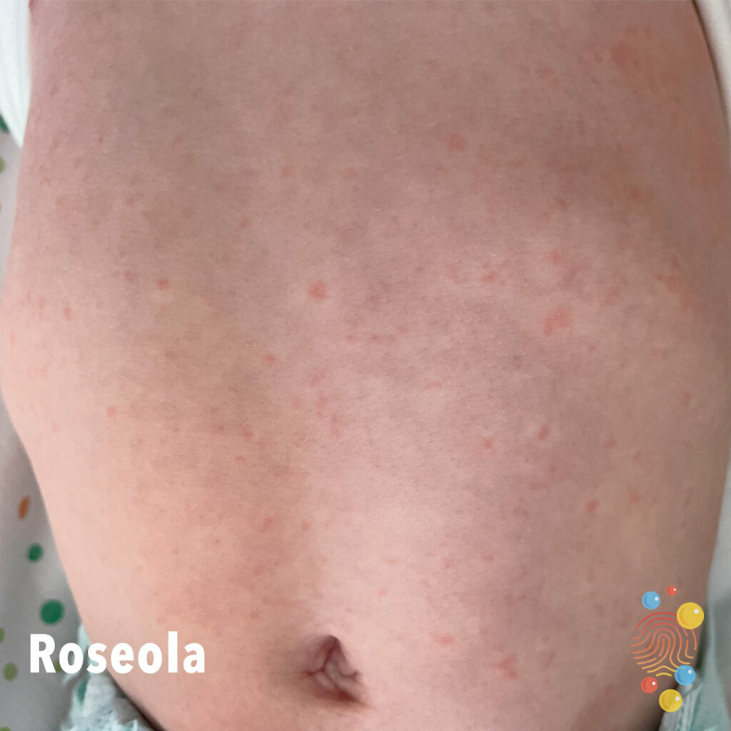
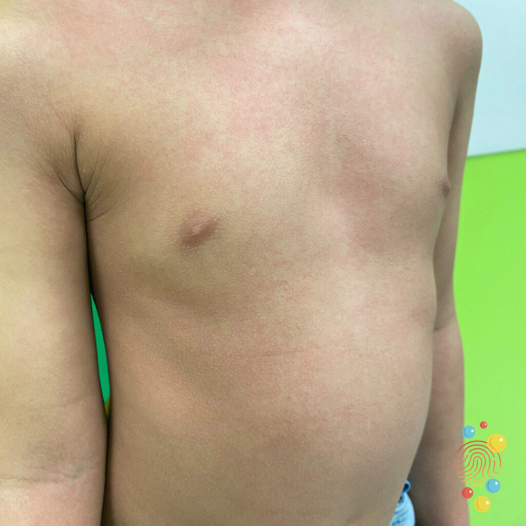
Fine macular erythema on trunk.
Learn more about roseola

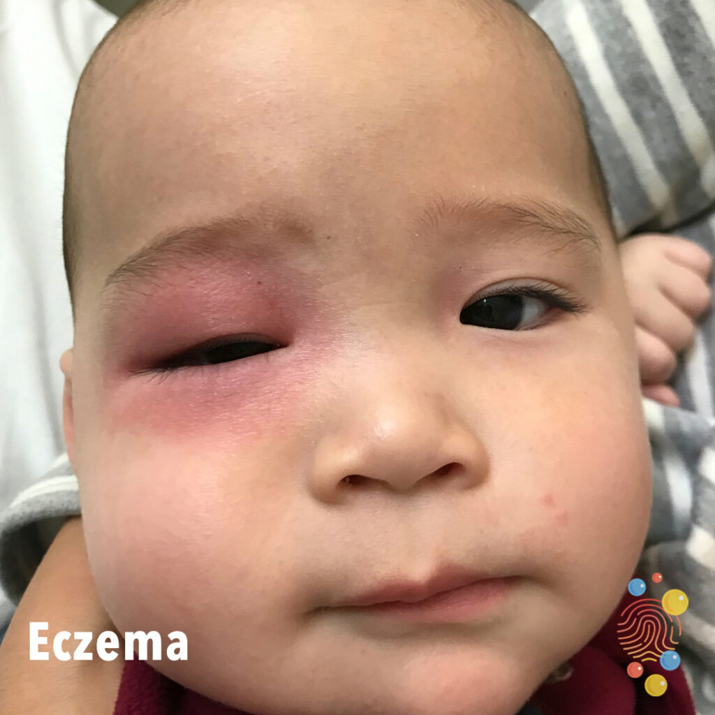
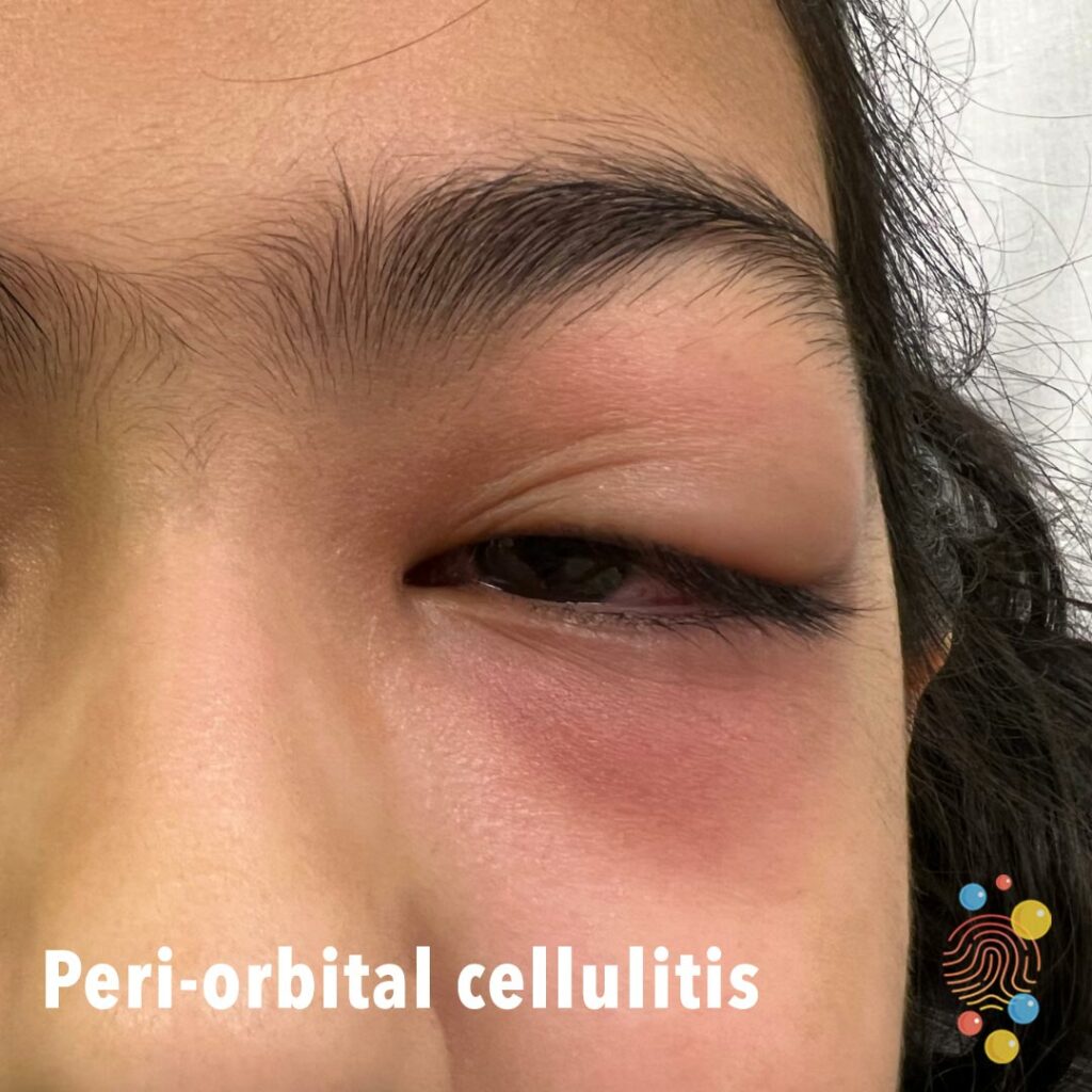
Asymmetrical, left sided orbital erythema and associated oedema. Associated mild crusting/skin peeling. Possible exotropia and secondary ptosis.
Learn more about periorbital cellulitis
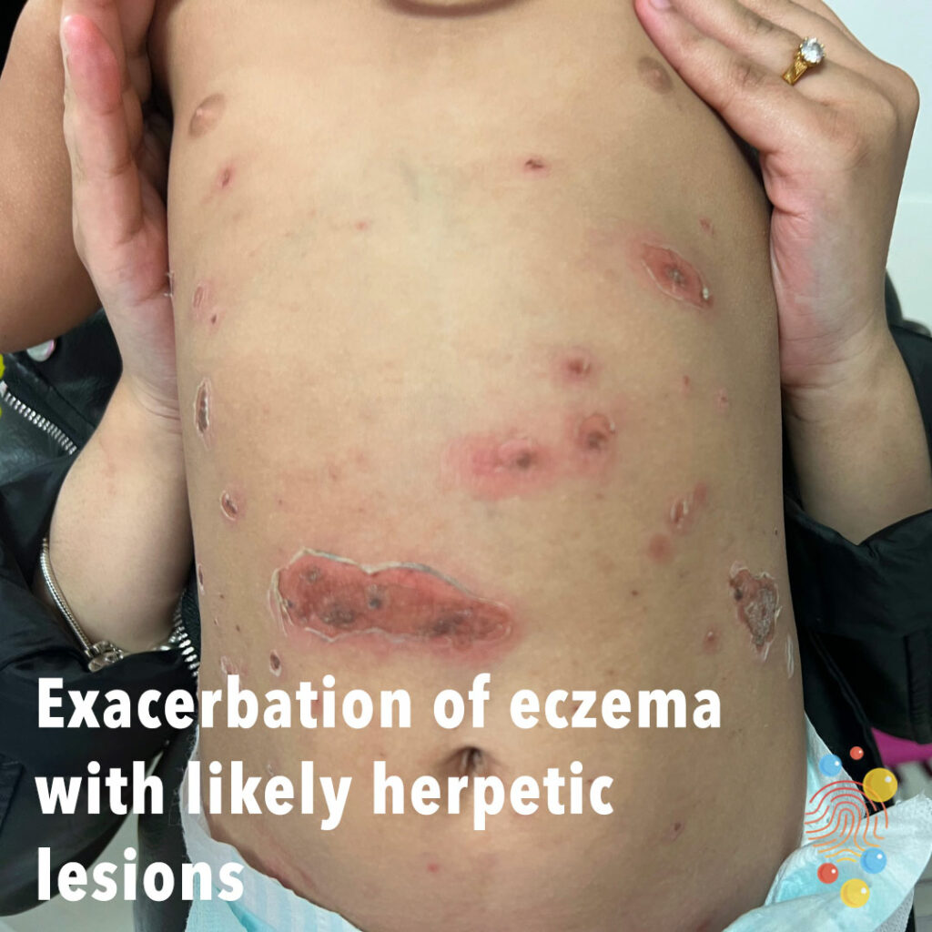
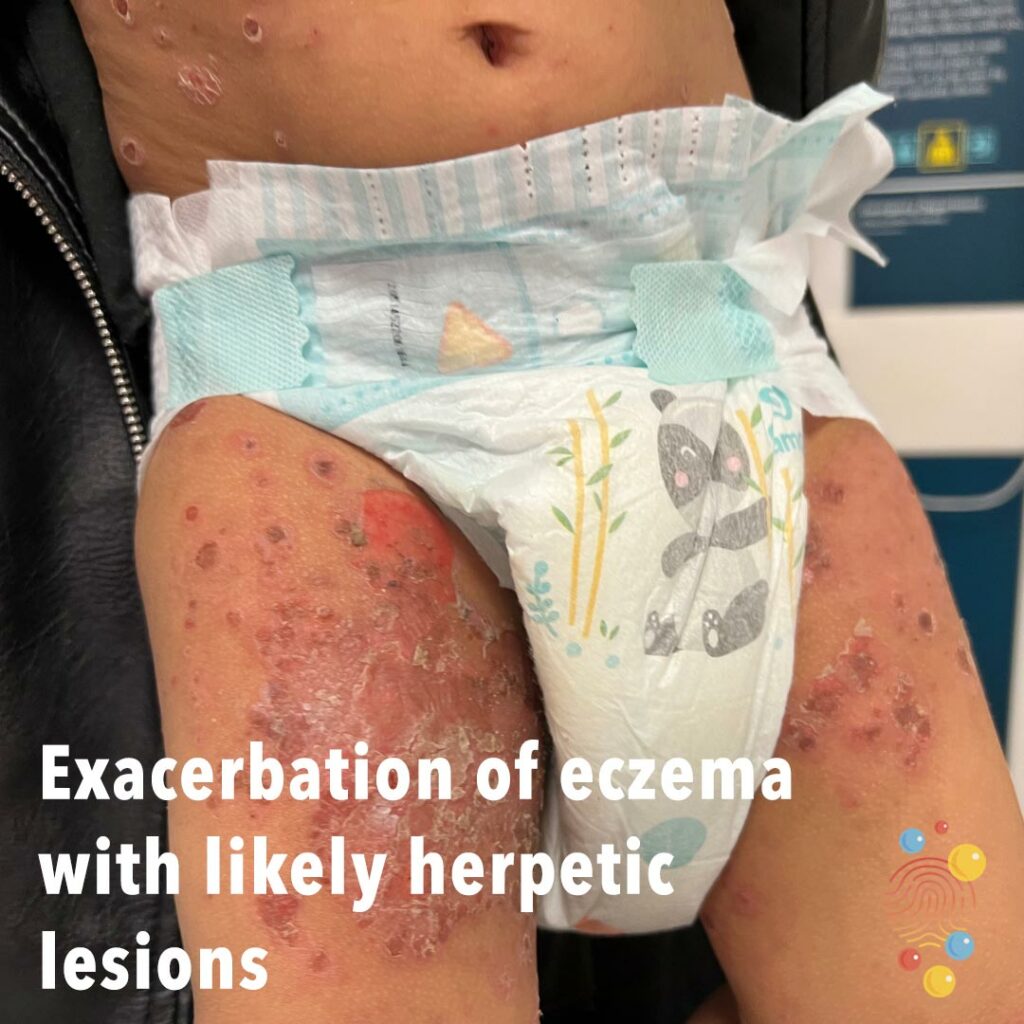
Exacerbation of eczema with likely herpetic lesions
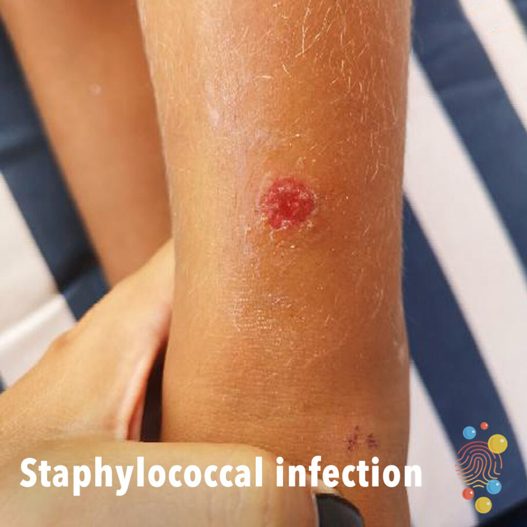
Solitary erosion lower leg +/- crust.
Learn more about staphylococcal infection
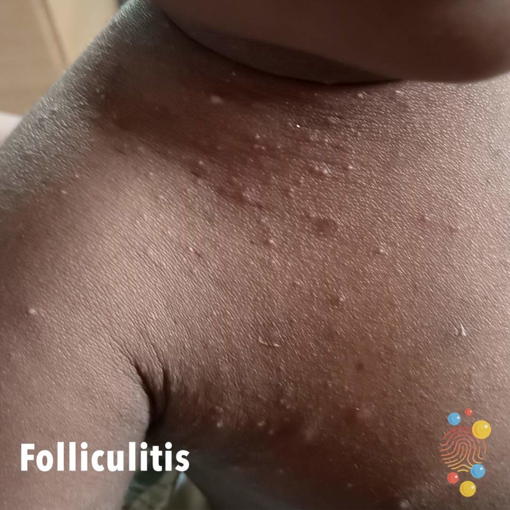
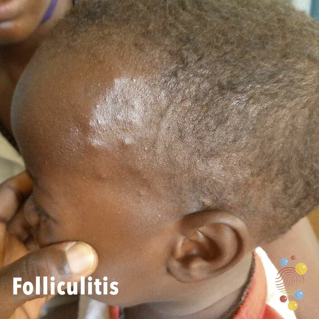
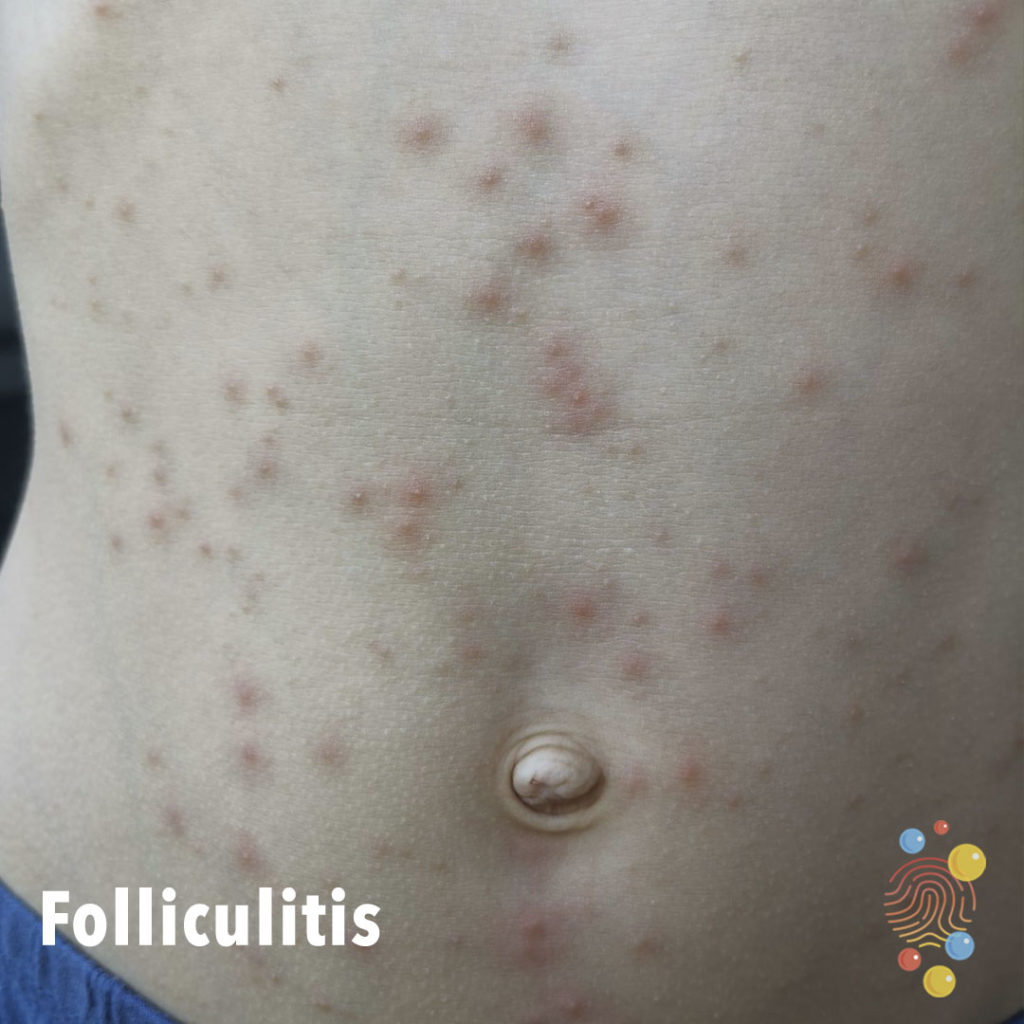
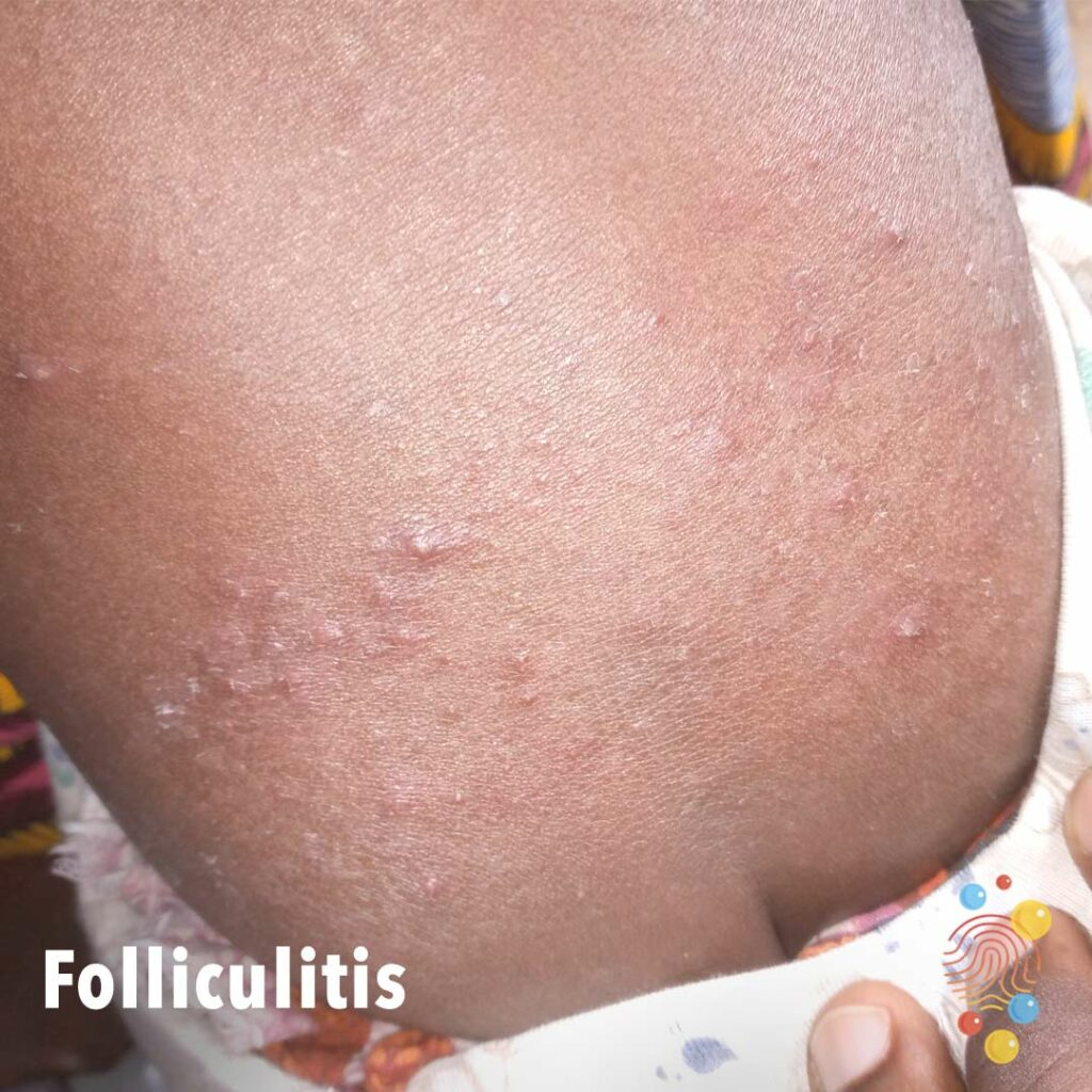
Widespread follicular rash upper chest, with papules and some small pustules.
Learn more about folliculitis
Learn more about gastrostomies
Numerous irregular ulcerating lesions on background of lichenification. Possible impetiginisation with golden crust overlying lesions.
Learn more about eczema

Erythema with lichenification of the dorsum of the hand focused over the MCP joints and wrist.
Learn more about eczema
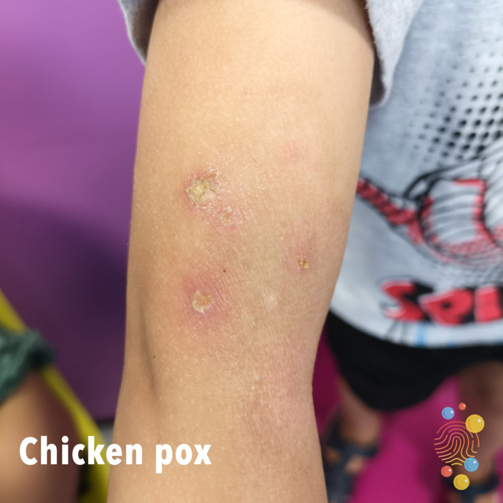
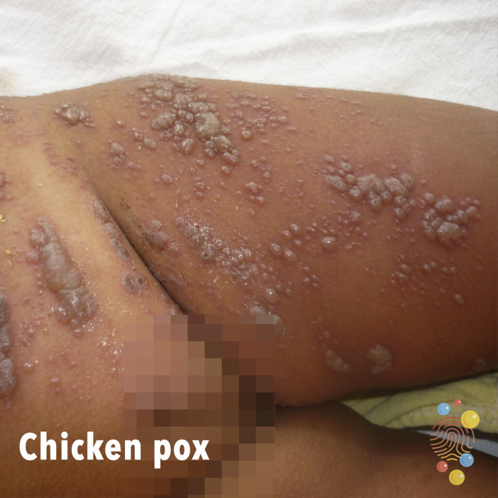
Multiple excoriated vesicles on an erythematous base, some crusted.
Learn more about chicken pox
Widespread facial erythema. Fine surface scale and some possible impetiginisation and crusting over the nose. Sparing of forehead and chin. Excoriations.
Learn more about eczema
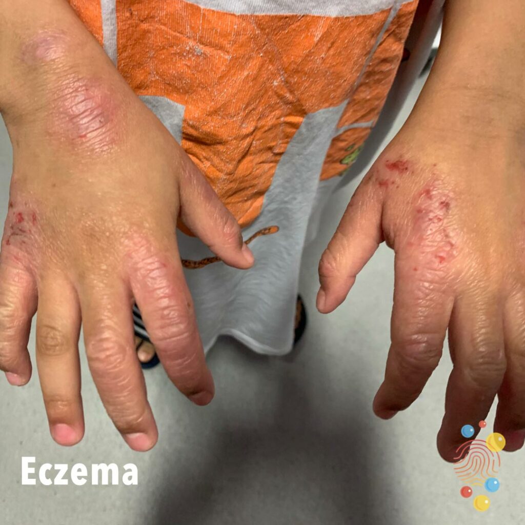
Erythema and lichenification of the dorsal hands, with excoriations and bleeding.
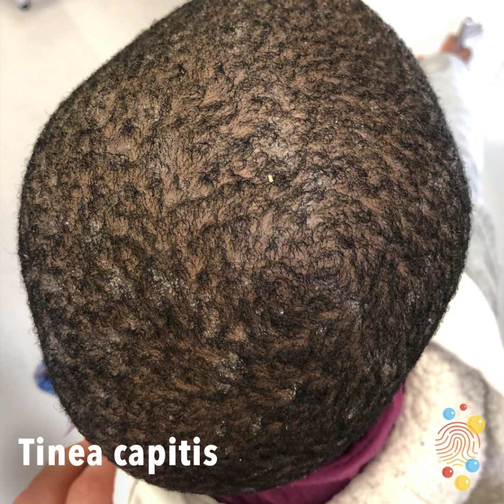
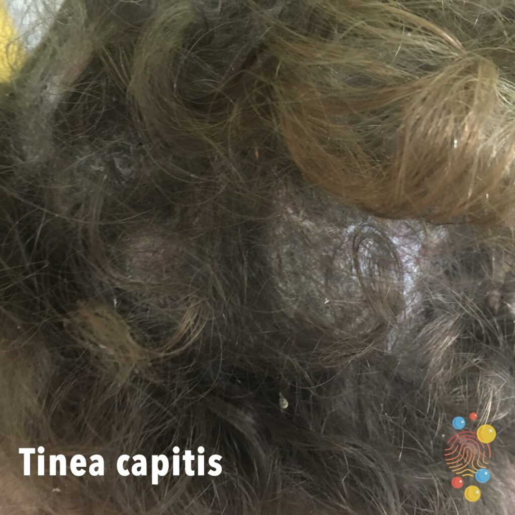
Extensive scalp scale.
Learn more about tinea capitis

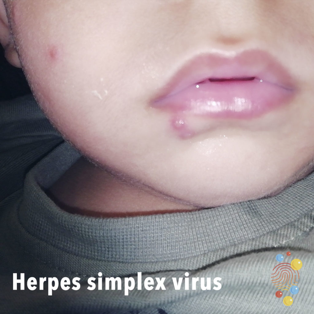
Eroded vesicles at corners of mouth and lower lip.
Learn more about herpes simplex virus
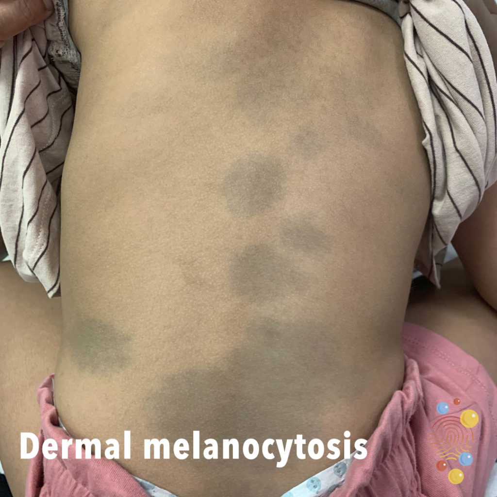
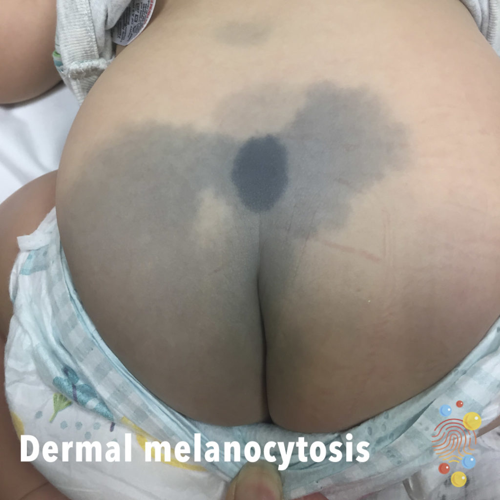
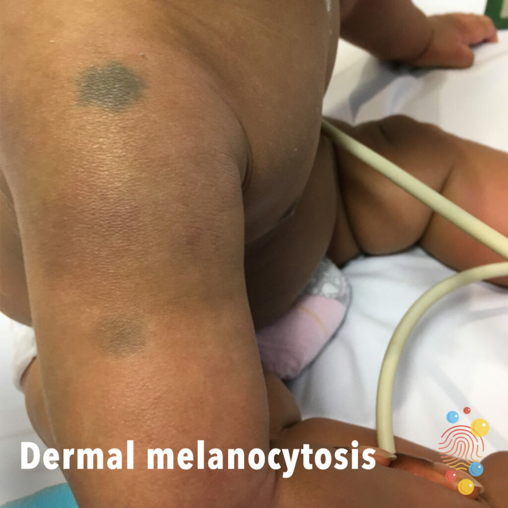
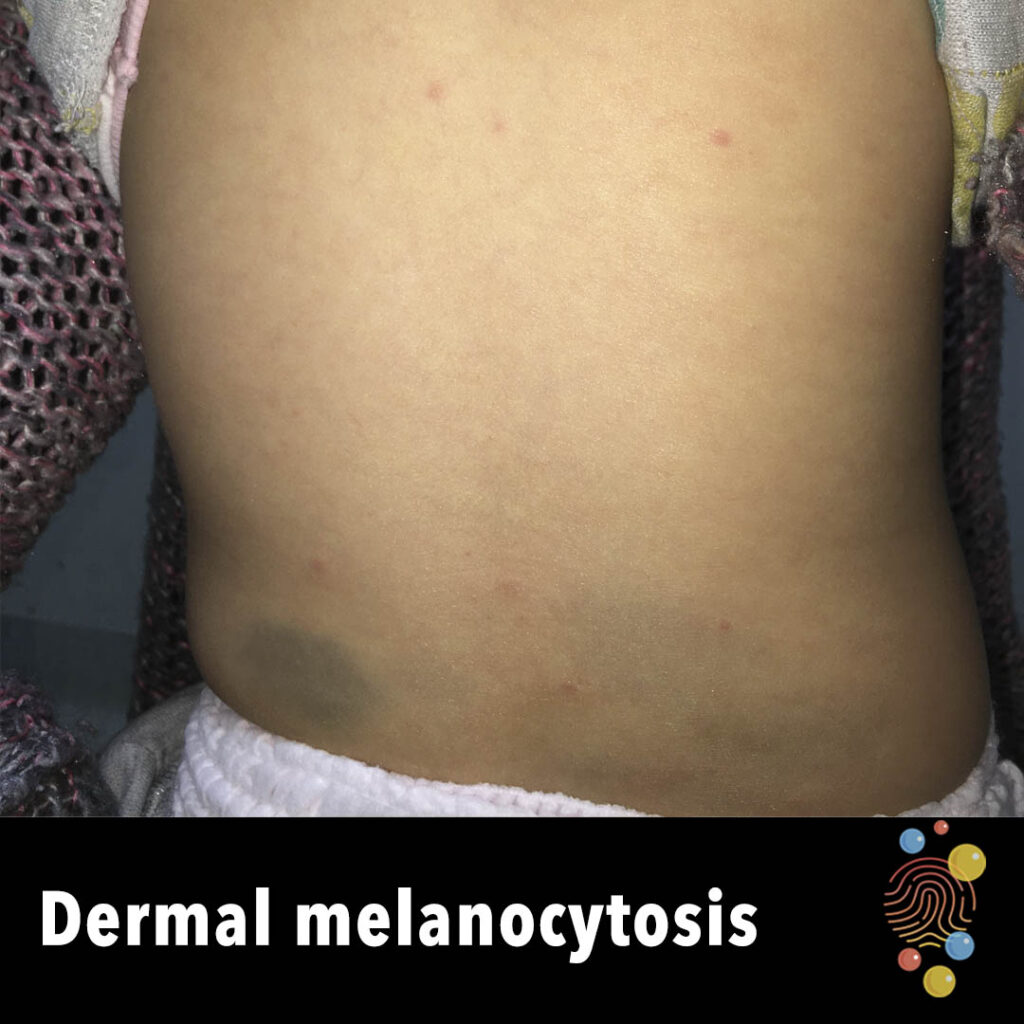
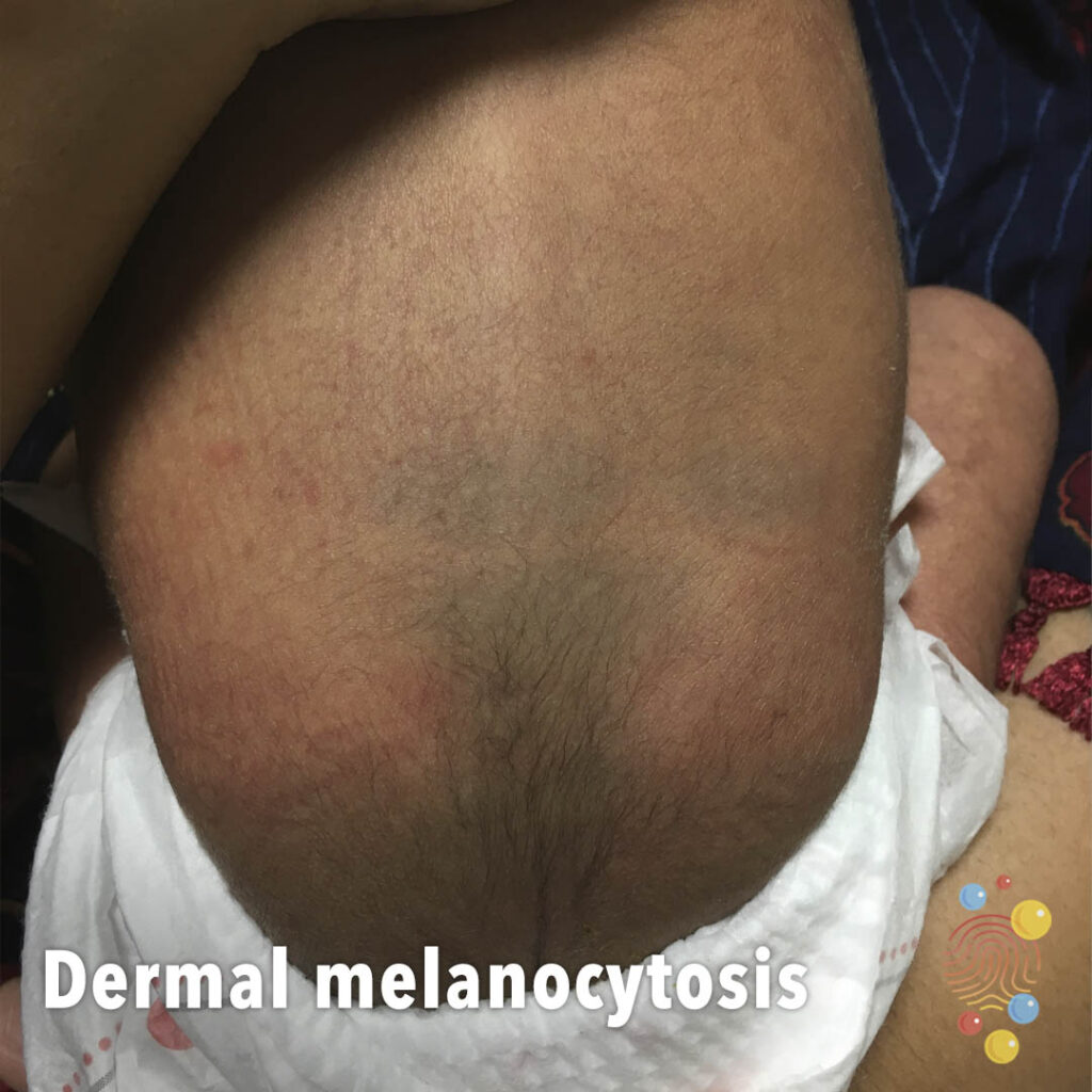
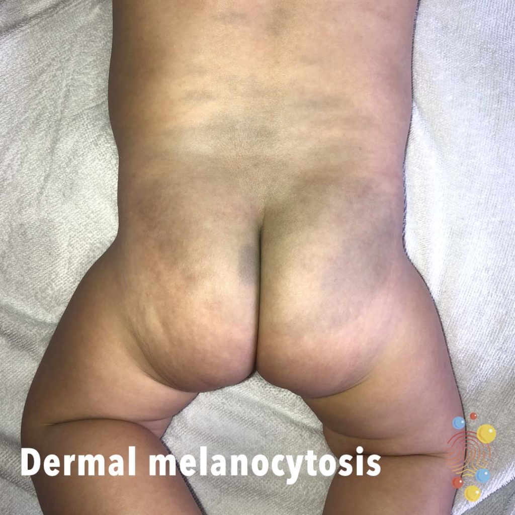
Flat blue/grey areas on the back.
Learn more about dermal melanocytosis

Multiple small facial pustules predominantly on forehead and peri-nasal areas.
Learn more about neonatal cephalic pustulosis
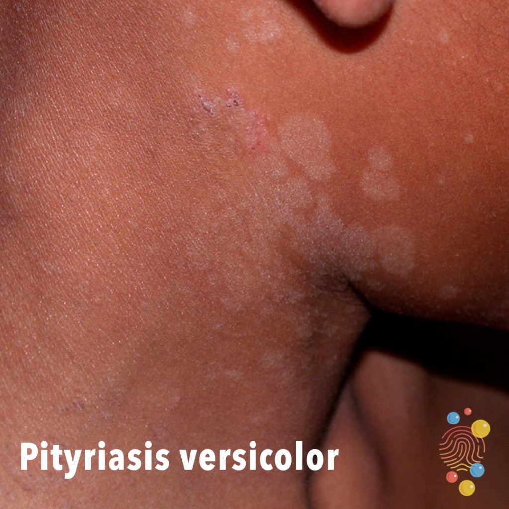
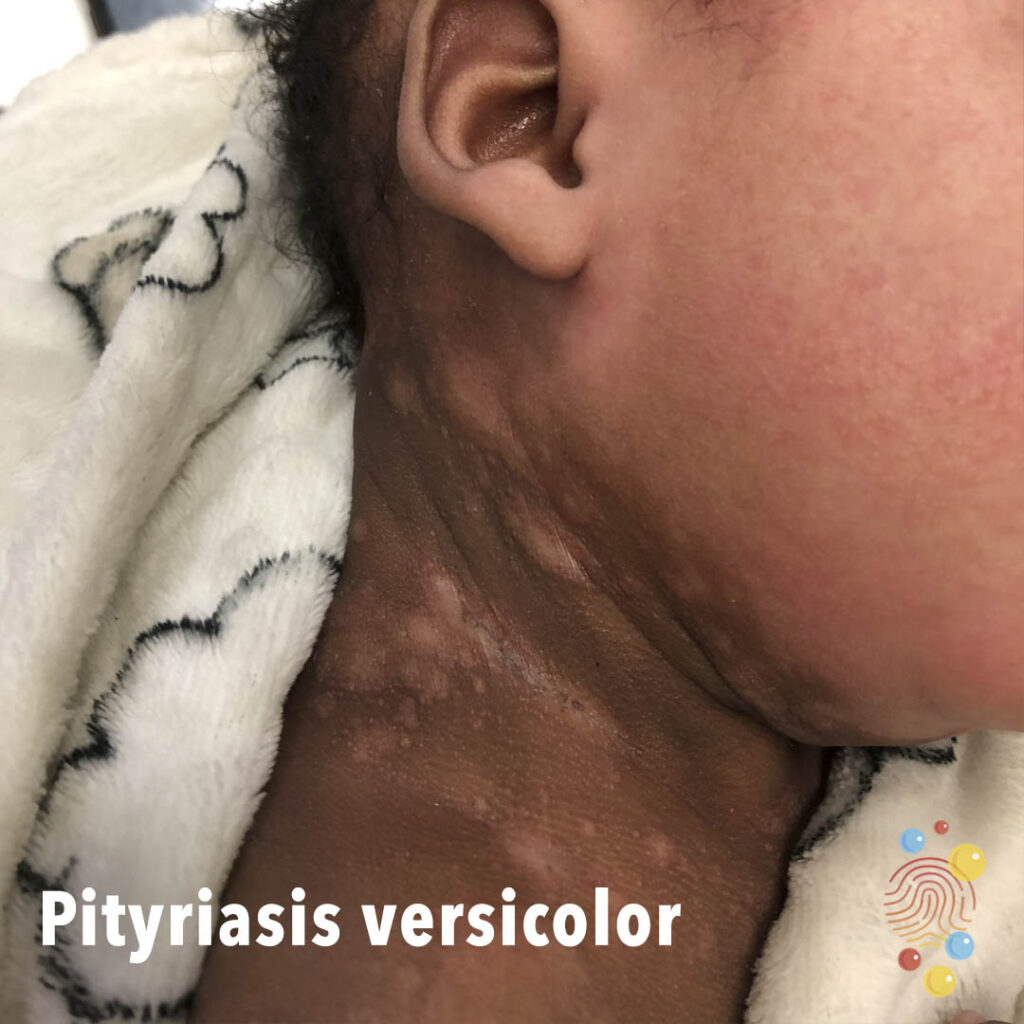
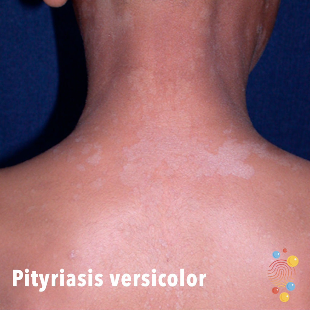
Scaly hypopigmented patches on the lateral neck.
Learn more about pityriasis versicolor

Severe lichenified eczema with induration and impetiginisation
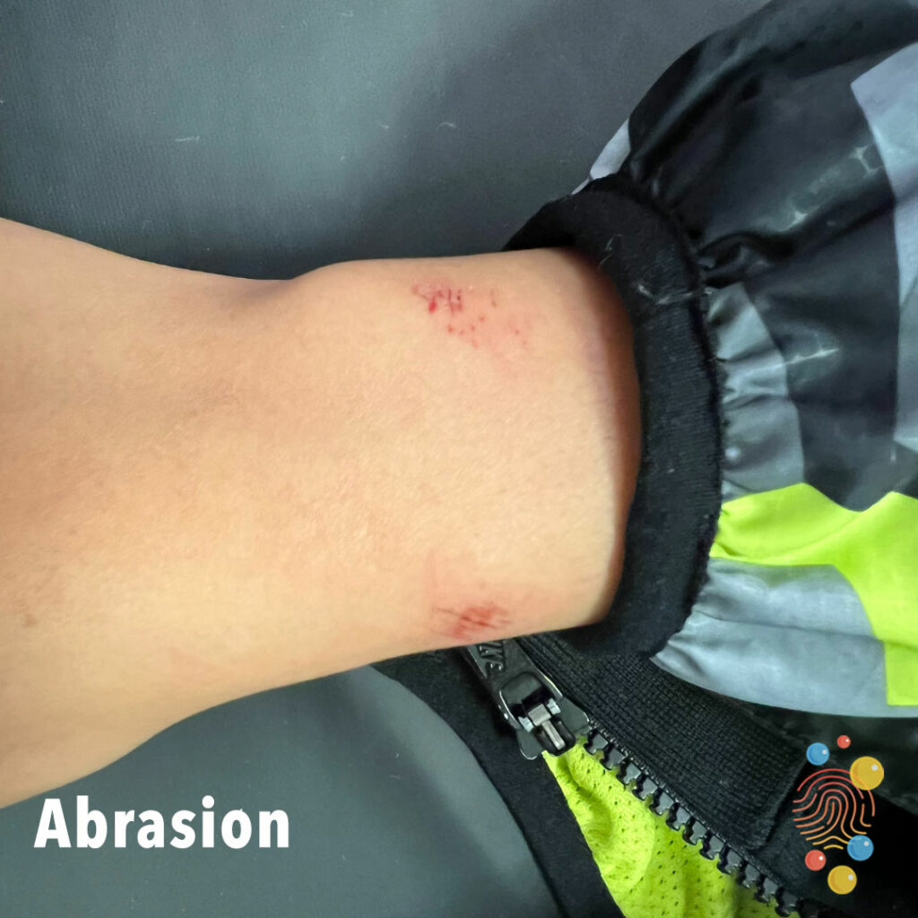
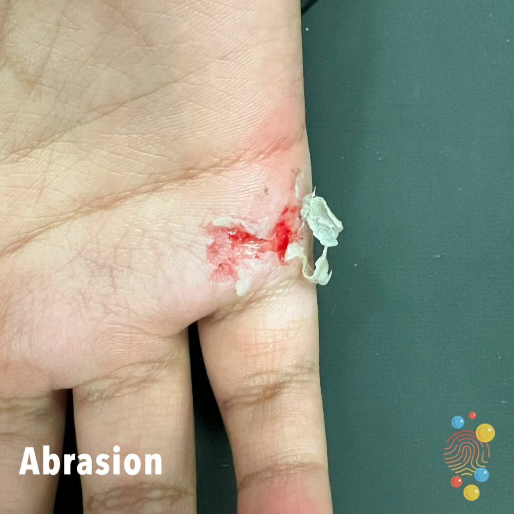
Abrasion
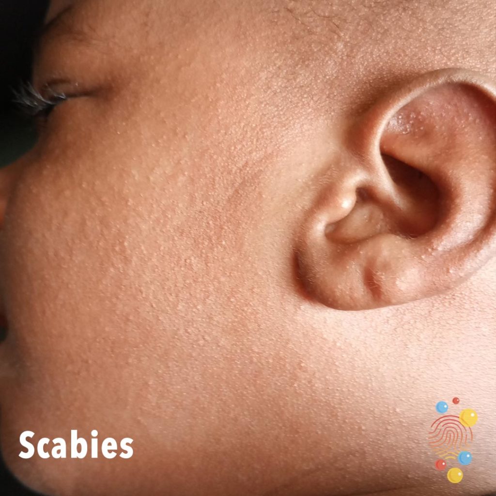
Widespread small papules across left side face, including inner ear. Small amount of scale in ear helix.
Learn more about scabies
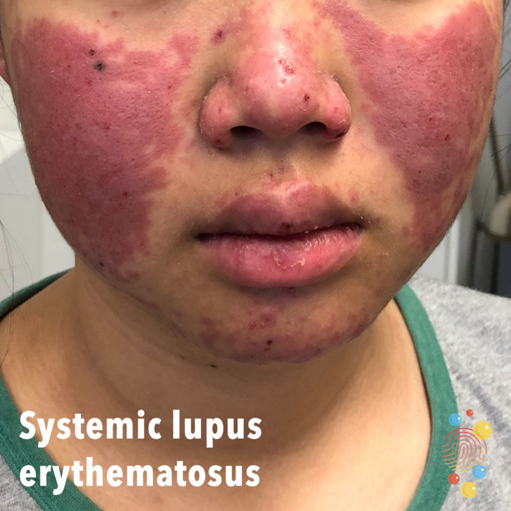
Butterfly or malar violaceous raised erythematous rash across the cheeks and bridge of the nose. There is mucosal involvement with lesions on the lips.
Learn more about systemic lupus erythematosus

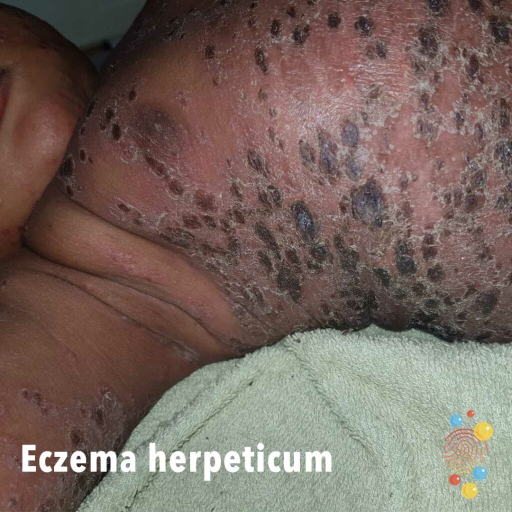
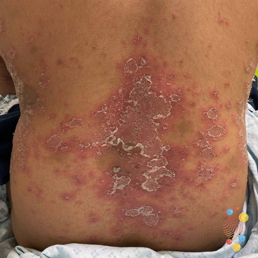
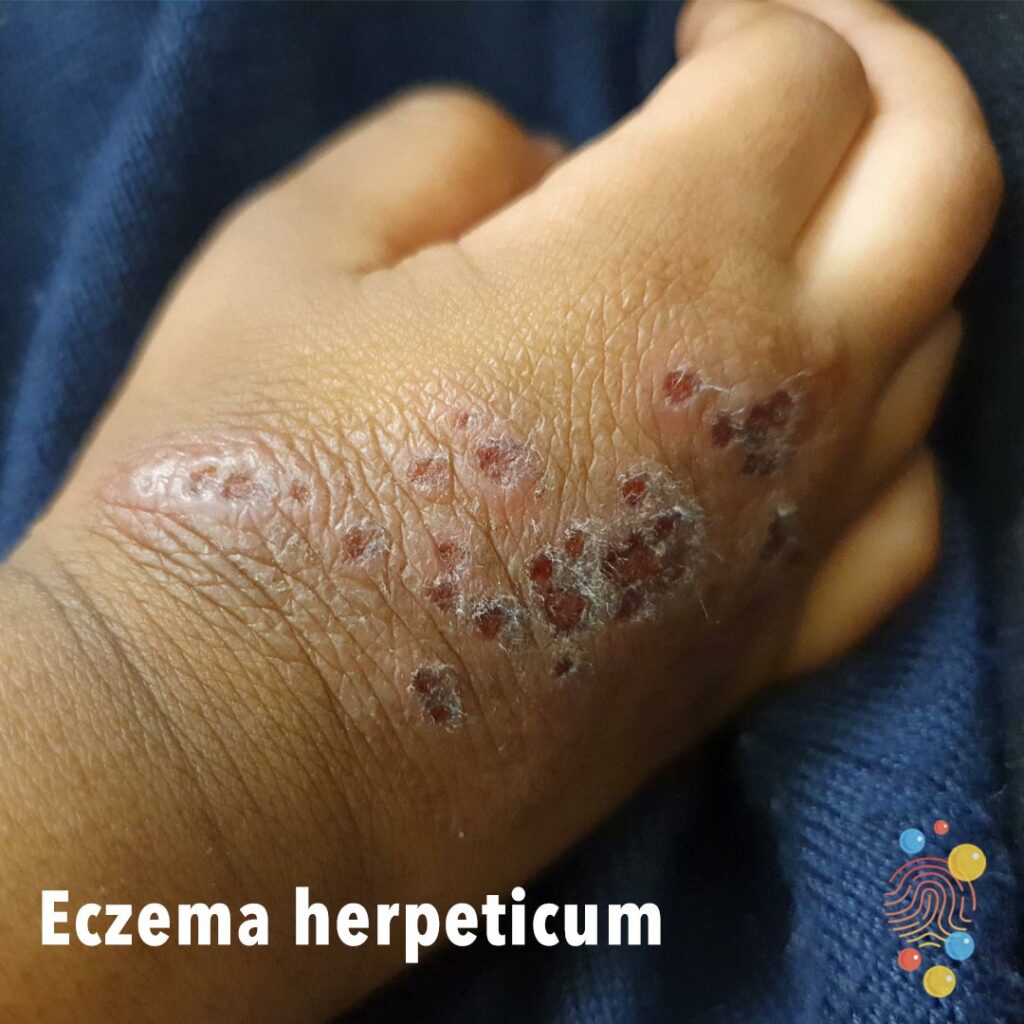
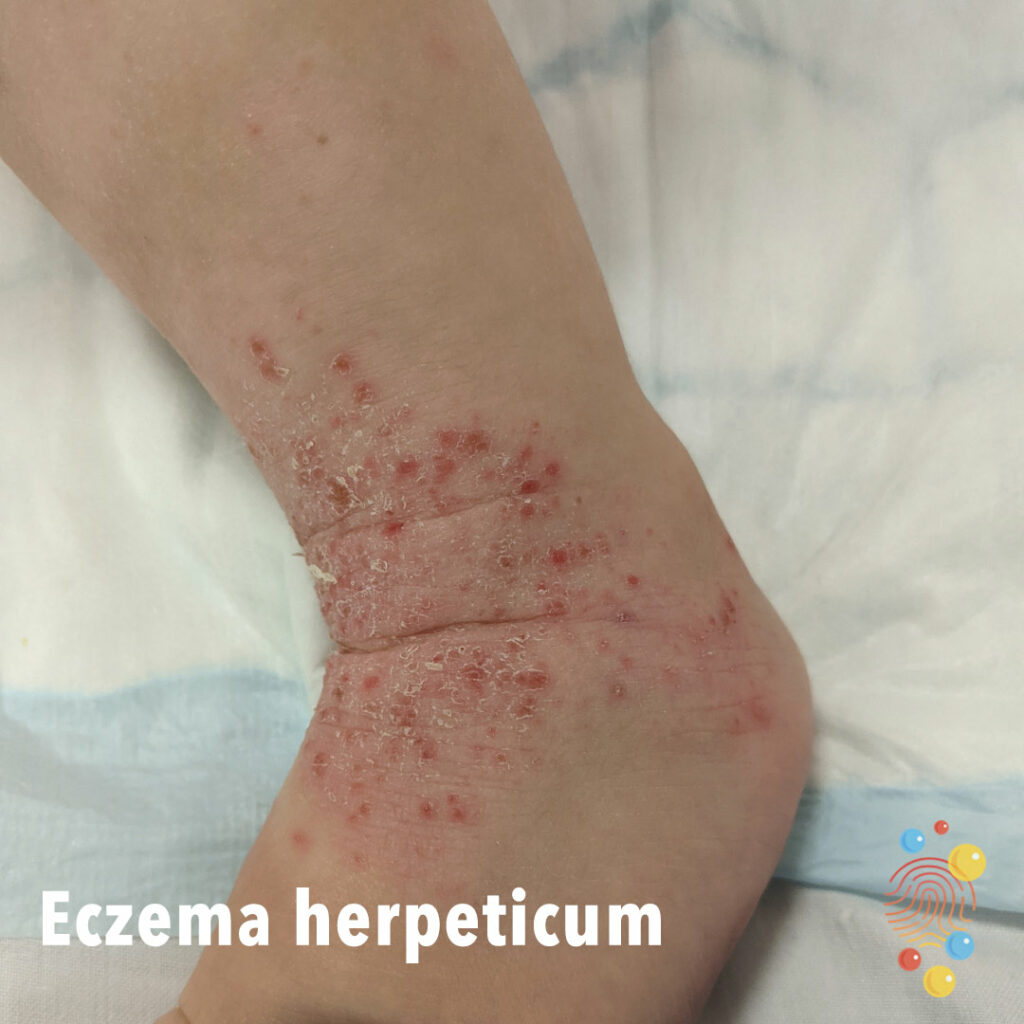
Eruption of dark red macules, vesicles, and erosions distributed across areas previously affected by atopic dermatitis, with relative sparing of the trunk
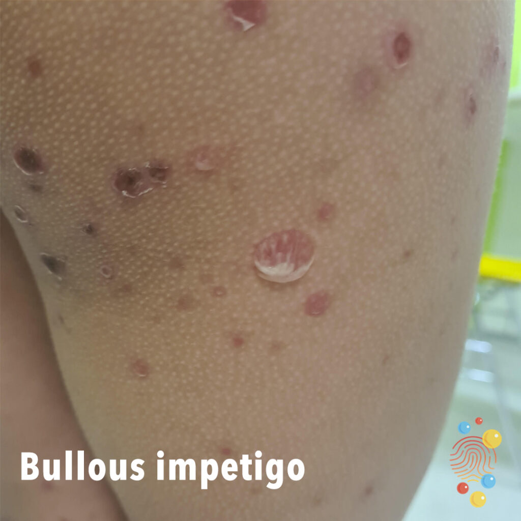
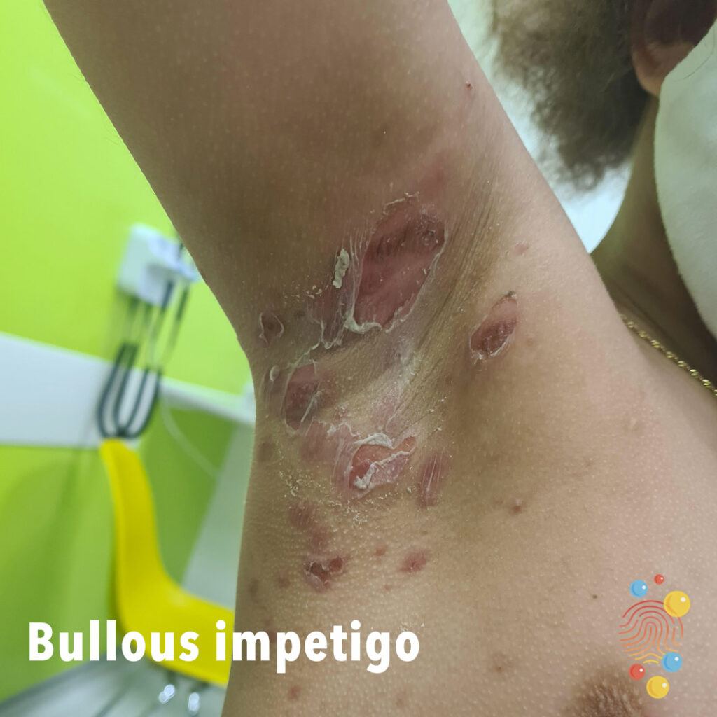
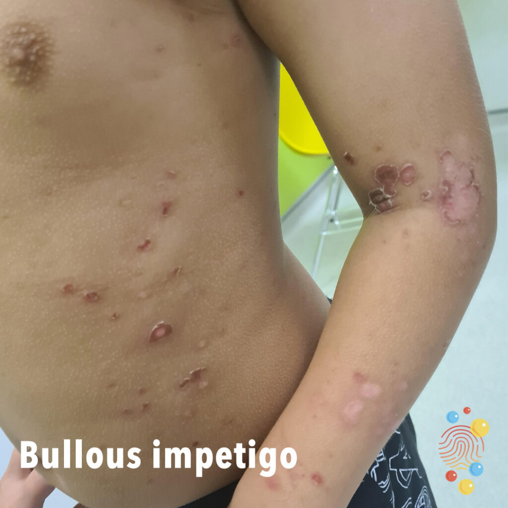
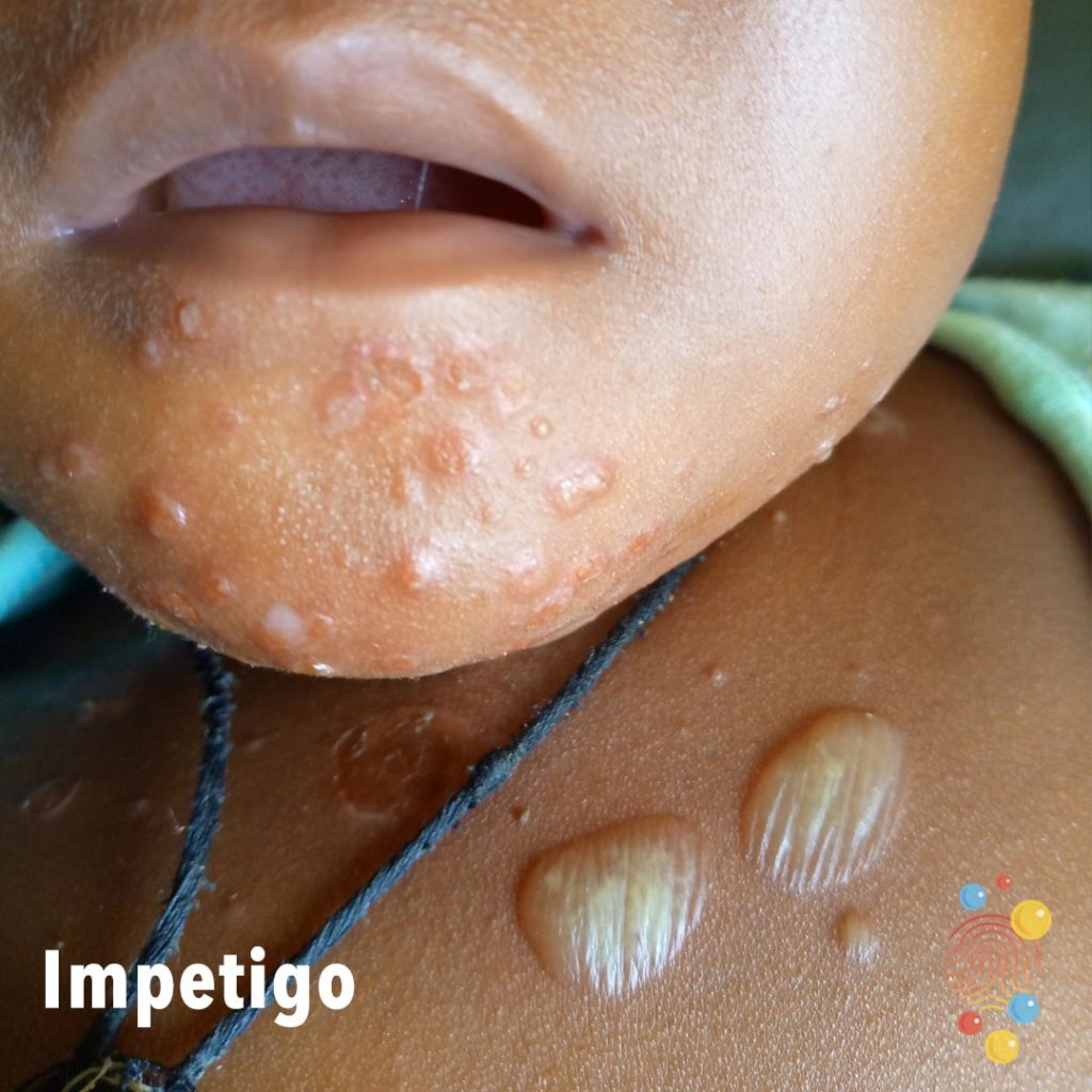
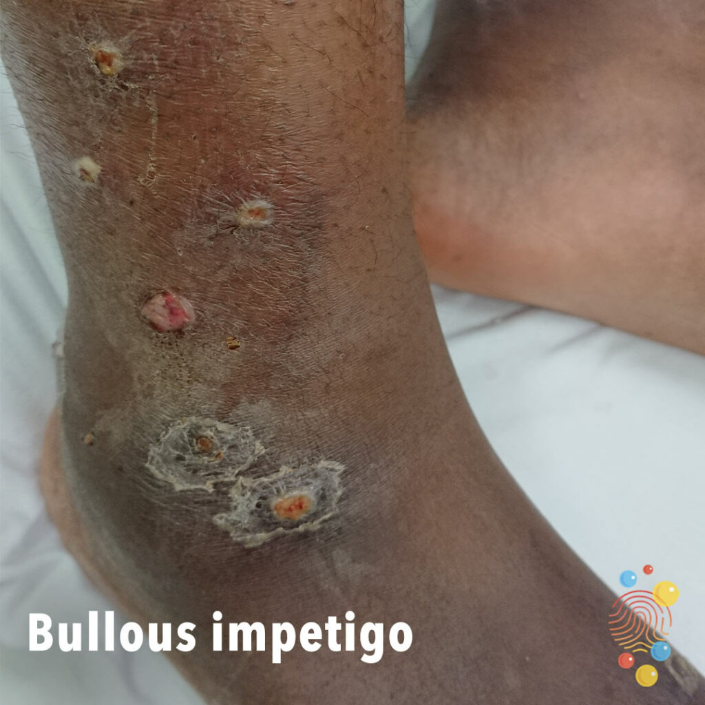
Confluent pustules and nodules with honey-colored crusts and ragged edge.
Learn more about bullous impetigo
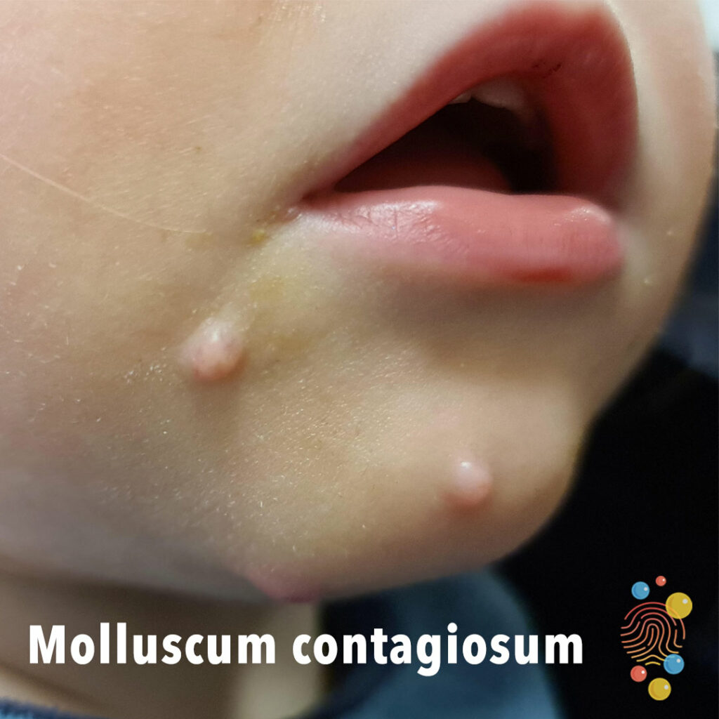
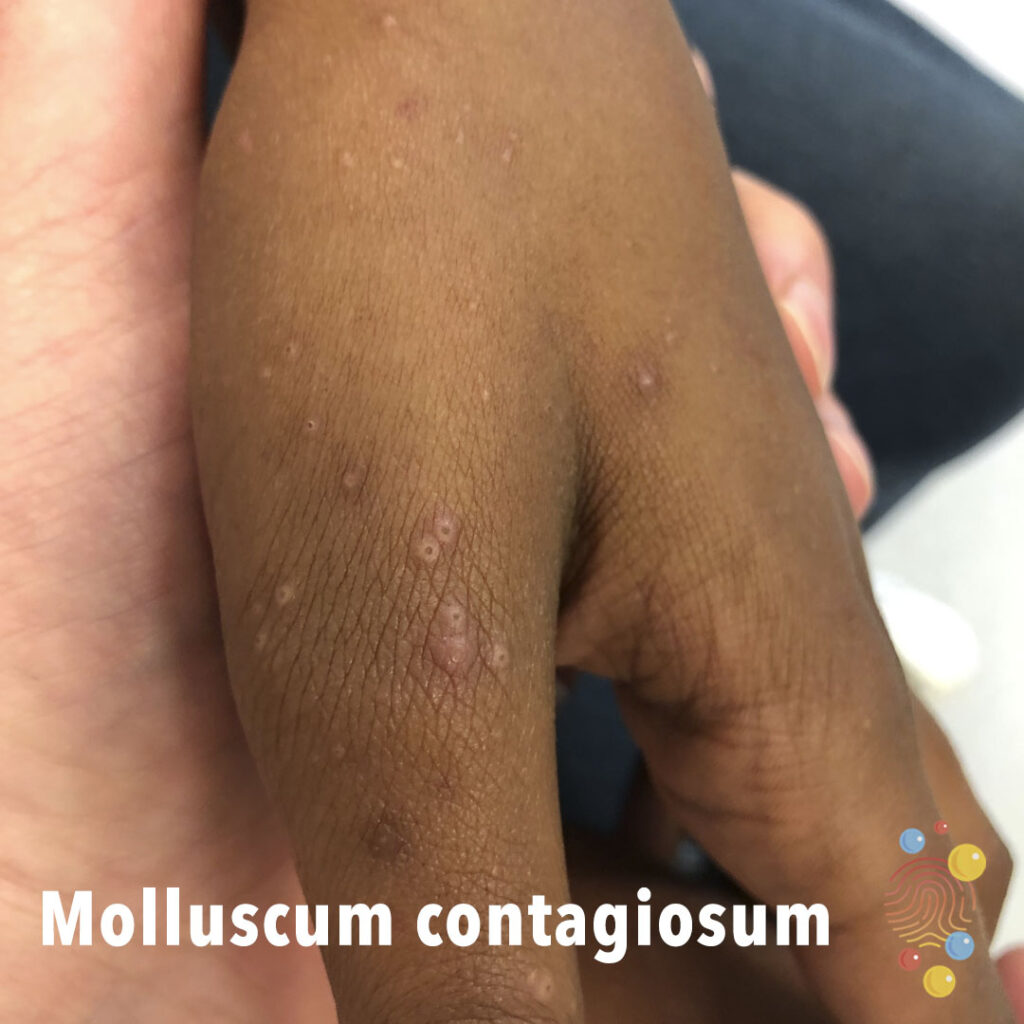
White smooth shiny papules with central umbilication.
Learn more about molluscum contagiosum

Bright red rash in symmetrical distribution on cheeks

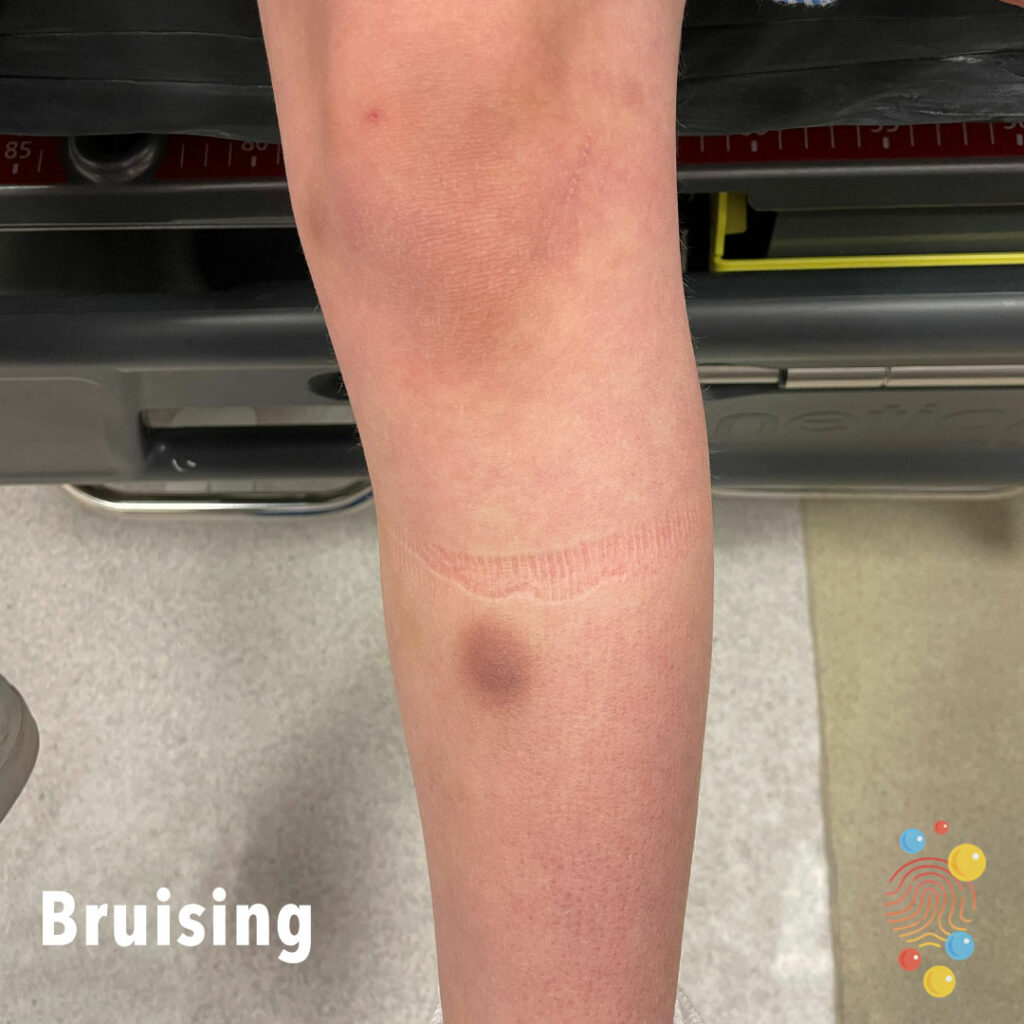
Accidental bruising to shin

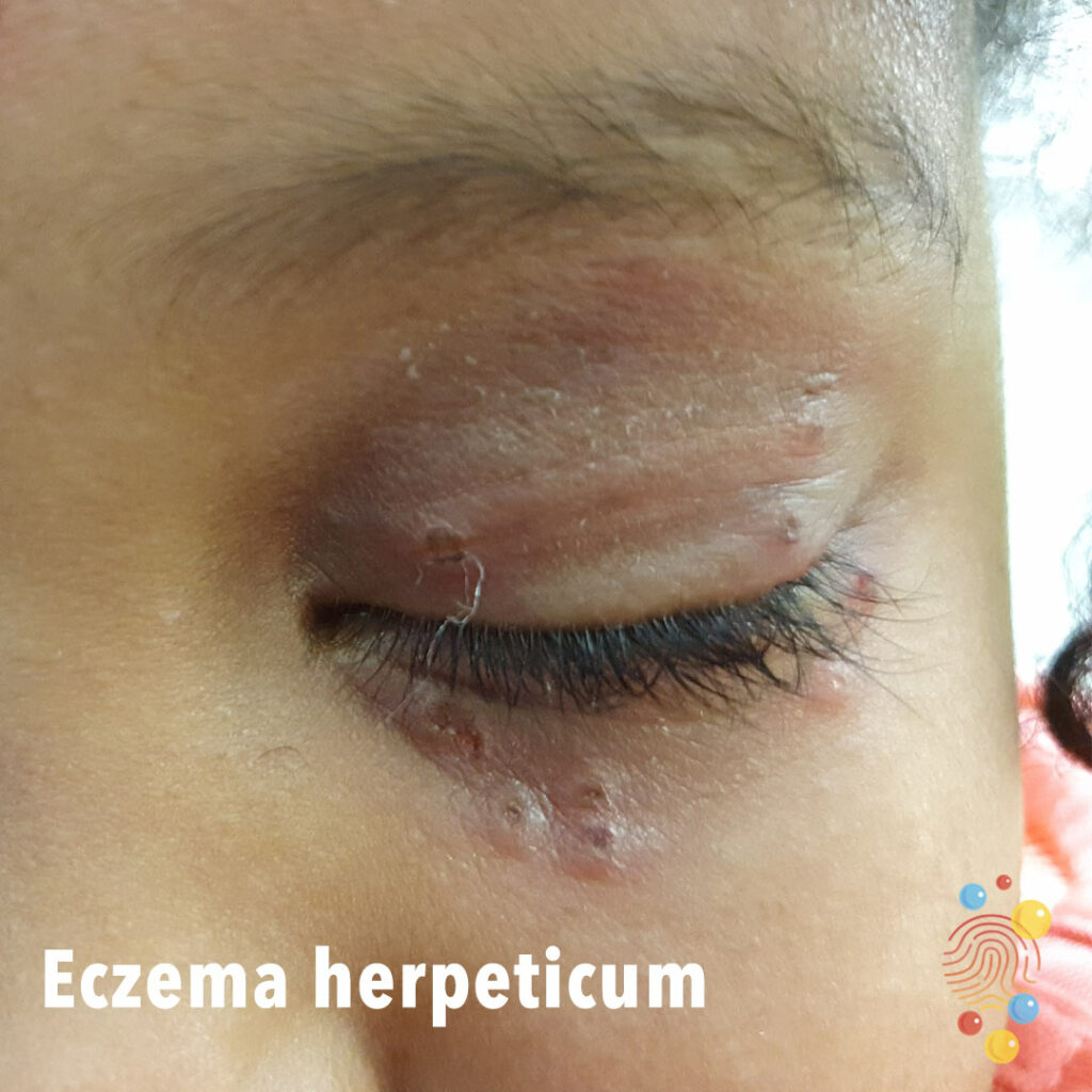
Excoriated vesicles around the eye.
Learn more about herpes simplex virus

Patch of hypopigmentation on the right flank with indistinct margins.
Learn more about hypopigmentation
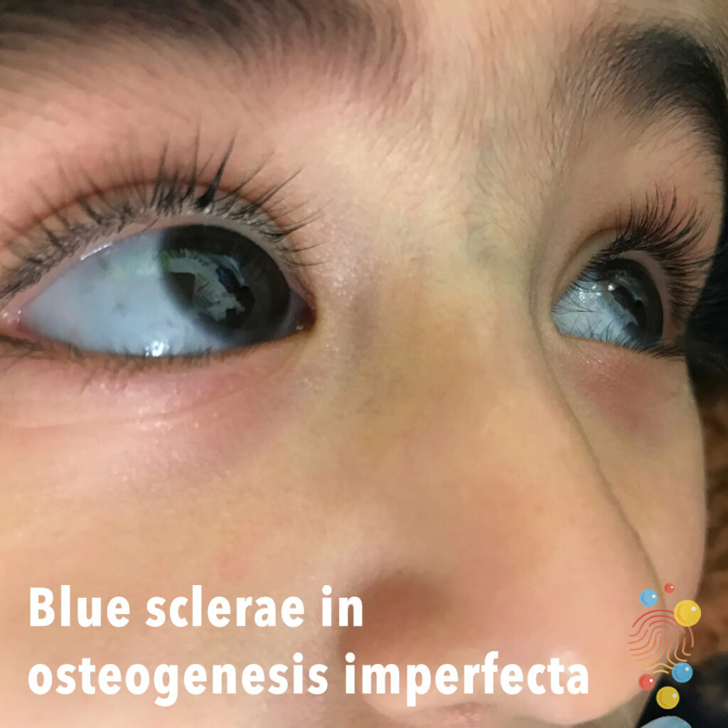
Blue sclera is associated with osteogenesis imperfecta.
Learn more about blue sclerae
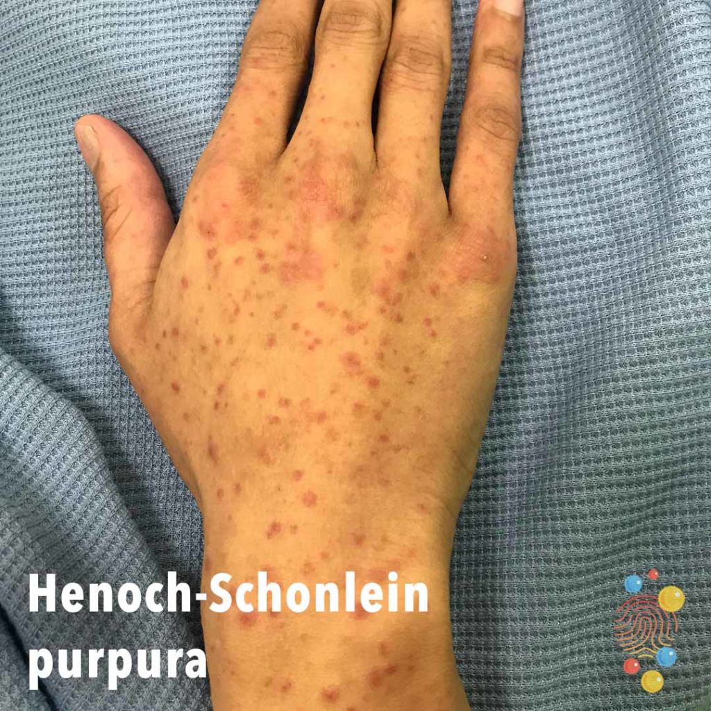
Non blanching symmetrical purpuric lesions on the dorsum of the hands.
Learn more about Henoch-Schonlein purpura
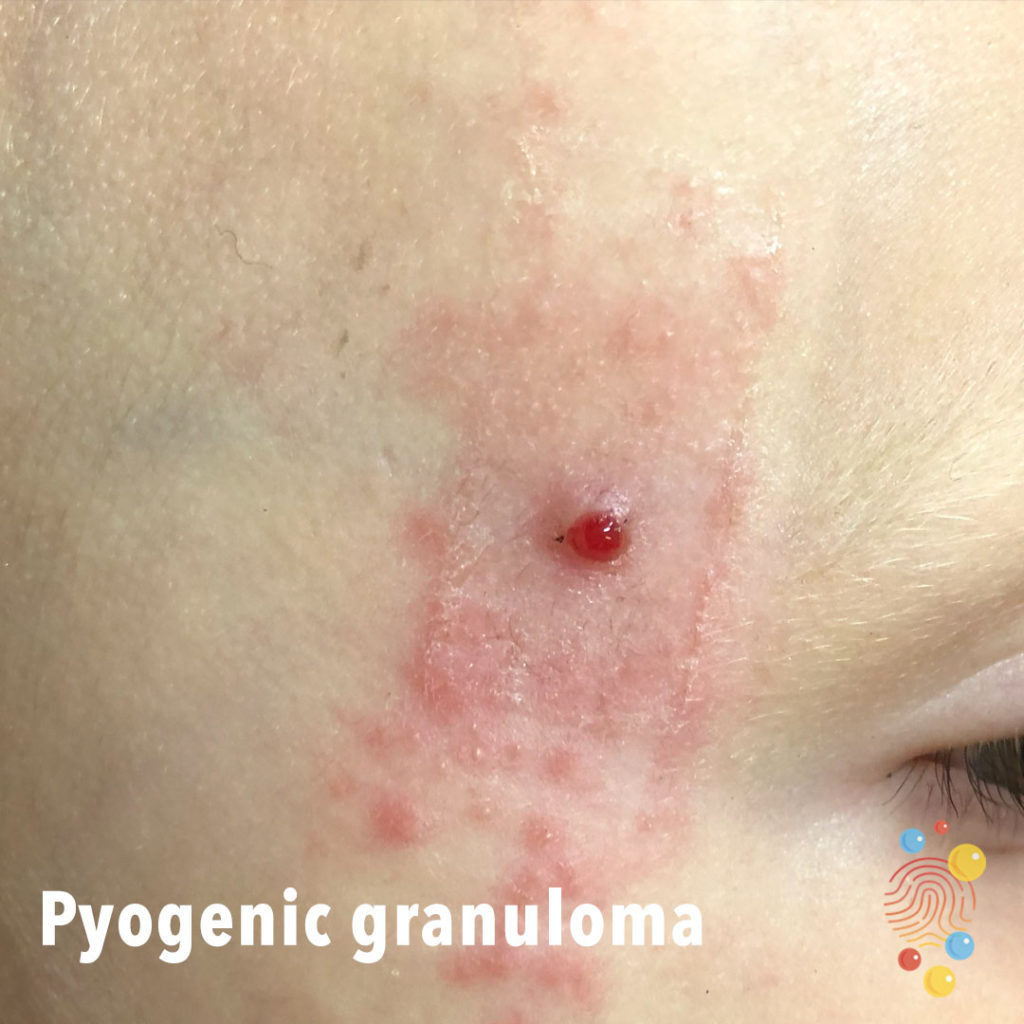
Small raised shiny cherry like lesion on the temple. There is an associated well-demarcated, erythematous, scaly rash suggestive of contact dermatitis secondary to tape placed over the lesion.
Learn more about pyogenic granulomas
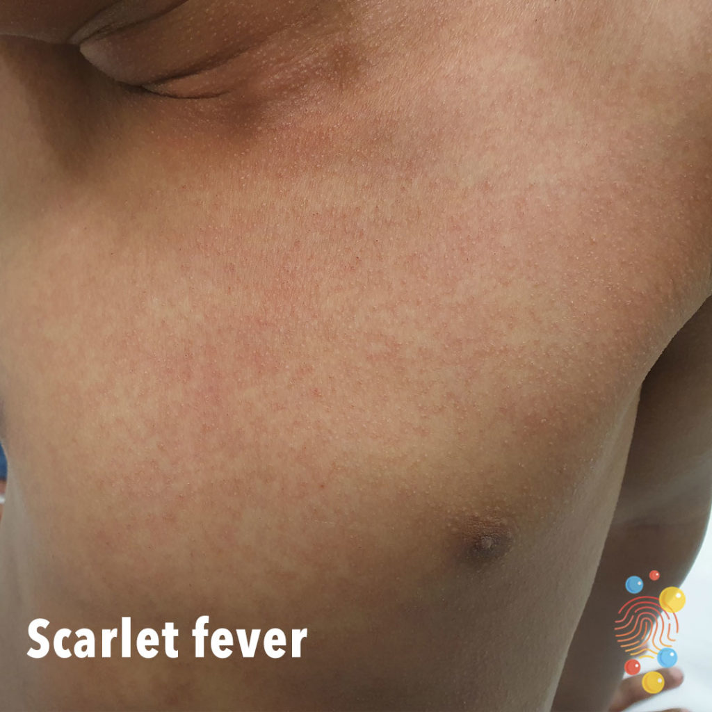
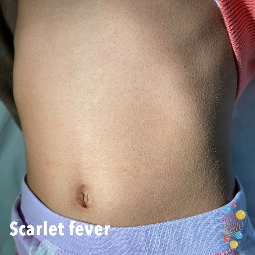
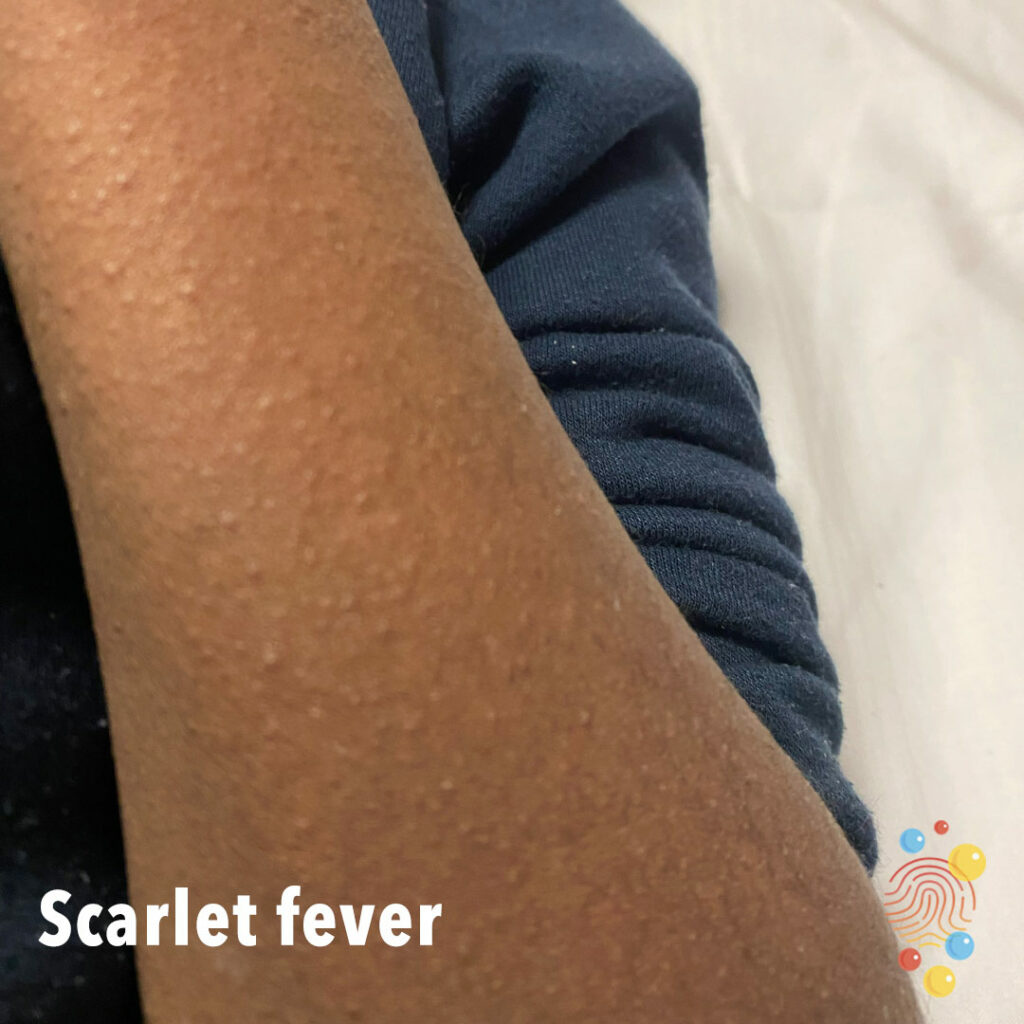
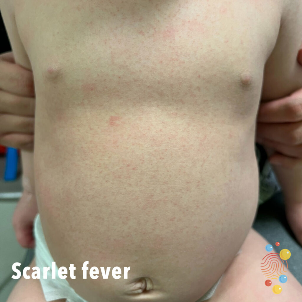
Bright red non-confluent eruption with very fine papules.
Learn more about scarlet fever
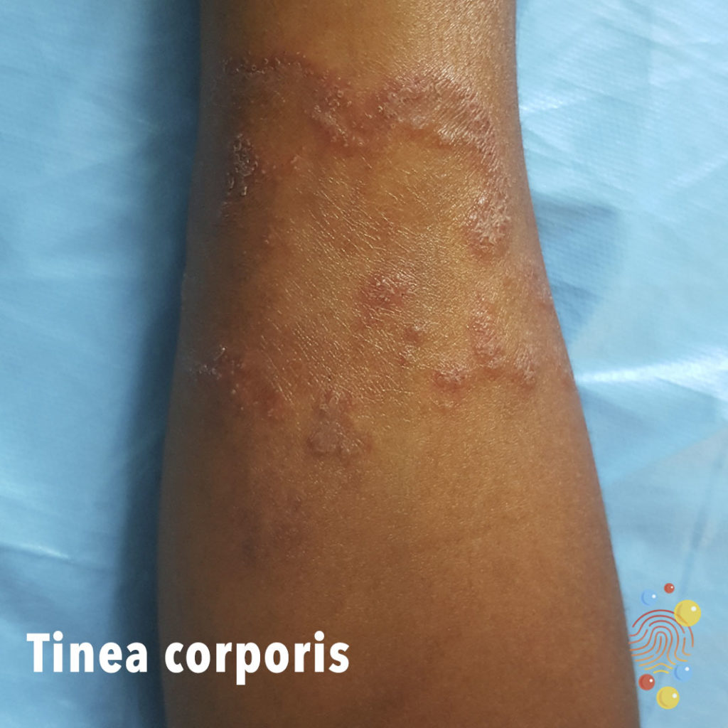
Annular plaques with some peripheral scaling on limb.
Learn more about tinea corporis

Widespread dusky erythema of the posterior trunk with no blistering
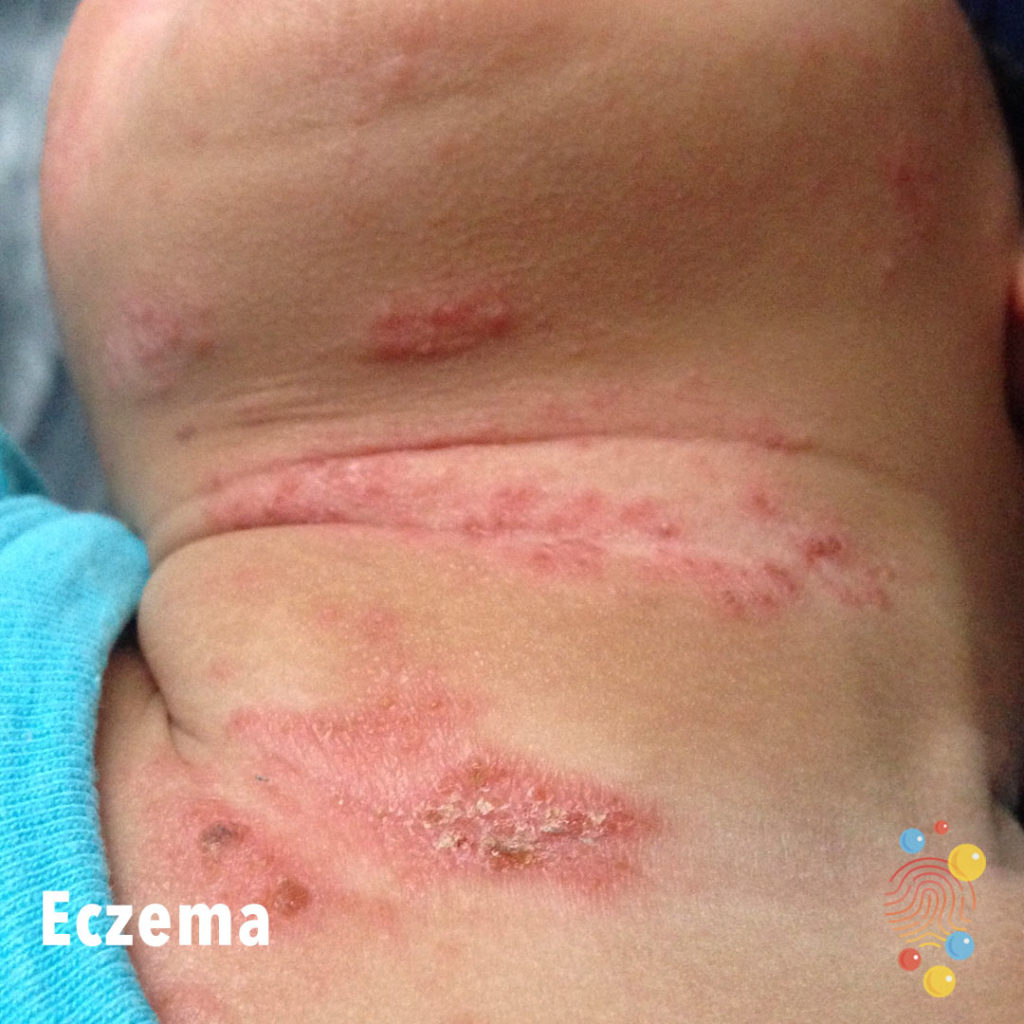
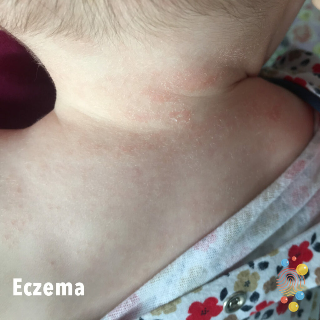
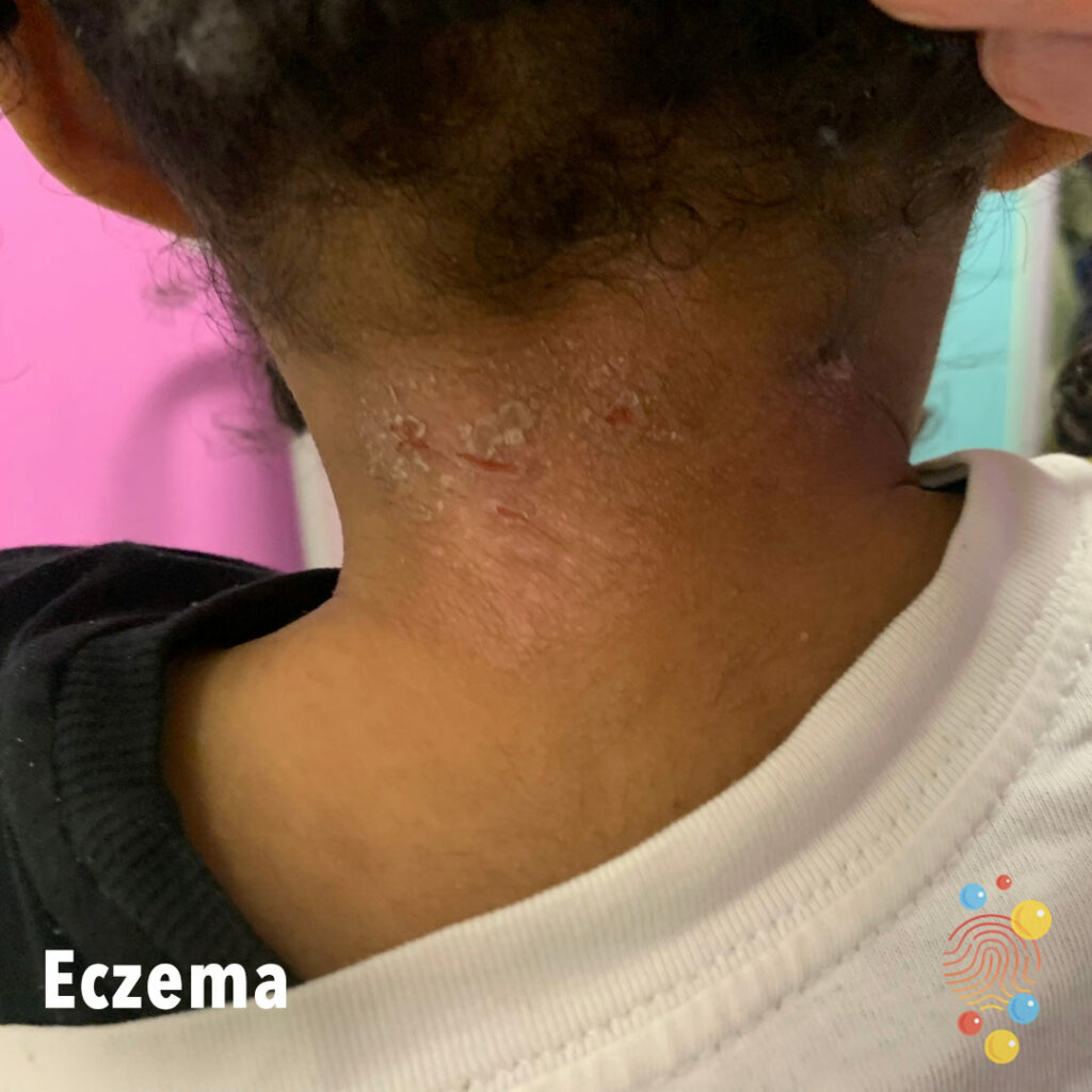
Multiple patches of eczema with post inflammatory hypopigmentation over flexural aspect of neck and likely secondary infection (crusting).
Learn more about eczema
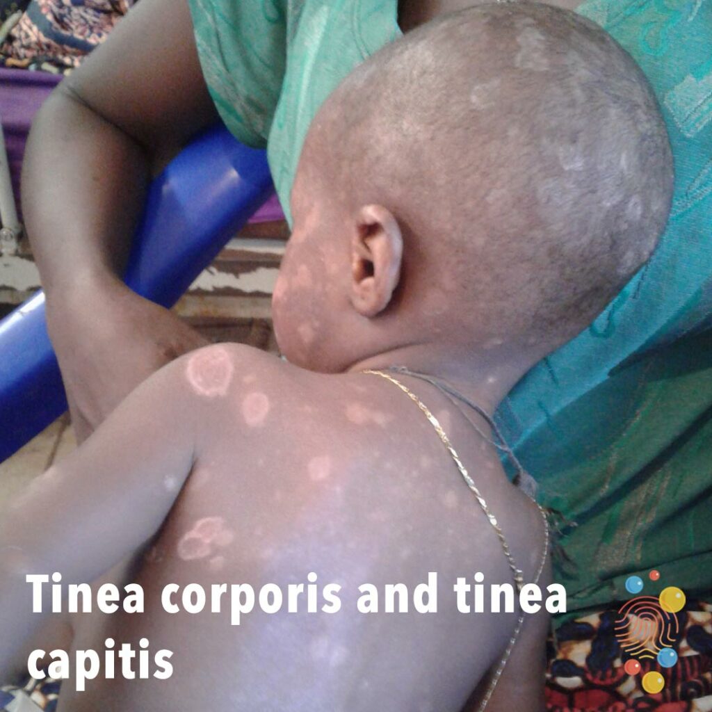
Multiple annular, scaly patches all over body, face, and scalp.
Learn more about tinea corporis

Everted portion of soft tissue.
Learn more about accessory digits

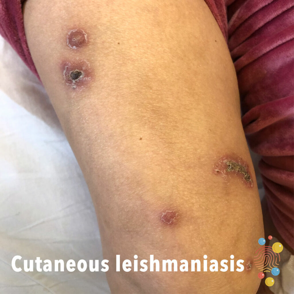
Multiple erythematous papules with peripheral scale some with a dry crusted scab over the right thigh and knee.
Learn more about leishmaniasis
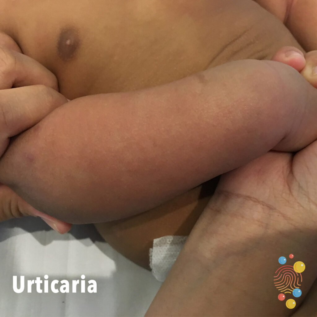
Confluent erythema affecting forearm with no associated angioedema.
Learn more about urticaria
Eczema Coxsackium

Rectangular patch of erythematous, inflamed skin.
Learn more about eczema
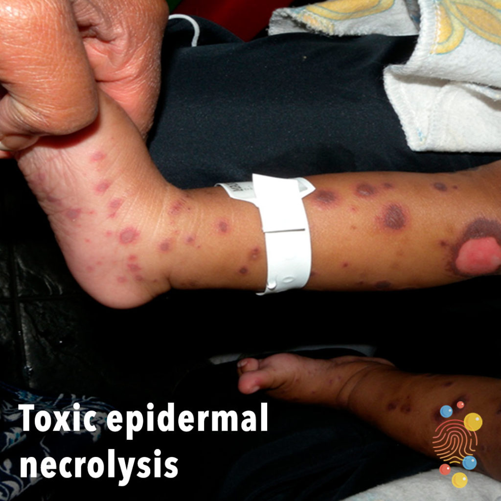
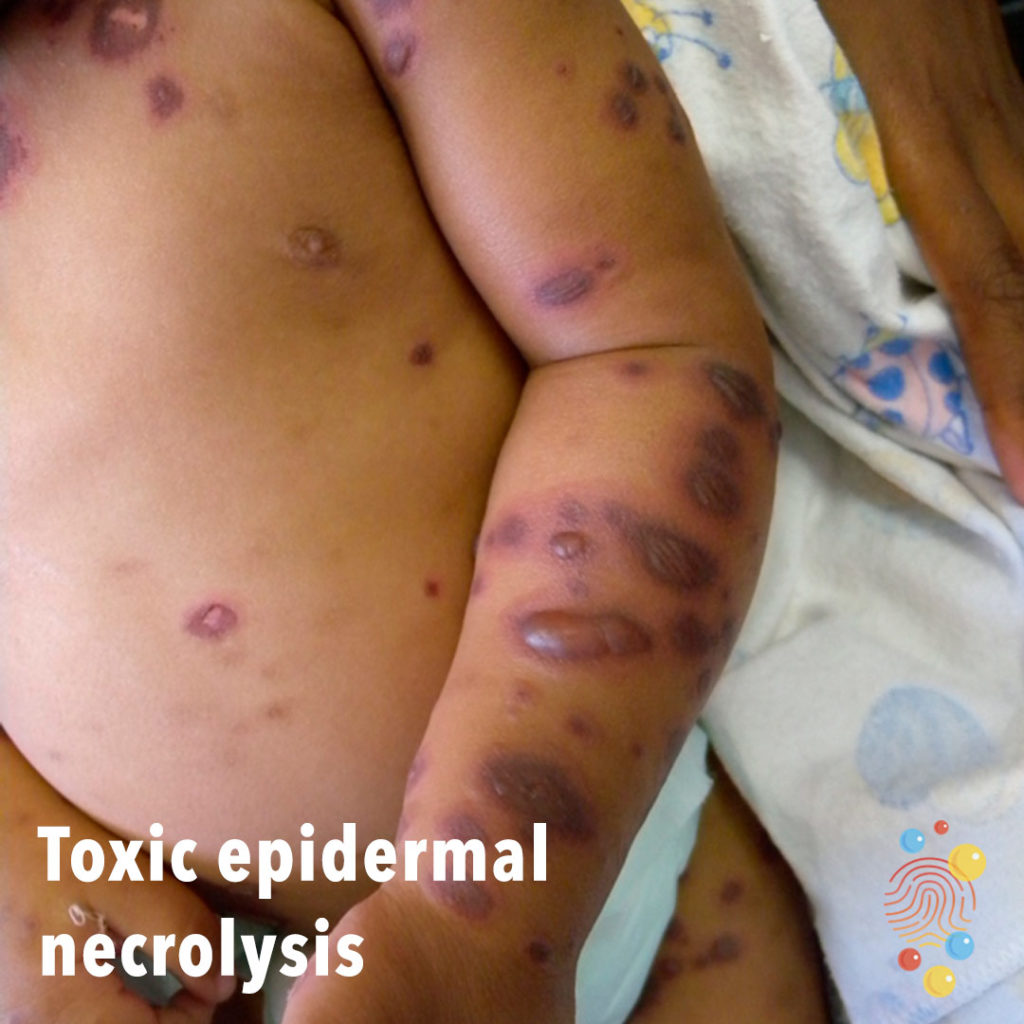
Dusky atypical target lesions with blisters and erosions.
Learn more about toxic epidermal necrolysis

Vesicular lesions on palm and fingers of left hand.
Learn more about hand, foot and mouth
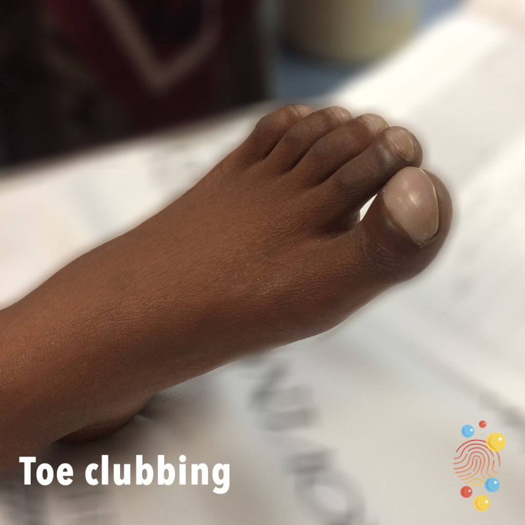
Clubbing noted at distal toes/nails.
Learn more about clubbing
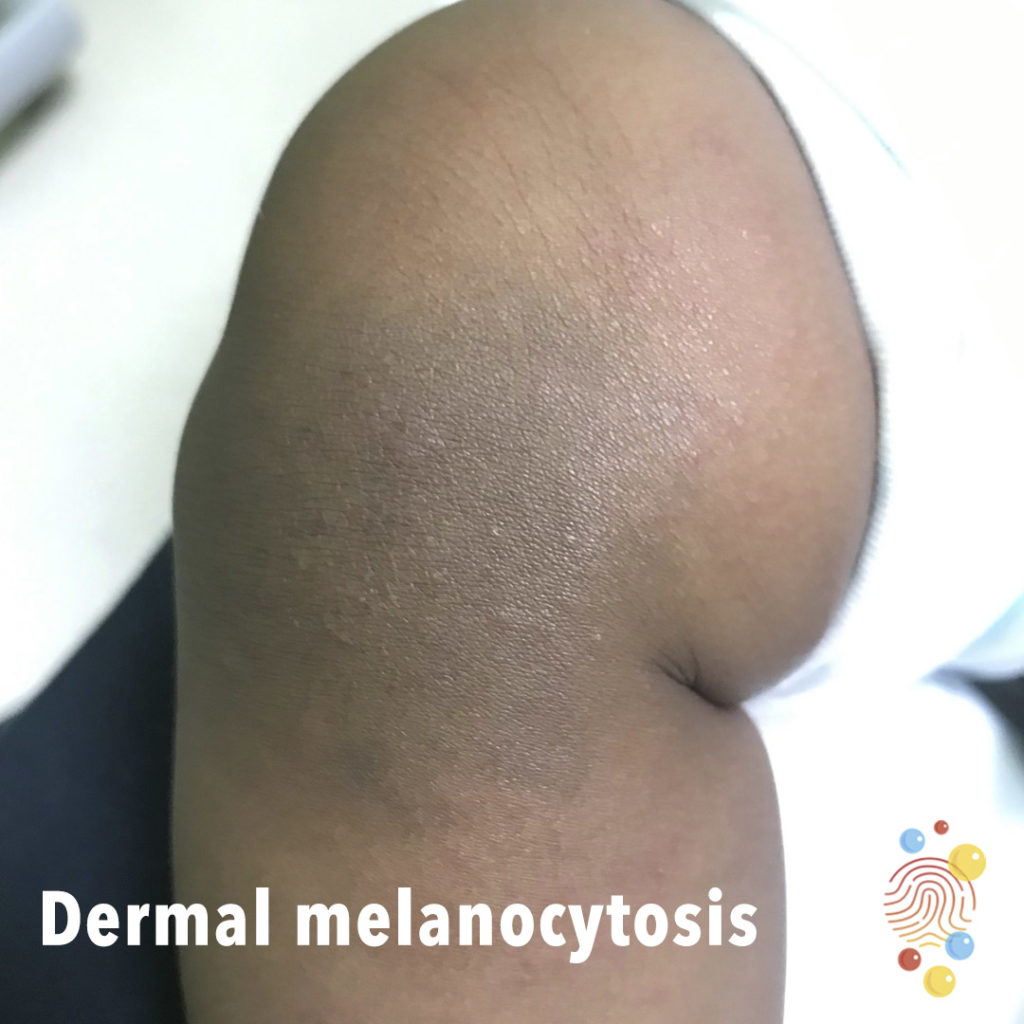
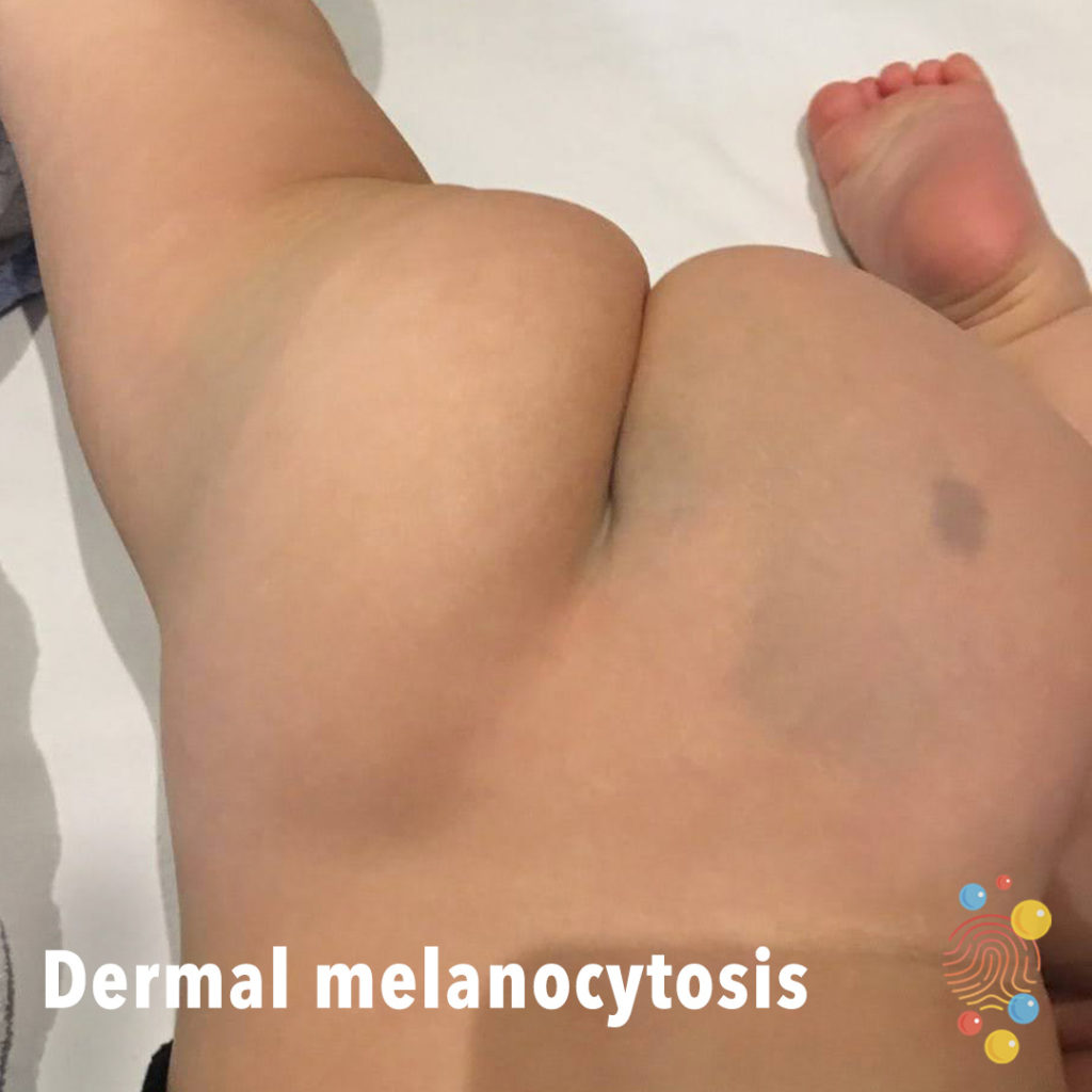
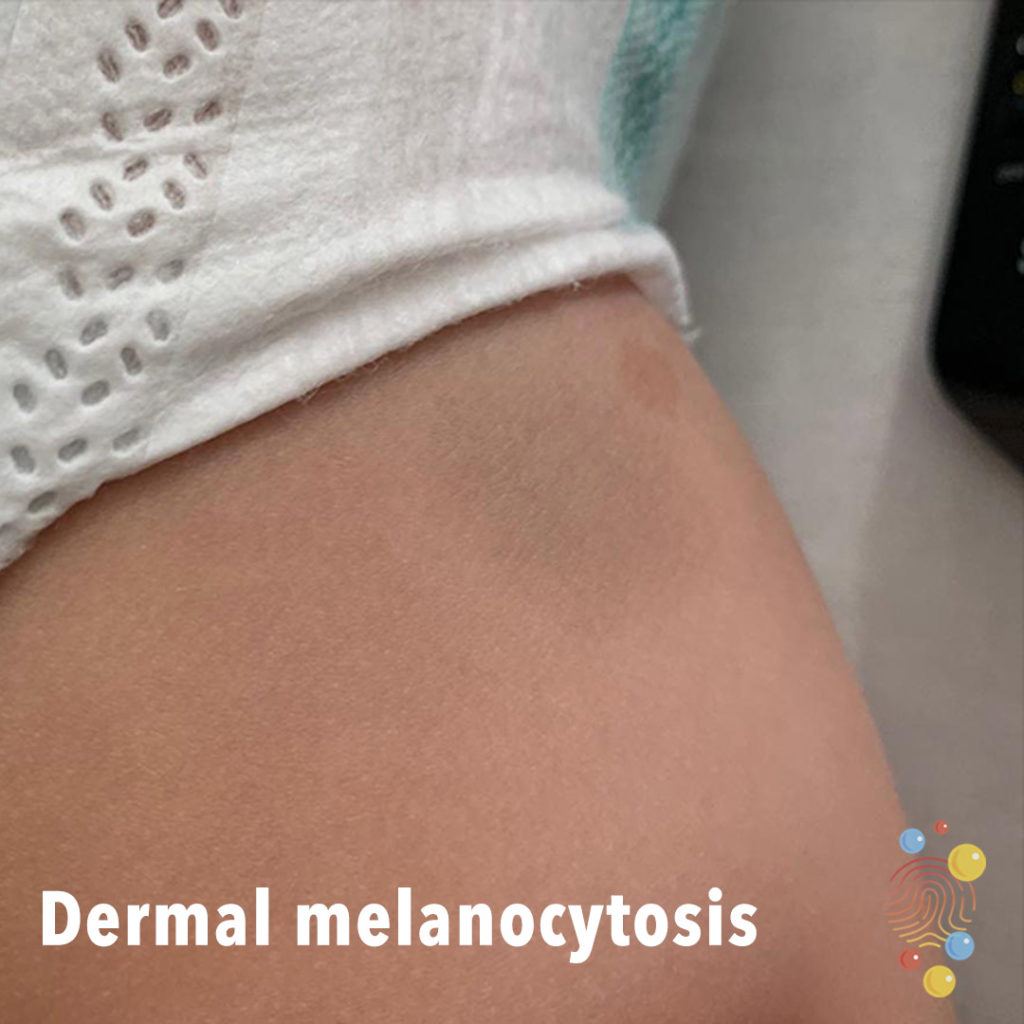
Hyperpigmented patch on the leg.
Learn more about dermal melanocytosis
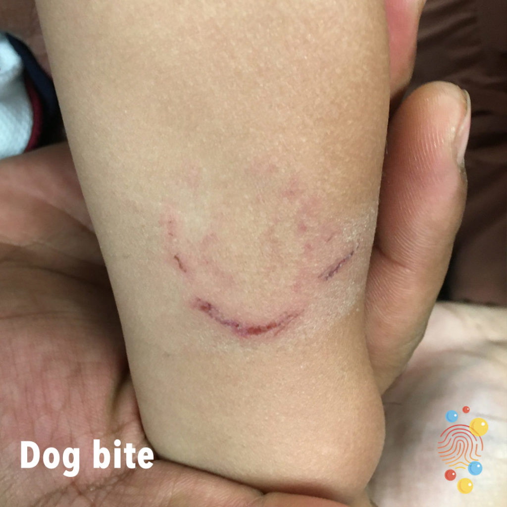
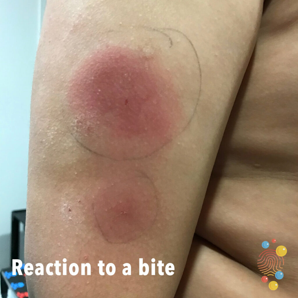
Learn more about bites
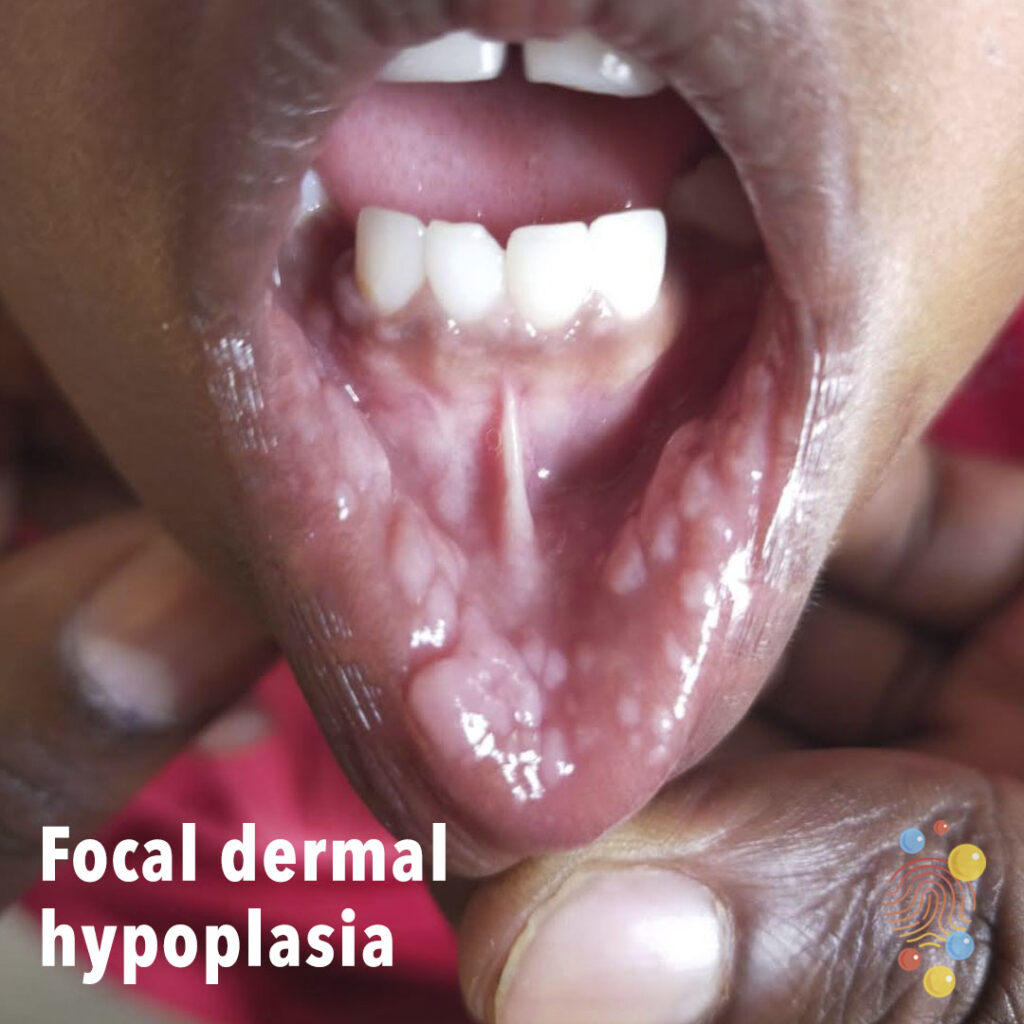
Pale, white, flat-topped papules on labial mucosa

Extensive blue-grey discolouration affecting the lower back.
Learn more about dermal melanocytosis
Erythematous patches on the posterior neck and back.
Learn more about eczema
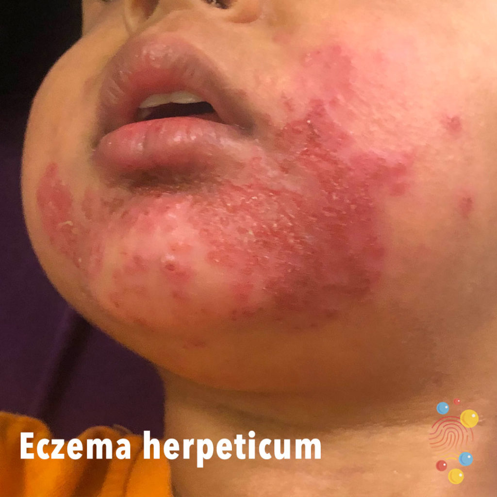
Erythema and punched-out erosions affecting the chin.
Learn more about eczema herpeticum
Papular rash on the abdomen.
Learn more about eczema
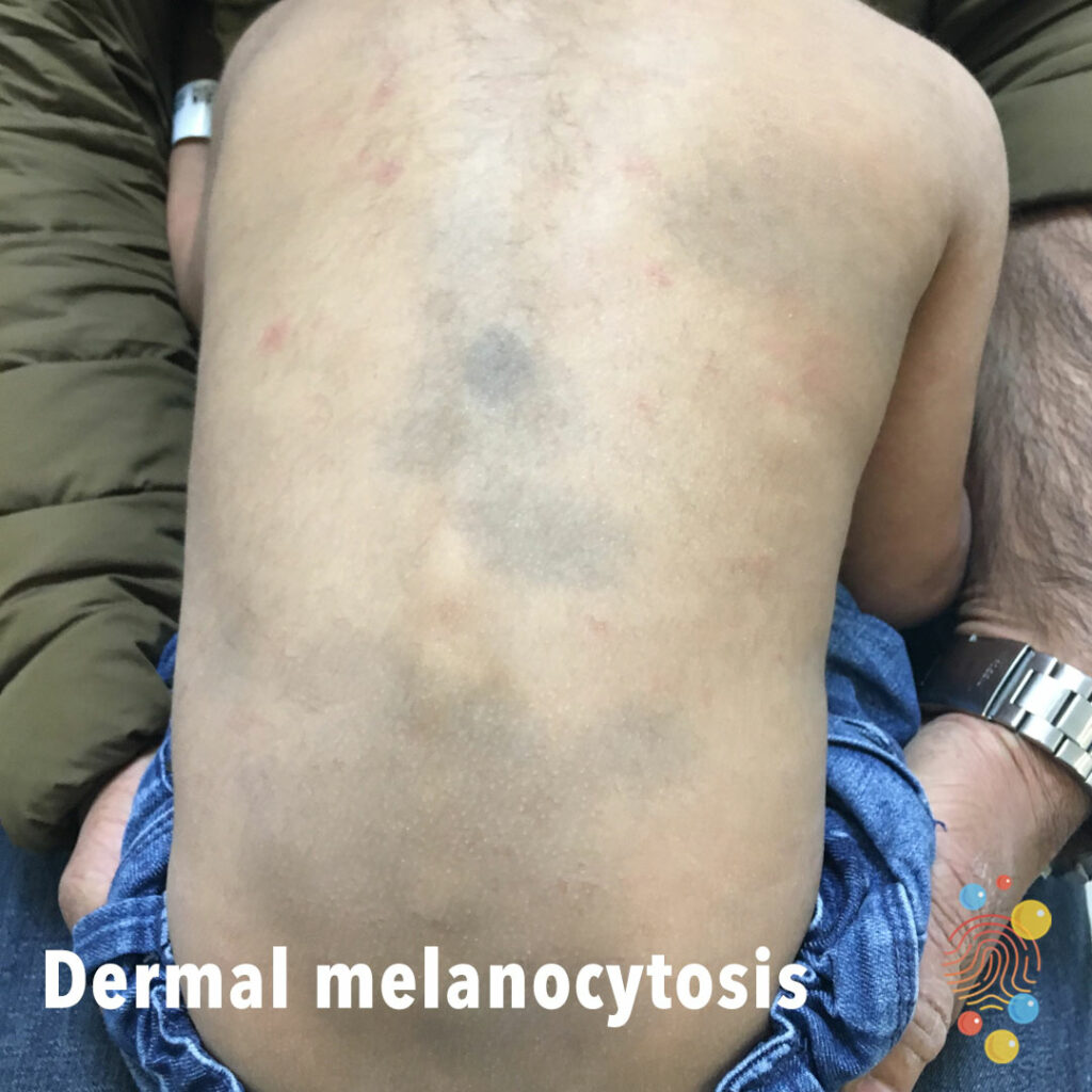
Multiple areas of deep blue dermal pigmentation on the back.
Learn more about dermal melanocytosis
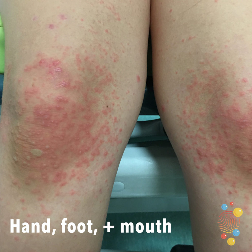
An erythematous rash with multiple and polymorphic vesicles and blisters, located on the dorsal skin of the knees.
Learn more about hand, foot and mouth

Localised induration with overlying erythema and oedema (peau d’orange) and areas of blistering and papulation within the erythema.
Learn more about cellulitis
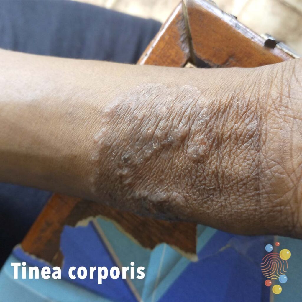
Raised red edge surrounding a central area of hyperpigmentation with accentuated skin markings.
Learn more about tinea corporis

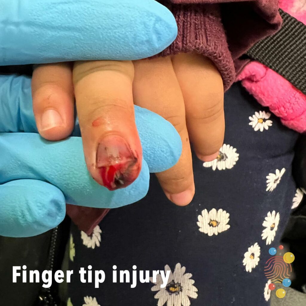
Avulsed Nail
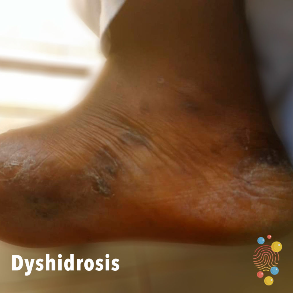
Small blisters on the sole of the foot, with post-inflammatory hyperpigmentation.
Learn more about dyshidrosis

Discrete tense bullae with surrounding erythema in linear “breakfast, lunch, and dinner” distribution.
Learn more about bites
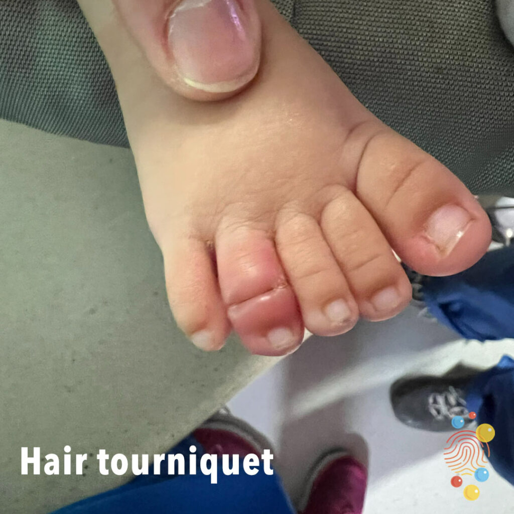
Hair Tourniquet
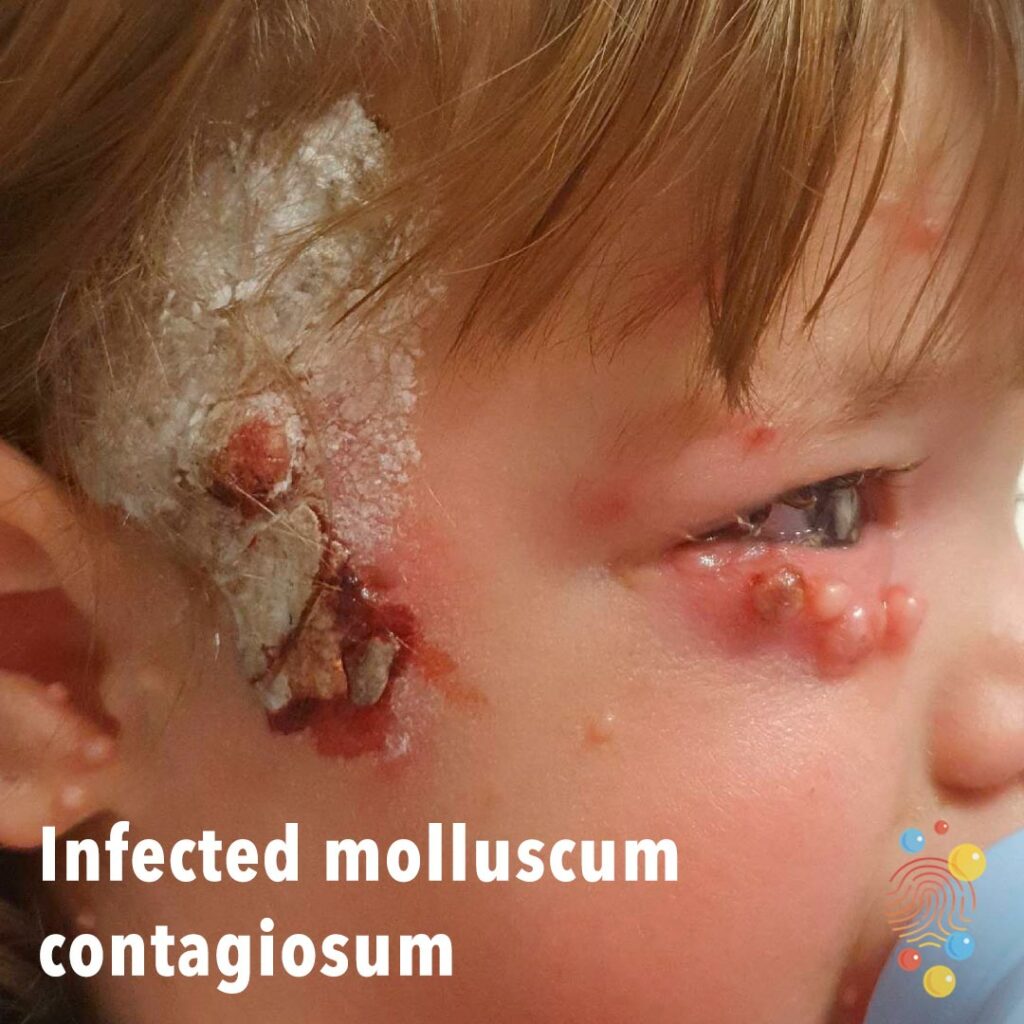
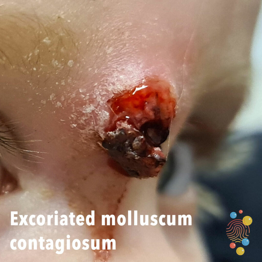
Molluscum contagiosum with secondary infection and ulceration. White material likely exogenous (?zinc cream).
Learn more about molluscum contagiosum
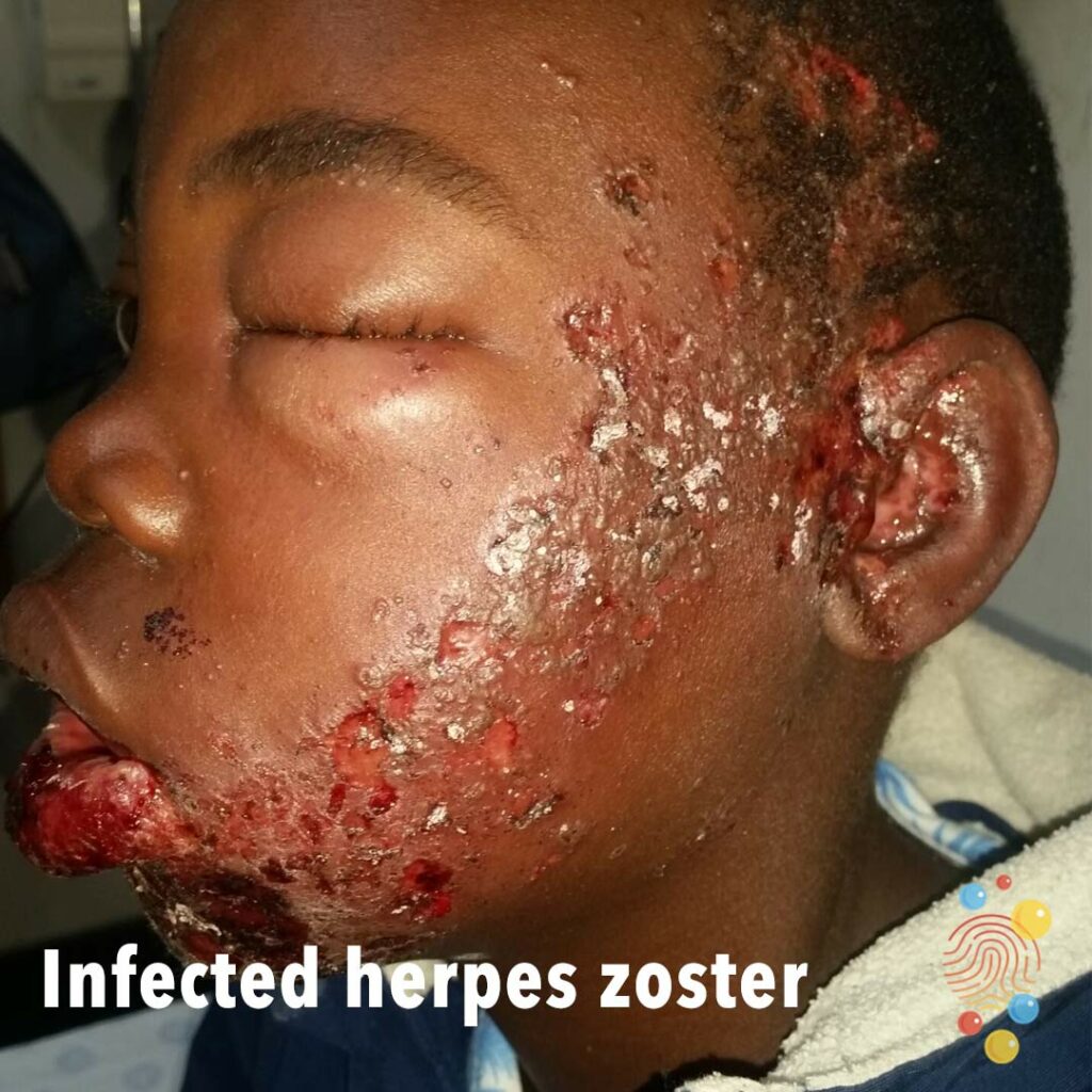
Extensive linear ulceration of the left side of the face extending from the chin to the temple including the tragus and ear, consistent with involvement of the maxillary and mandibular branches of the trigeminal nerve.
Learn more about herpes zoster
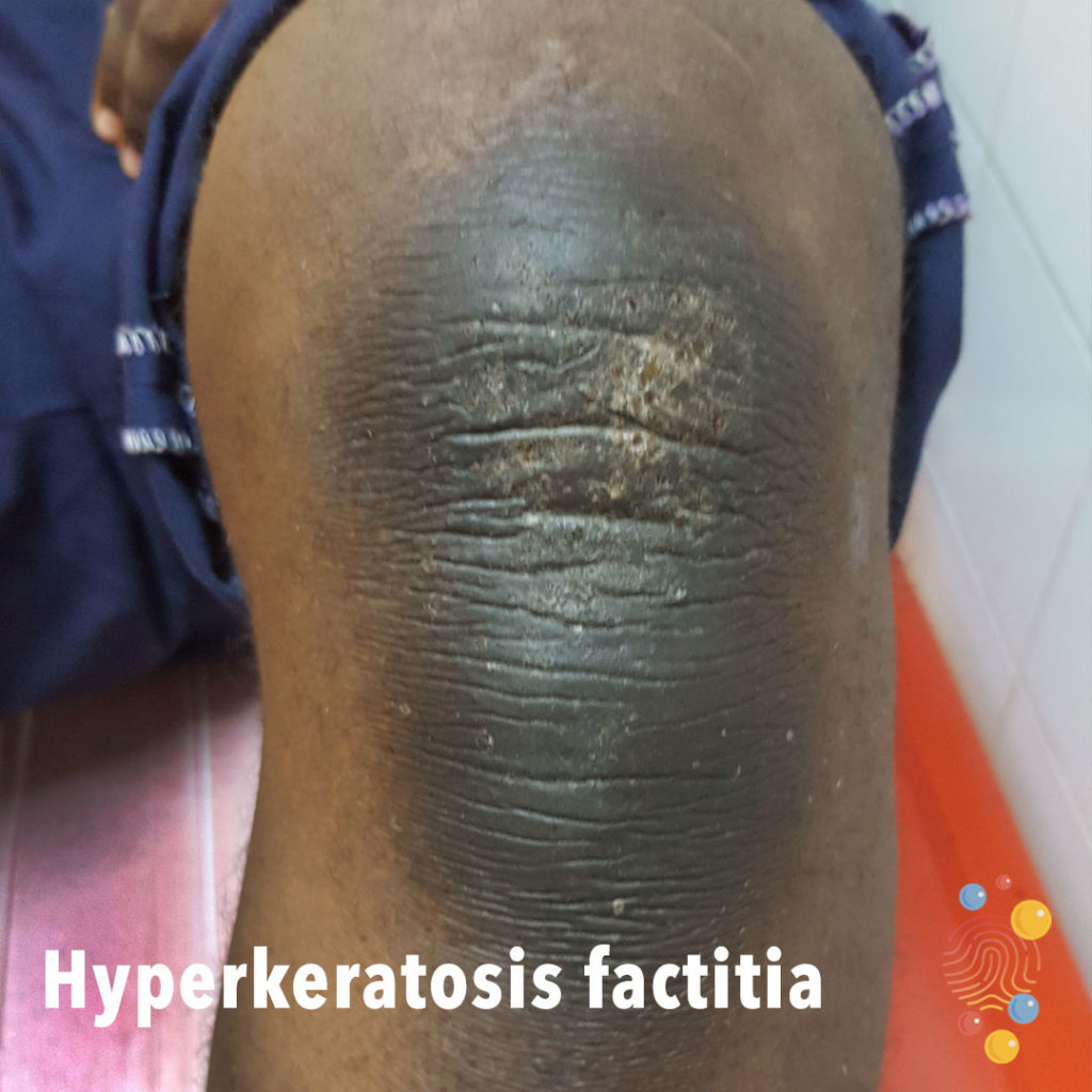
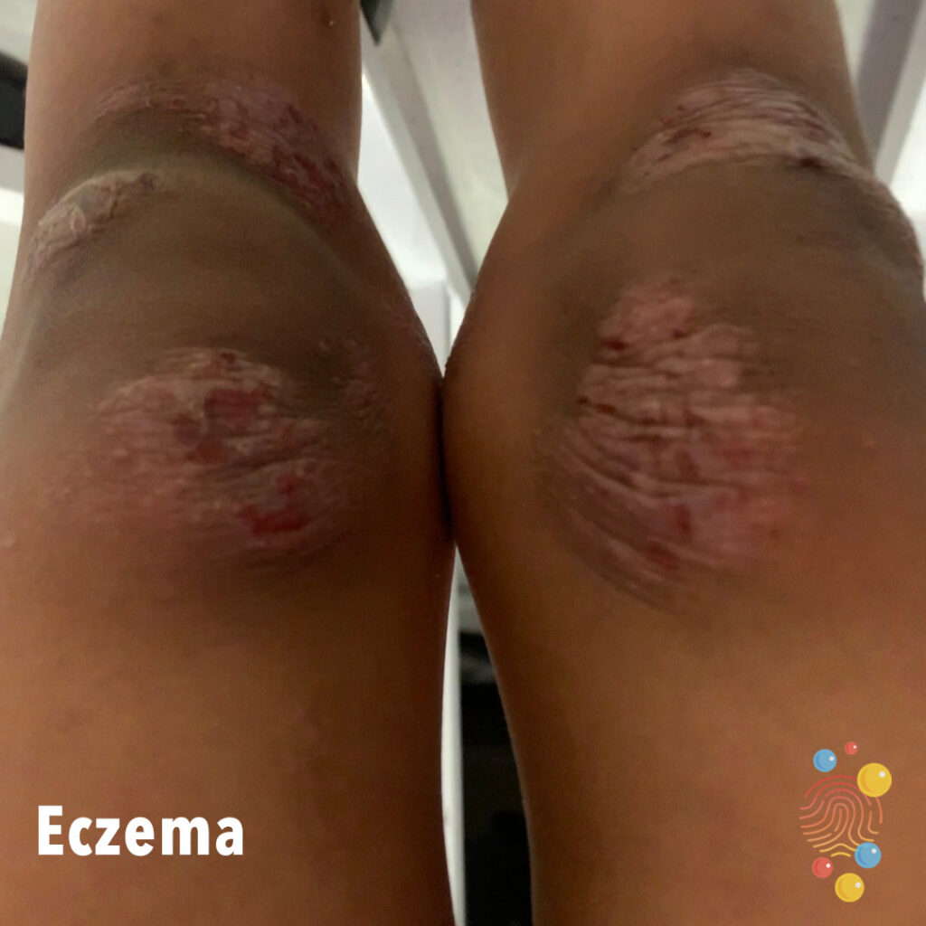
Frictional darkening of the extensor surfaces of the knees with thickened skin.
Learn more about hyperkeratosis factitia
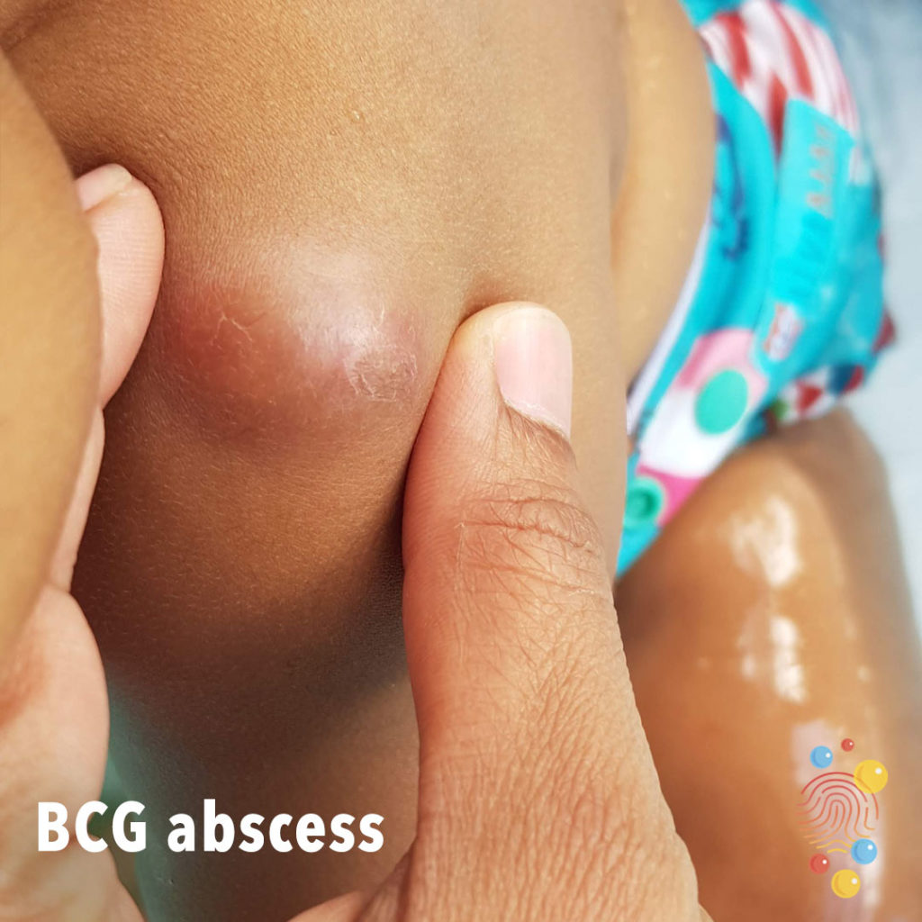
Discrete dermal nodule with associated erythema and hyperpigmentation.
Learn more about BCGs
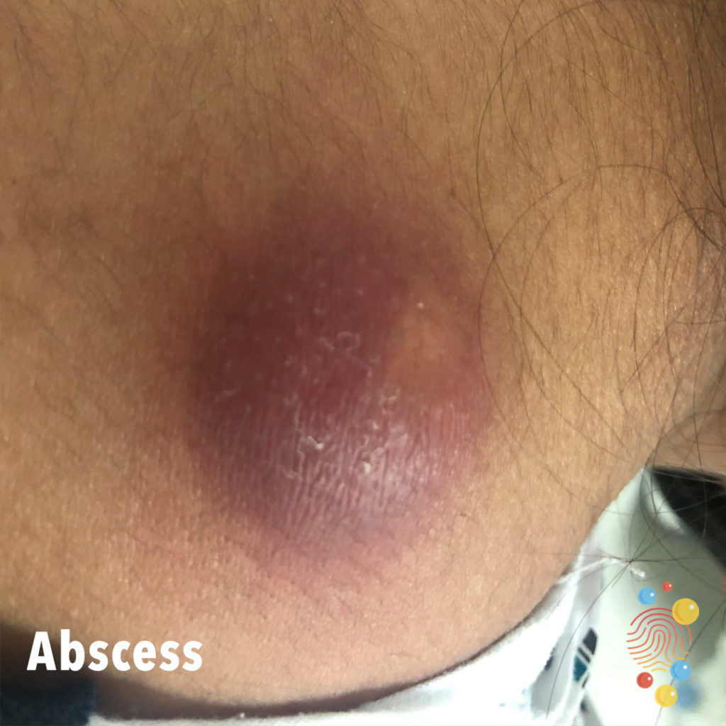
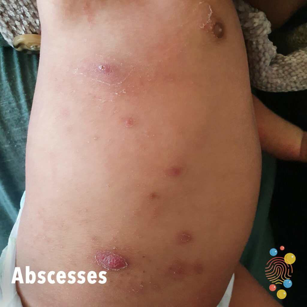
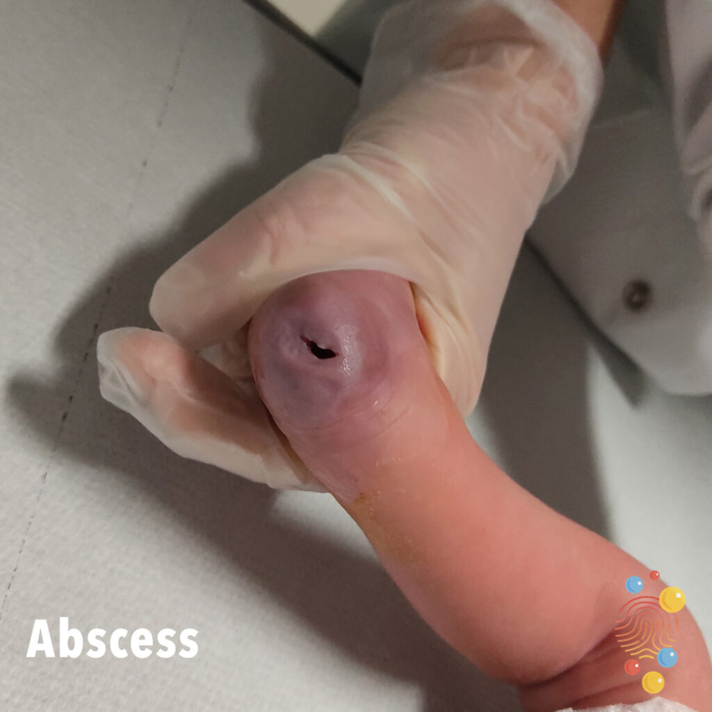
Large raised firm swelling with associated erythema.
Learn more about abscesses
Clubbing noted at distal fingers/nails.
Learn more about clubbing


Papulosquamous eruption affecting the dorsa of the fingers.
Learn more about syphilis
Facial atopic dermatitis with secondary infection and crusting.
Learn more about eczema

Vesicular eruption on palms.
Learn more about hand, foot, + mouth disease

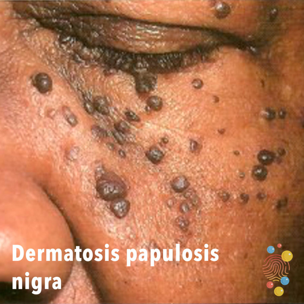
Multiple black papules on the cheeks.
Learn more about dermatosis papulosis nigra
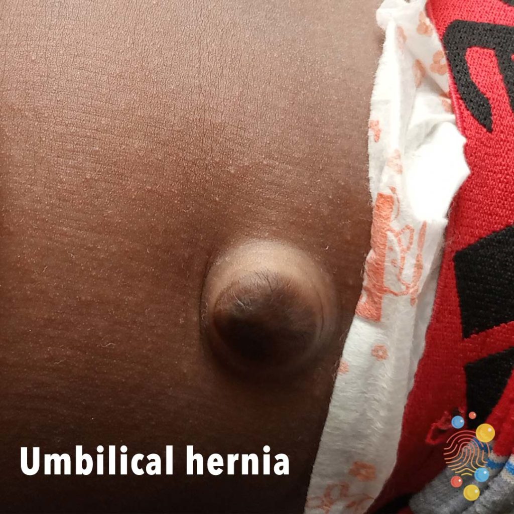
Skin-coloured protrusion emerging from the umbilicus.
Learn more about umbilical hernias
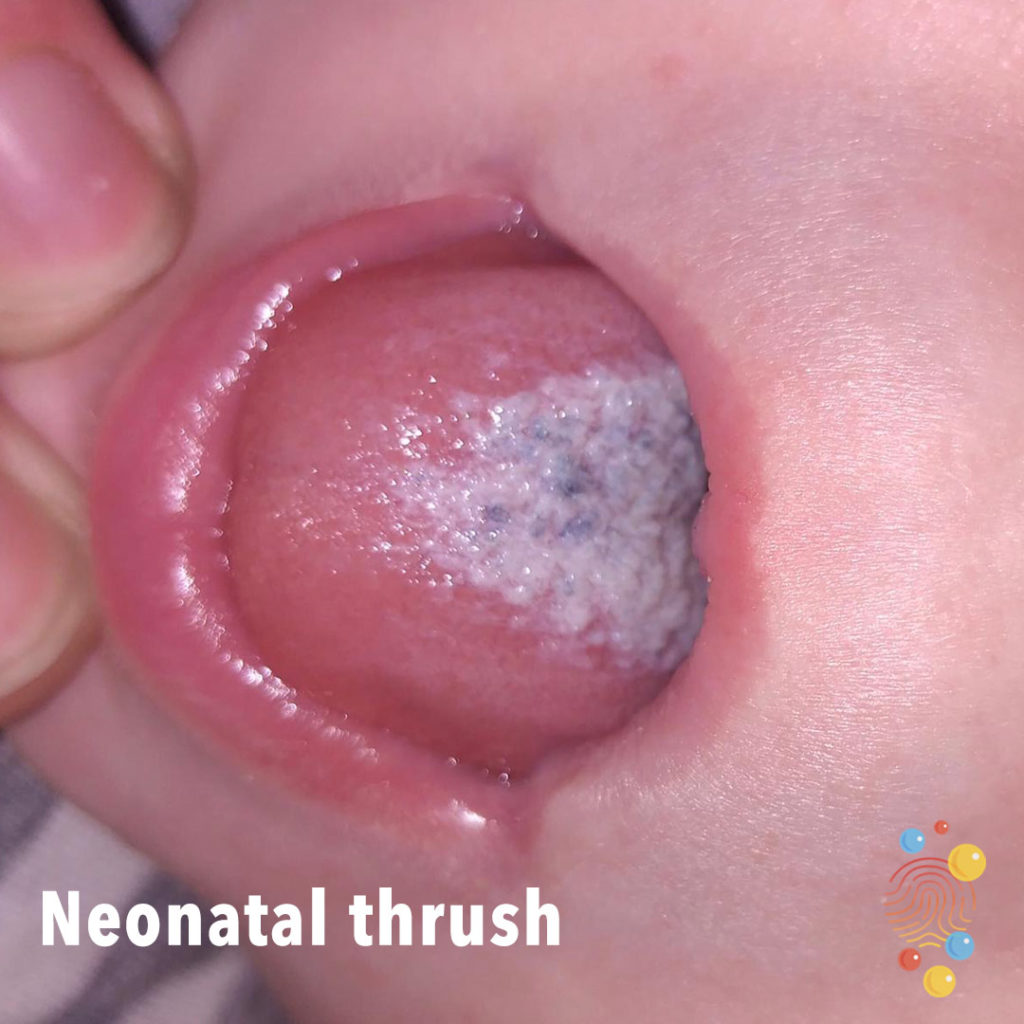
White coating on tongue.
Learn more about neonatal thrush

Vesicles on an erythematous base with some crusted lesions.
Learn more about chicken pox
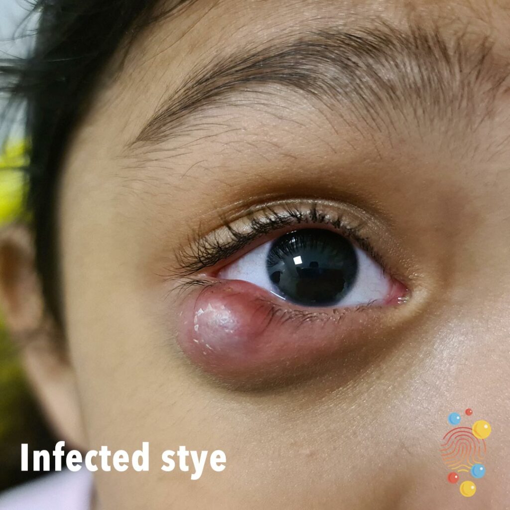
Infected stye
Oedema and erythema of the toes circumferentially.

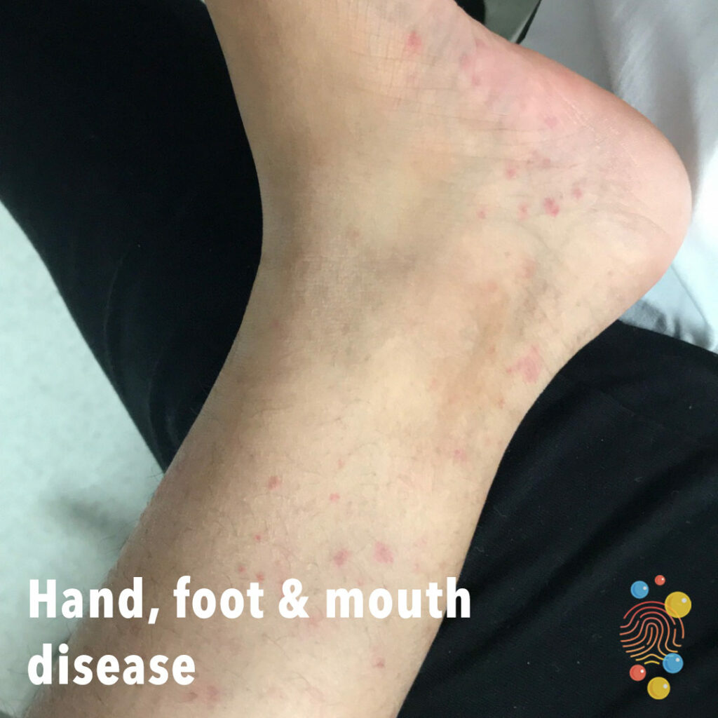
Acral erythematous macules. No vesicles/blisters or target lesions apparent.
Learn more about hand, foot and mouth
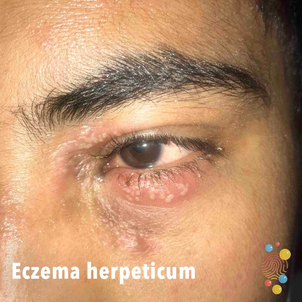
Clusters of peri-ocular pustules on a background of erythematous patches. Numerous vesicles and erythematous changes across the face.
Learn more about eczema herpeticum
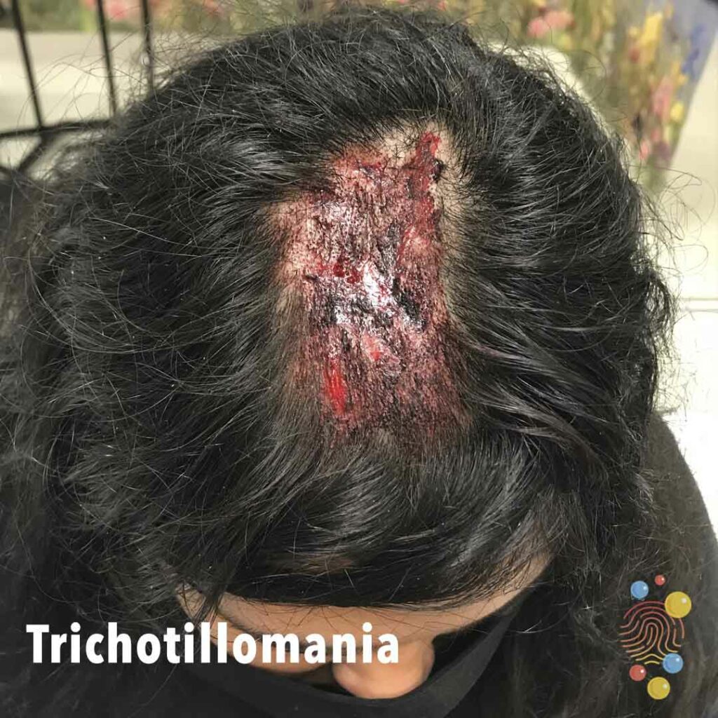
Linear superficial excoriations/erosions with associated loss of hair.
Learn more about trichotillomania
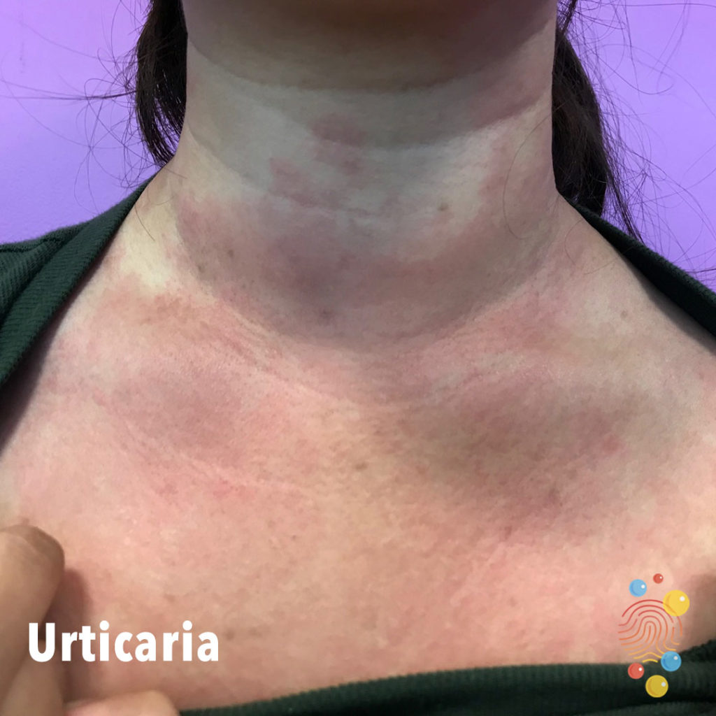
Widespread erythema across upper chest anteriorly extending to neck. Well defined border at neck. Some wheals.
Learn more about urticaria
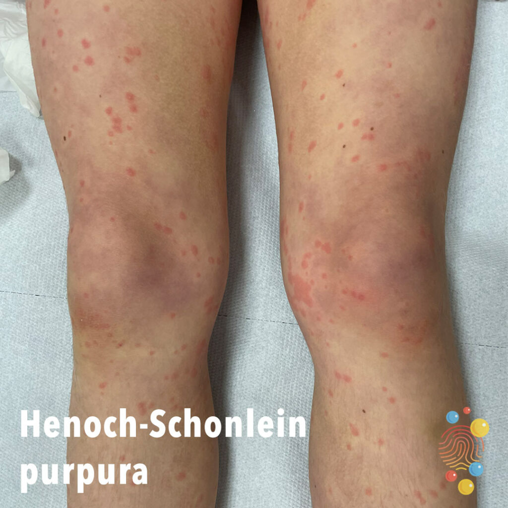
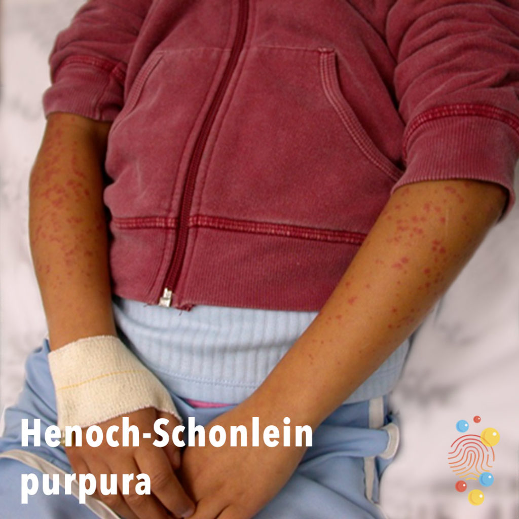
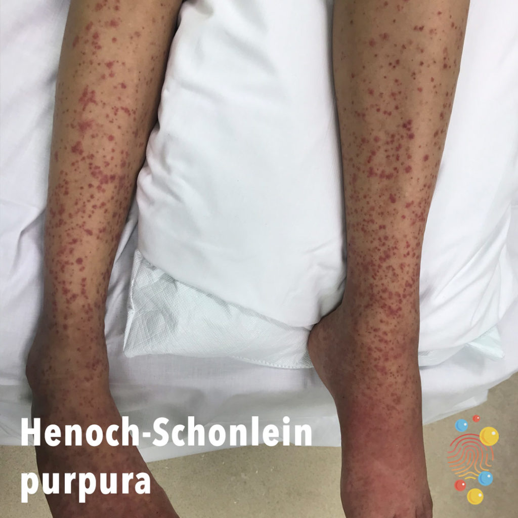
Erythematous papules symmetrically distributed on legs with surrounding pallor.
Learn more about Henoch-Schonlein purpura

Erythema with indistinct margins and sparing of pressure points (underwear).
Learn more about scombroid poisoning
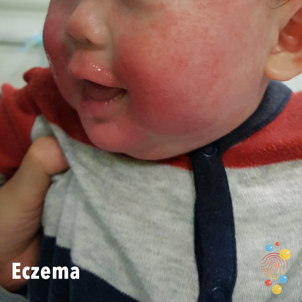
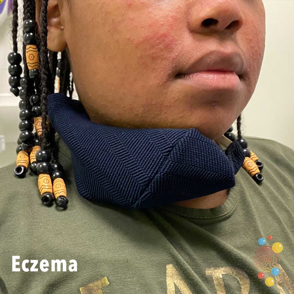
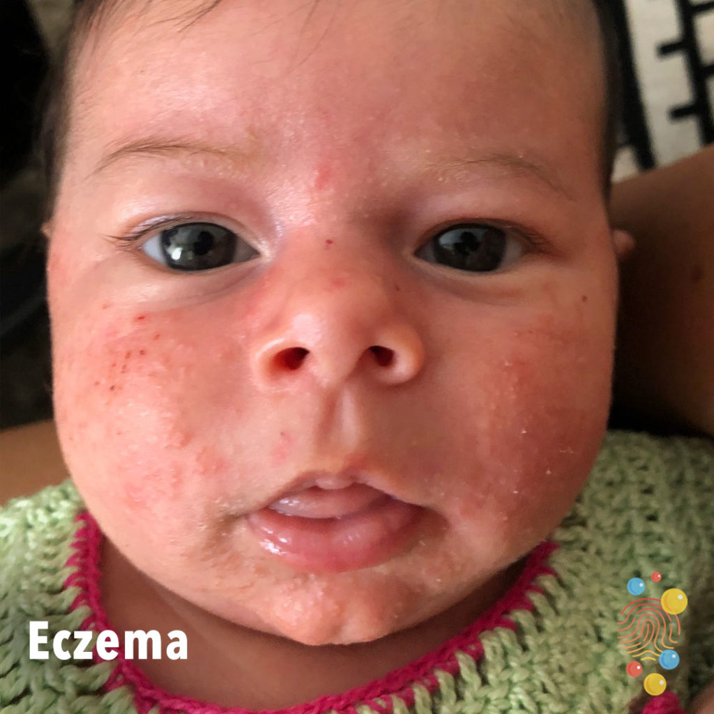
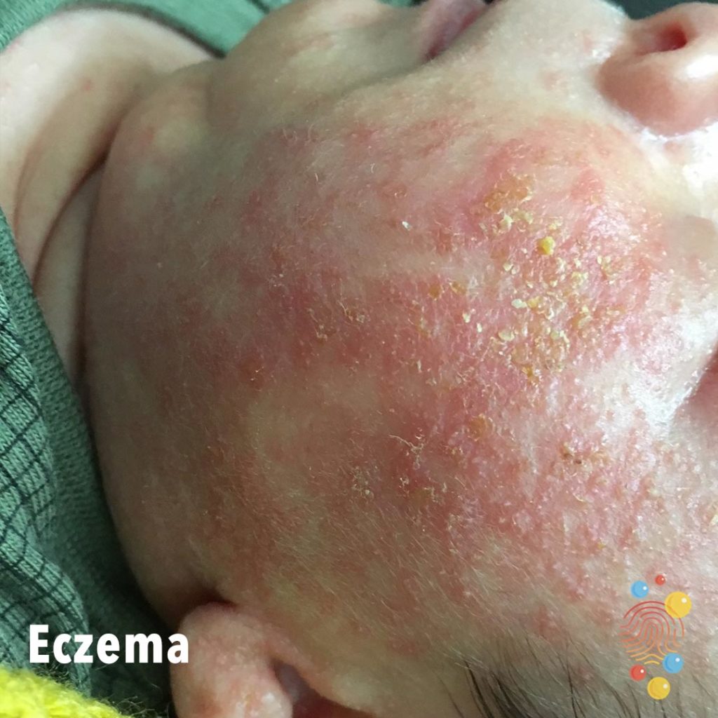
Erythematous scale over cheeks, mouth and chin. Atopic with irritant dermatitis.
Learn more about eczema


Linear patch of alternating light and dark brown pigmentation with hypertrichosis.
Learn more about beckers naevus
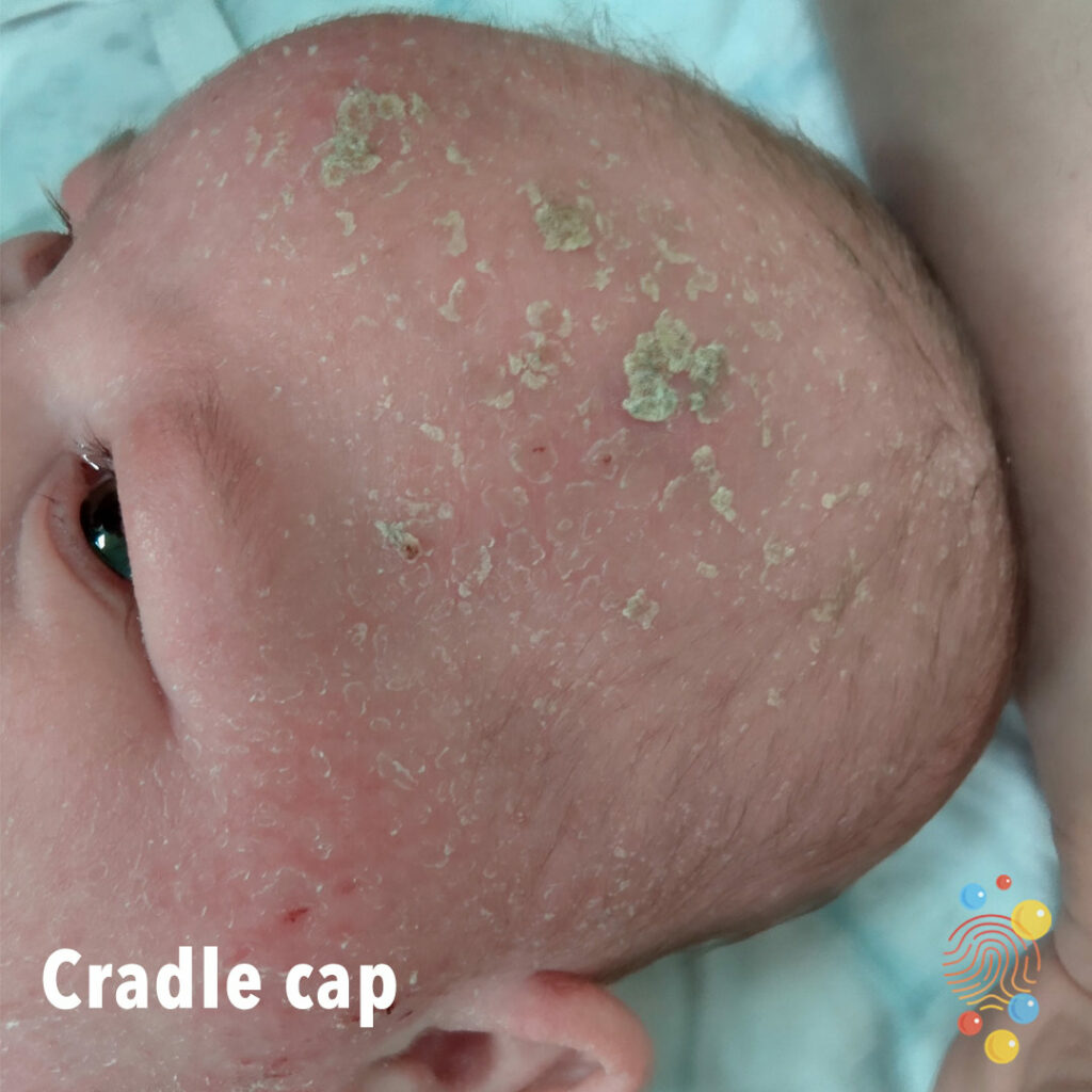
Cradle Cap
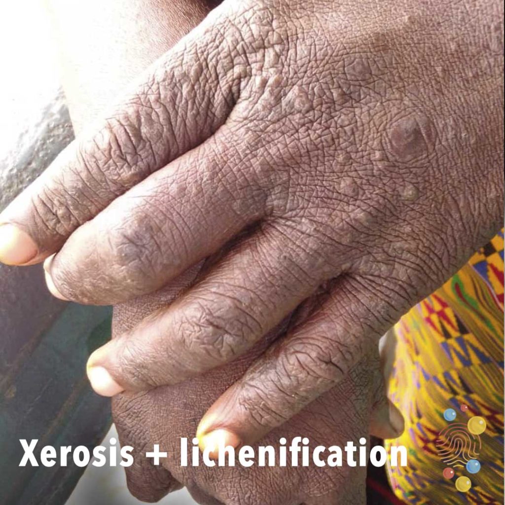
Scaly skin on dorsal hand with increased skin markings + scattered skin coloured papules.
Learn more about xerosis lichenification
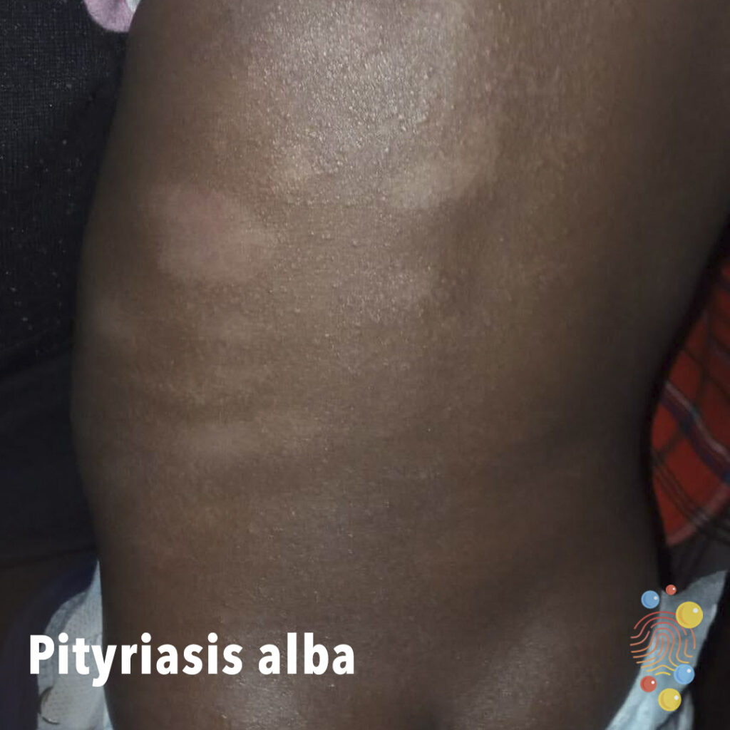
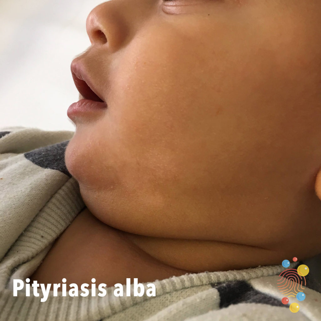
Pink and white oval patches on the trunk due to post-inflammatory hypopigmented marks on atopic skin.
Learn more about pityriasis alba

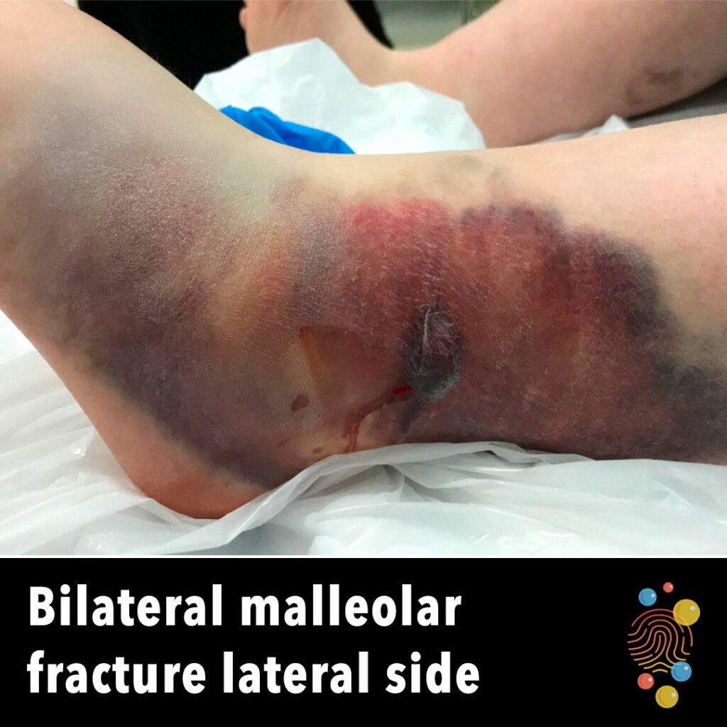
Extensive bruising and haematoma formation around left ankle.
Learn more about ecchymosis
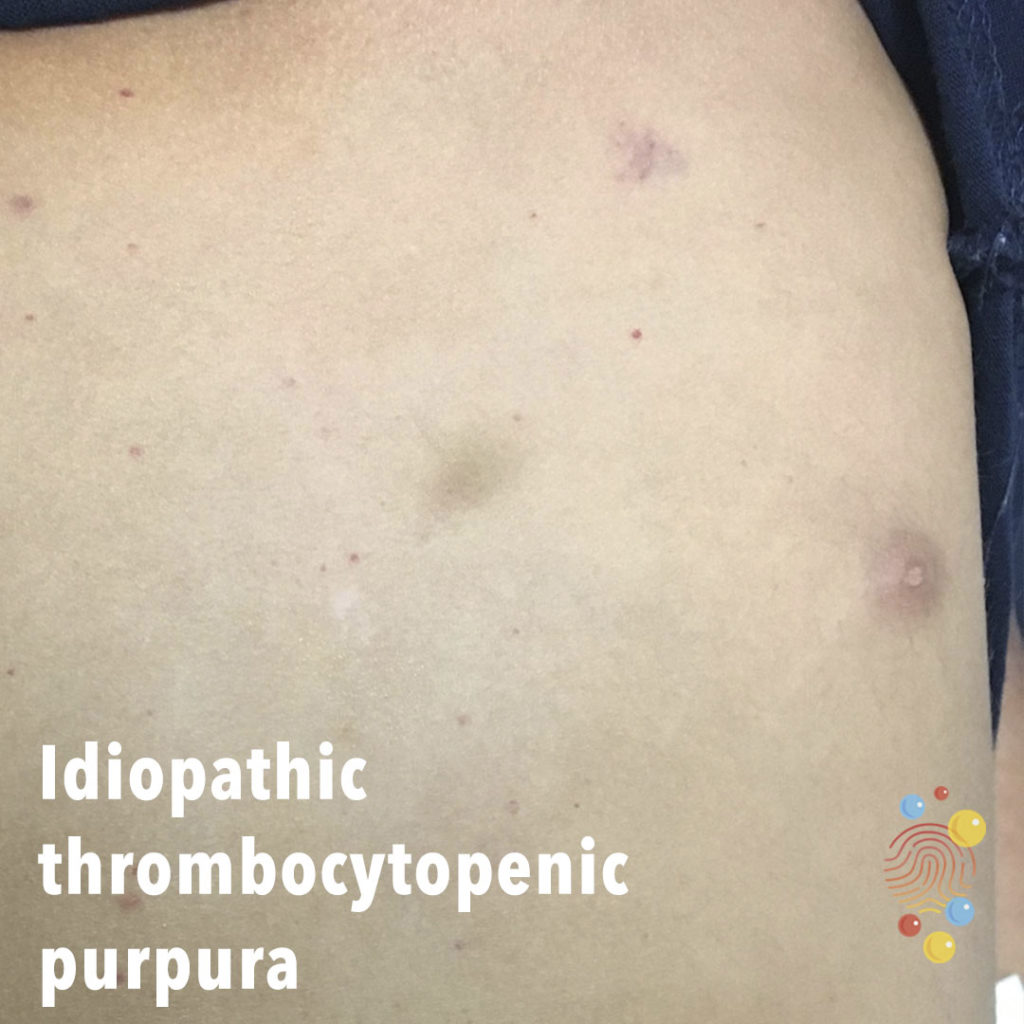
Scattered small lesions of non-blanching erythema/purpura. Two ecchymoses evident
Learn more about idiopathic thrombocytopenic purpura
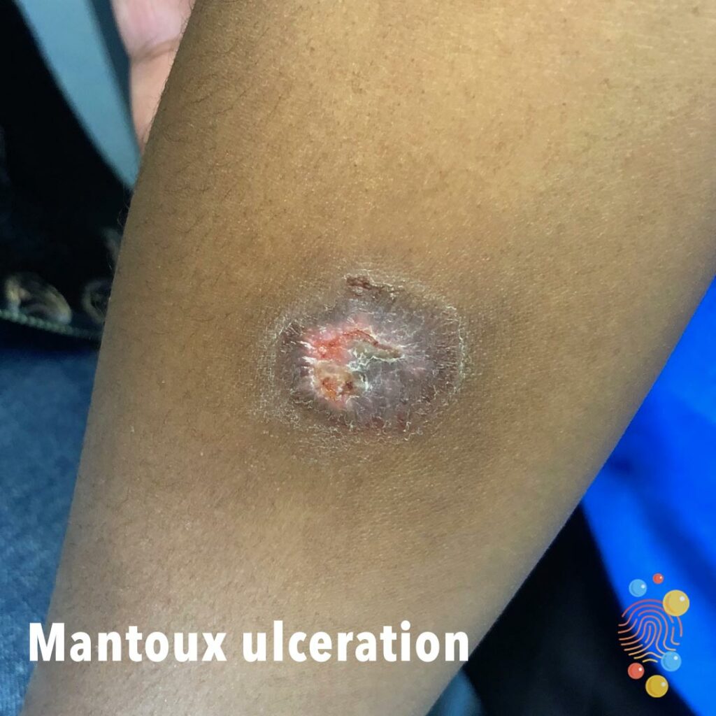
Single annular lesion with central ulceration. Thickened, pink edge to lesion.
Learn more about Mantoux ulceration

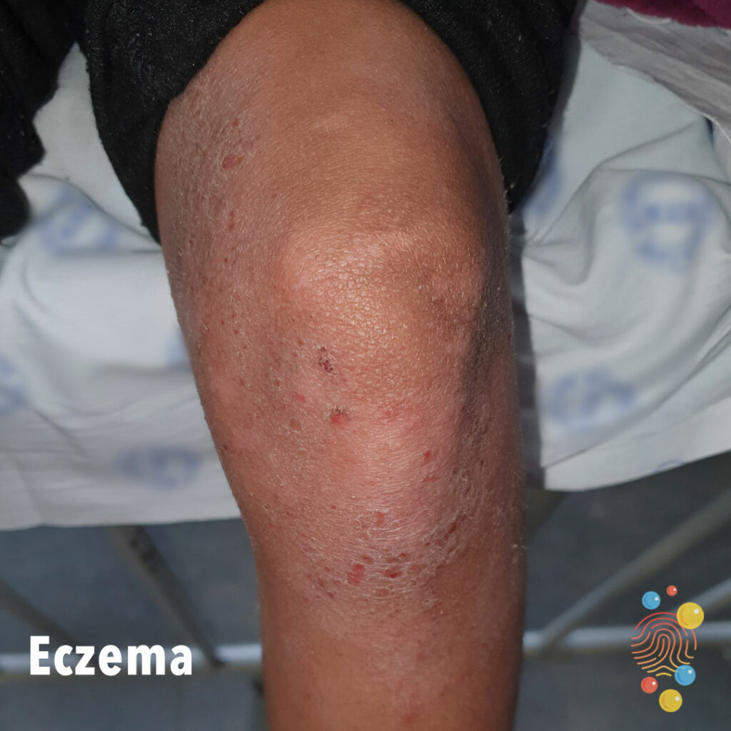
Excoriated erythematous papules on legs with surrounding dry skin.
Learn more about eczema
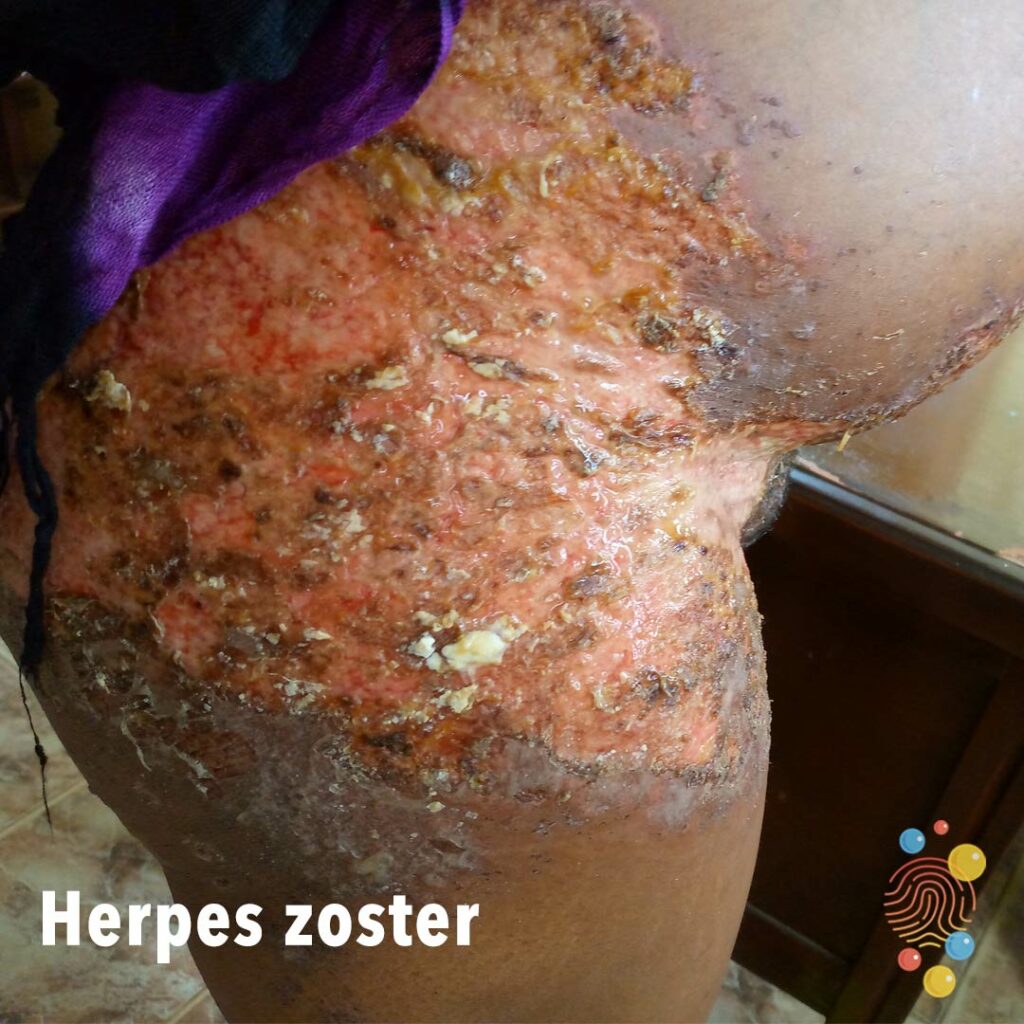
Blistering and desquamation with ulceration L1/2 distribution.
Learn more about herpes zoster
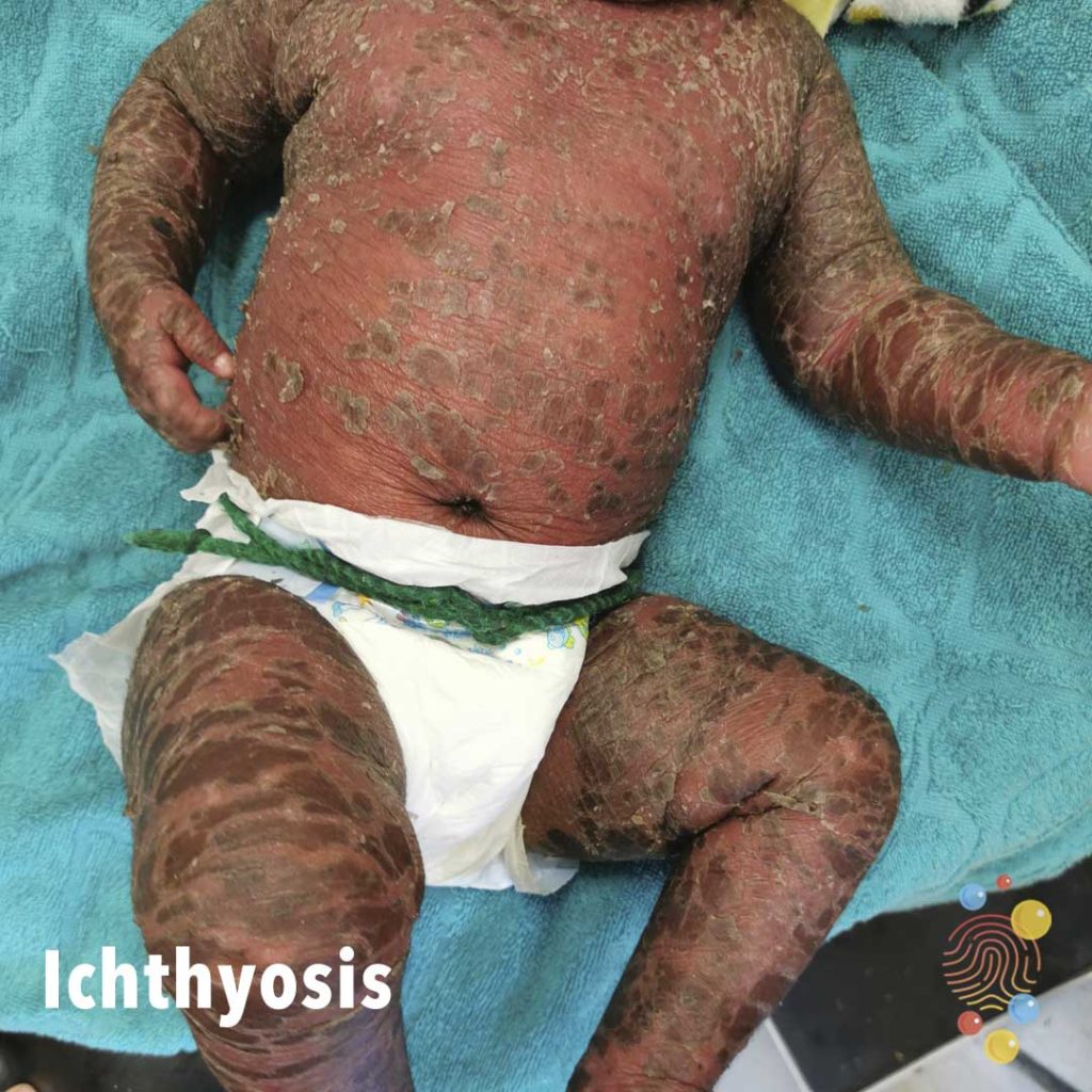
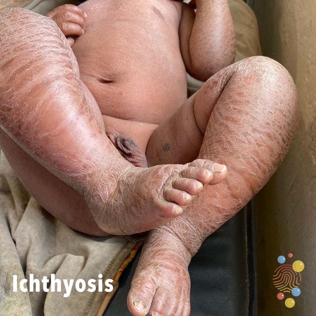
Erythematous skin with overlying scale.
Learn more about ichthyosis
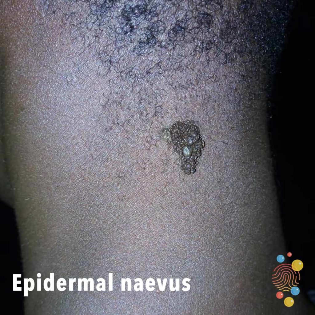
Single verrucous plaque on posterior neck.
Learn more about epidermal naevus
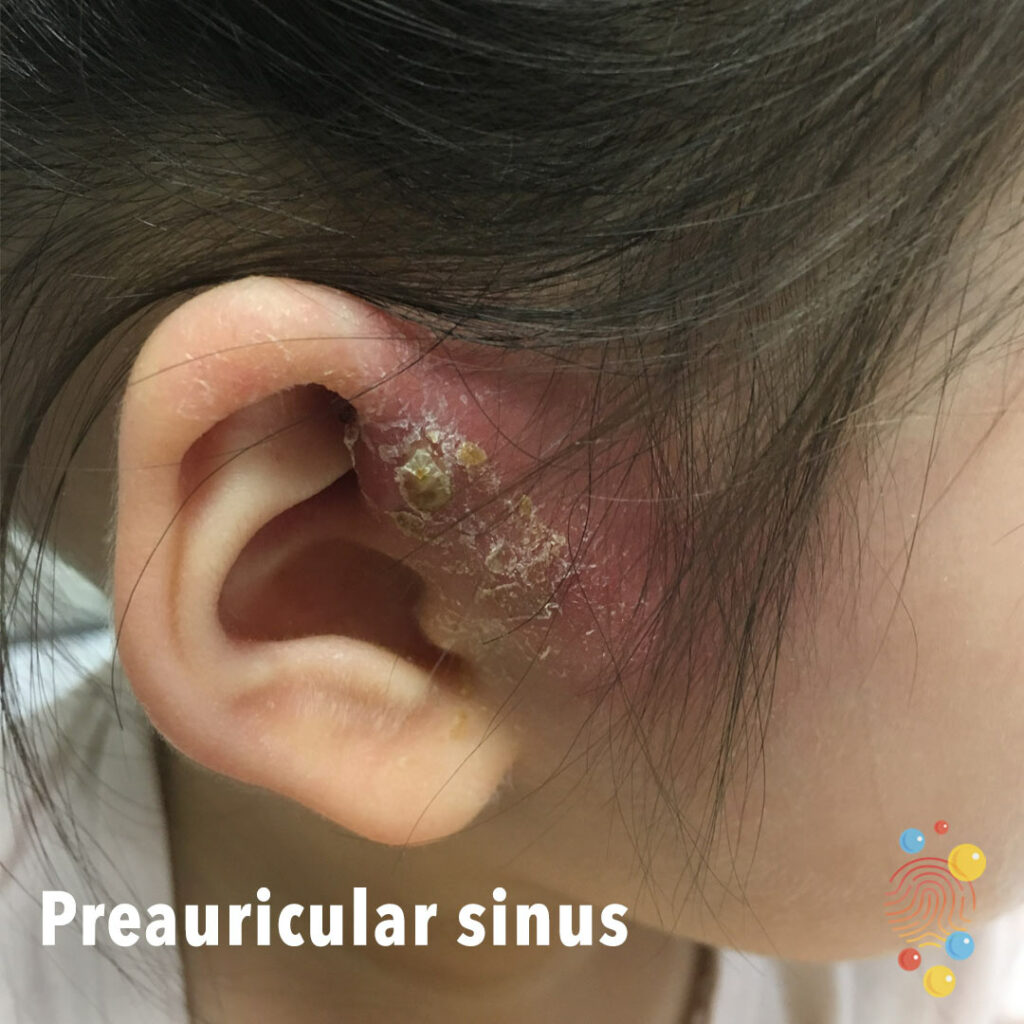
Erythematous and crusted pre-auricular lesion. Inflammation and swelling with surface scale.
Learn more about sinuses
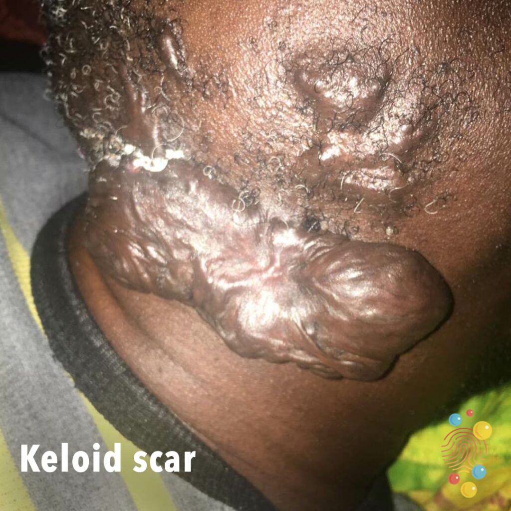
Keloid scar (scar extends outside the margin of the original scar).
Learn more about keloid scars.
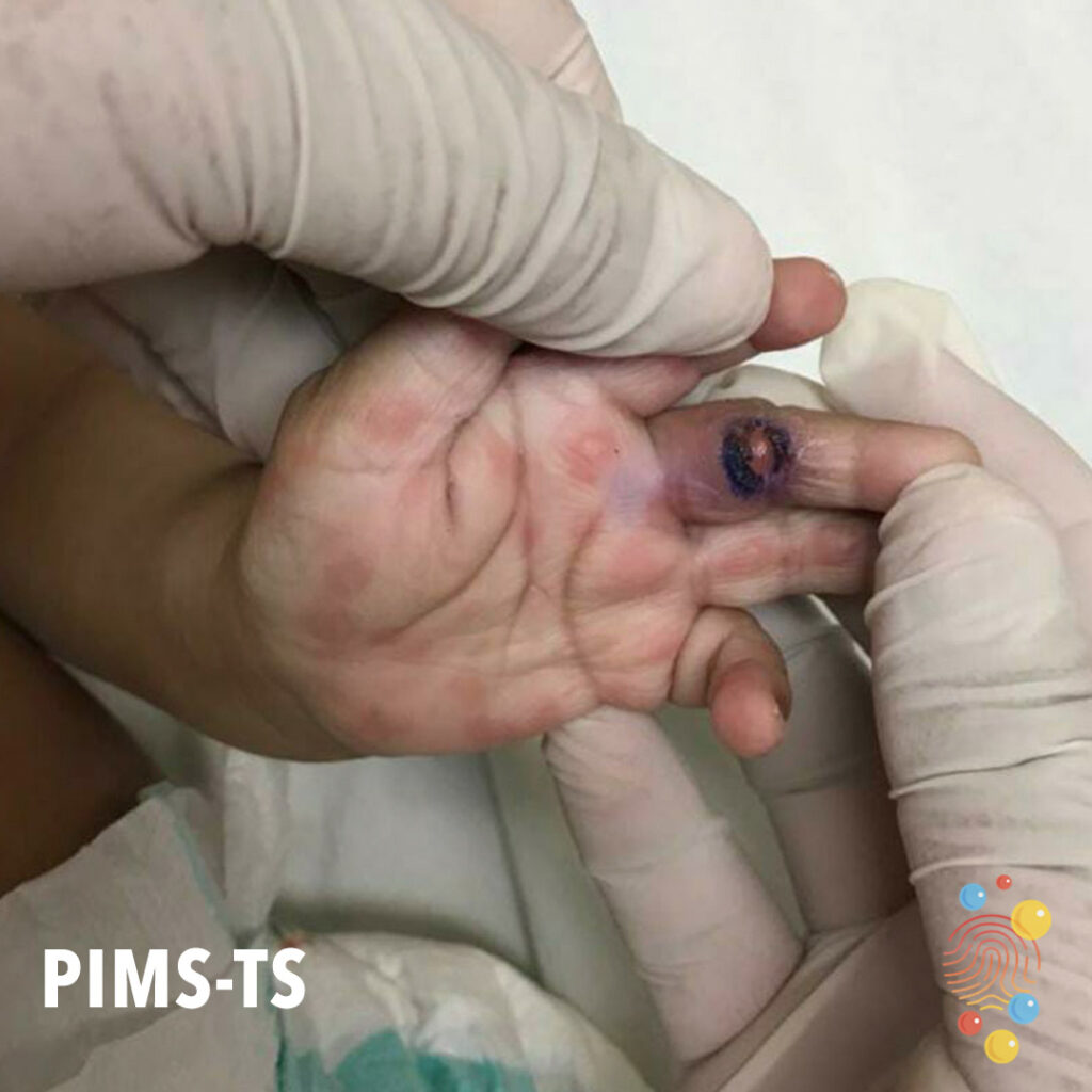
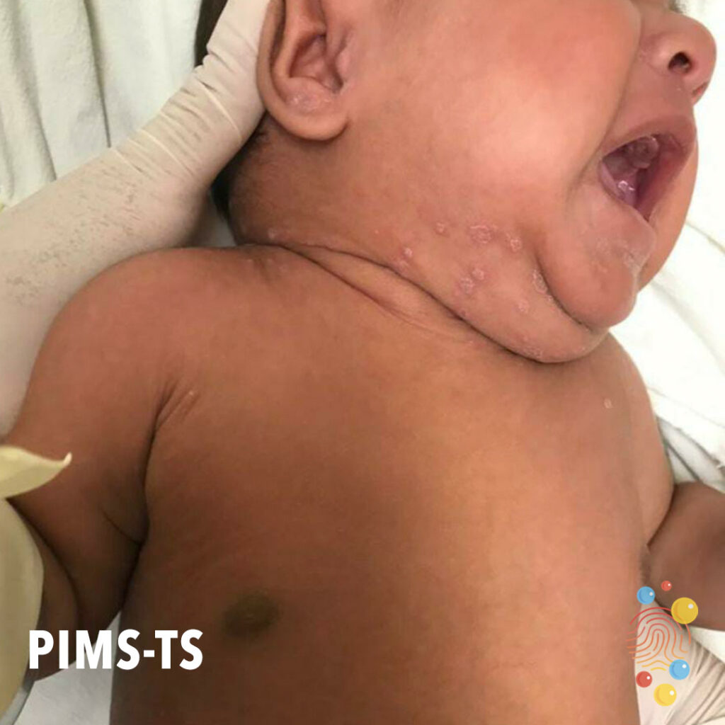
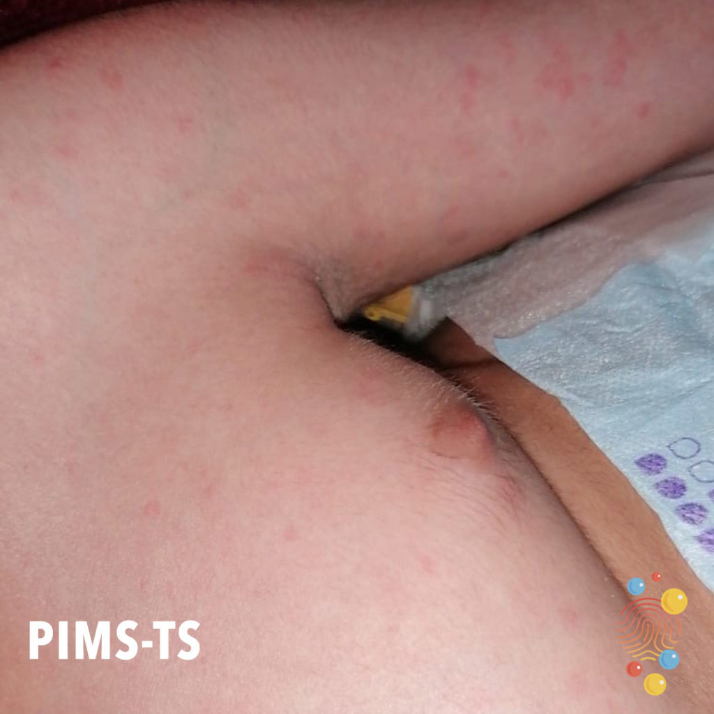
Palmar erythema as manifestation of PIMS-TS (area on middle finger circled in blue ink by patient relative).
Learn more about PIMS-TS
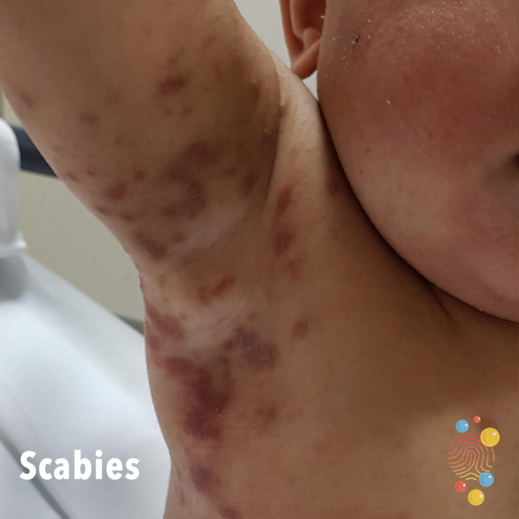
Erythematous papules coalescing into serpiginous plaques on a background of hazy erythema and hyperpigmentation in the right axilla. On the right cheek there is erythema and fine scale.
Learn more about scabies
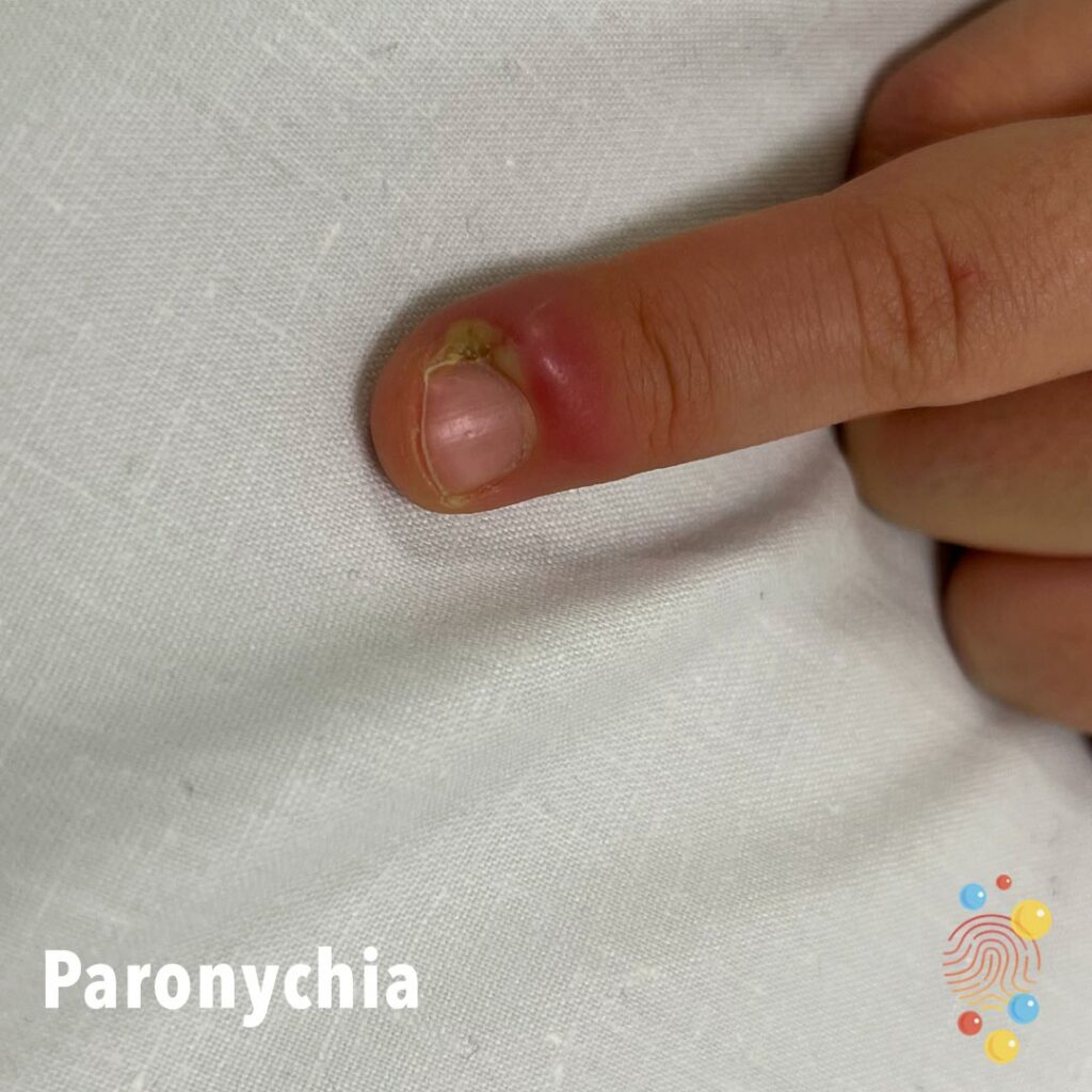
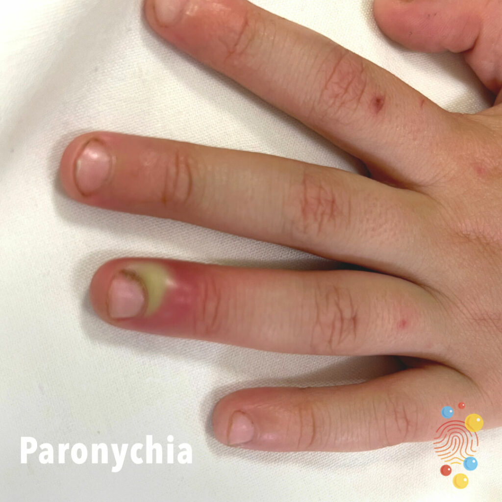
Paronychia

Mouth Injury
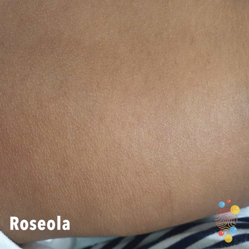
Fine predominantly macular erythematous eruption on the trunk.
Learn more about roseola
Eczema Coxsackium
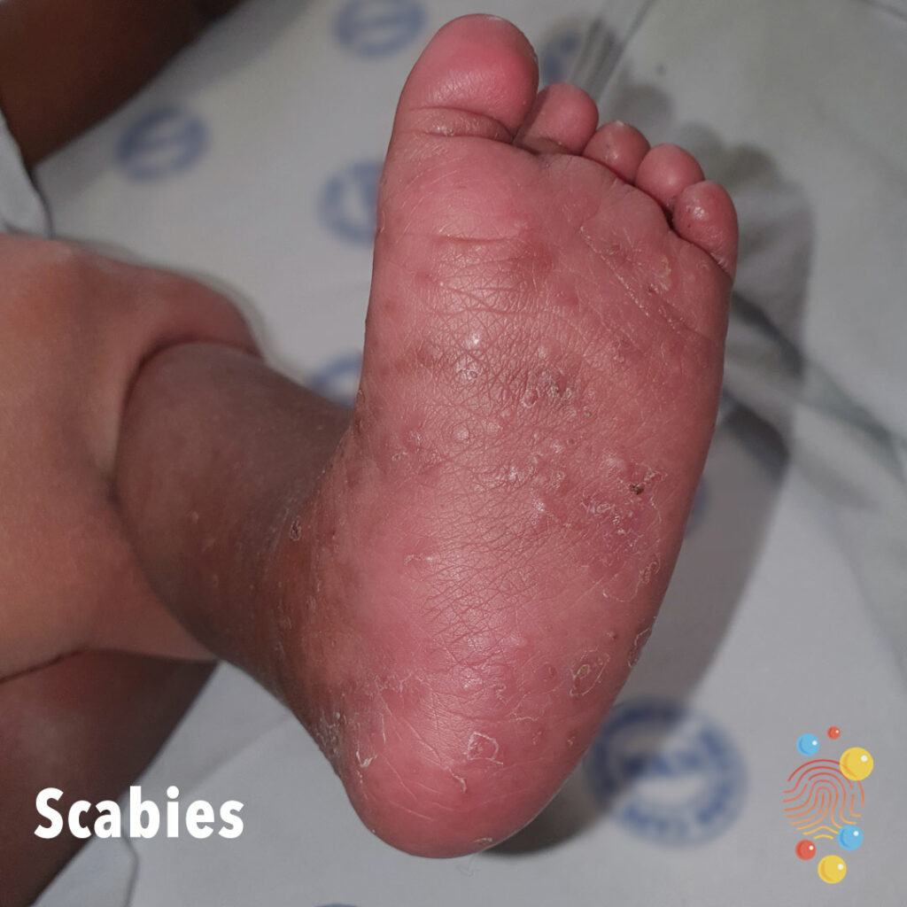
Multiple papules on the plantar surfaces of the feet with evidence of burrows.
Learn more about scabies
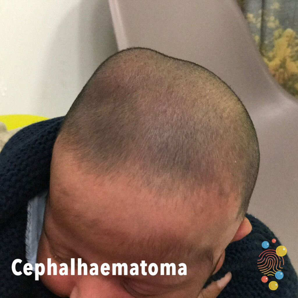
Asymmetric bulge over the right scalp.
Learn more about cephalhaematoma


Symmetric swelling of lower limbs associated with hyperkeratosis, plantar keratoderma, and dystrophic toenails
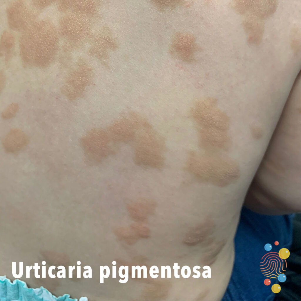
Generalised raised pink/brown macular papular rash on the back of varying sizes.
Learn more about urticaria
Vascular pedunculate lesion with underlying visible veins.
Learn more about haemangiomas.

Scattered erythematous papules on the left side of abdomen with some early pustulation seen in a few.
Learn more about folliculitis
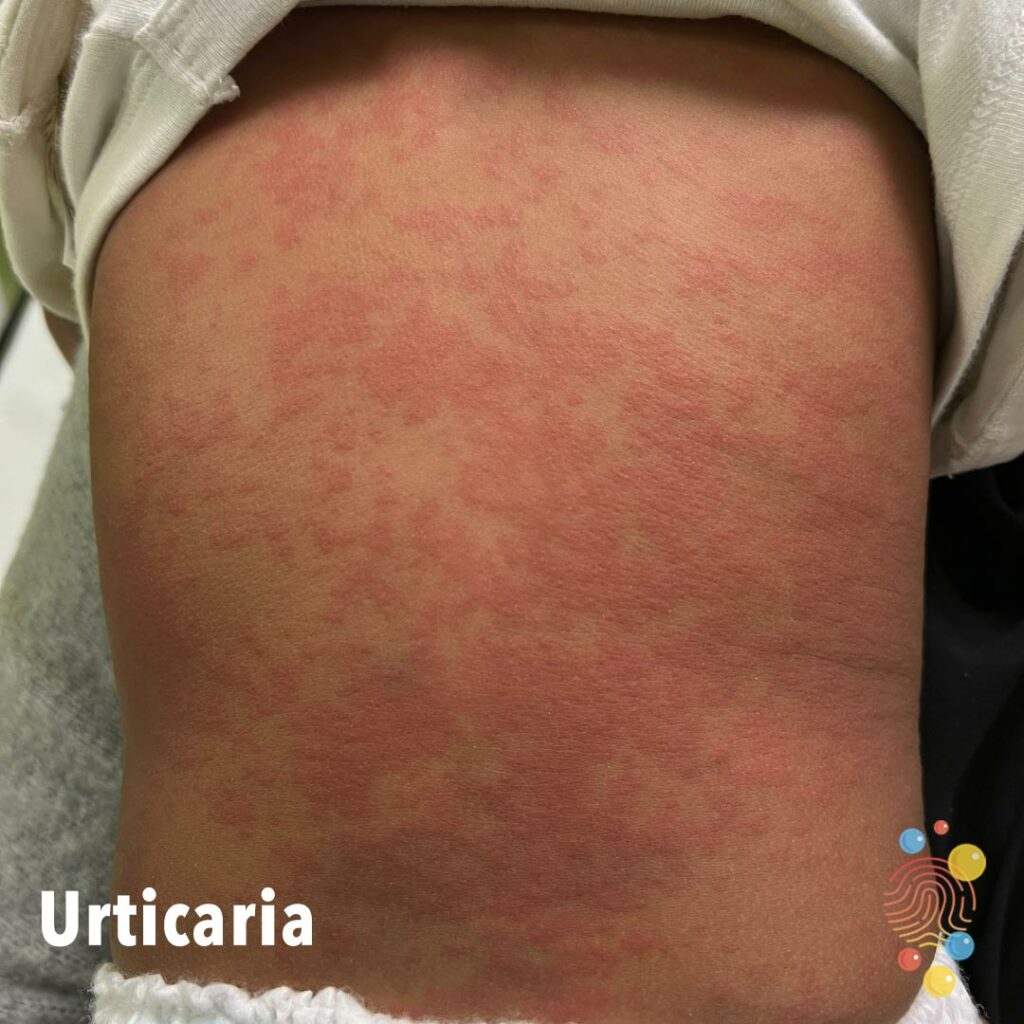
Urticaria
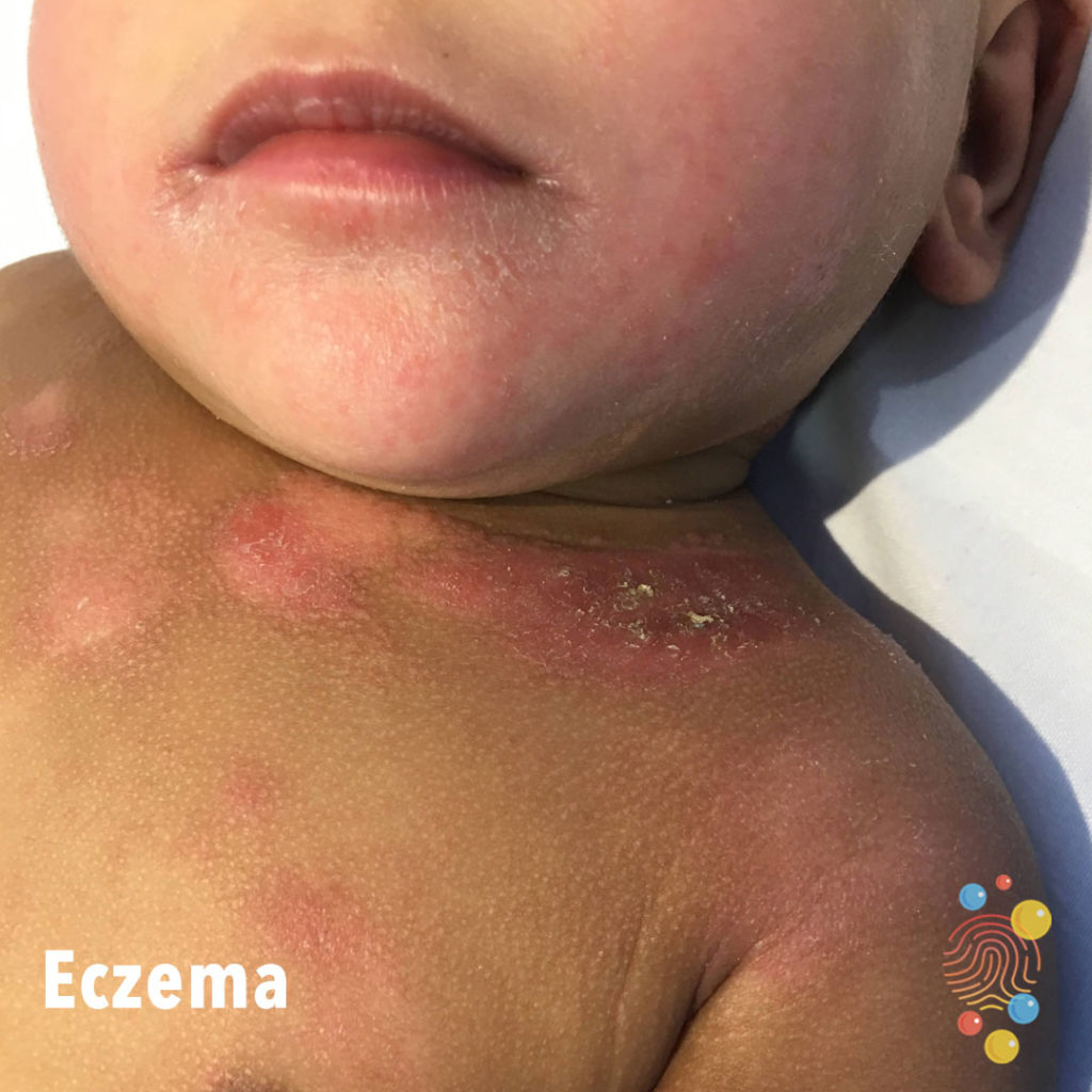
Eczematous patches on the neck and upper chest with some secondary mild impetiginsation. Mild lichenifaction around the mouth. Some areas are hypopigmeted.
Learn more about eczema
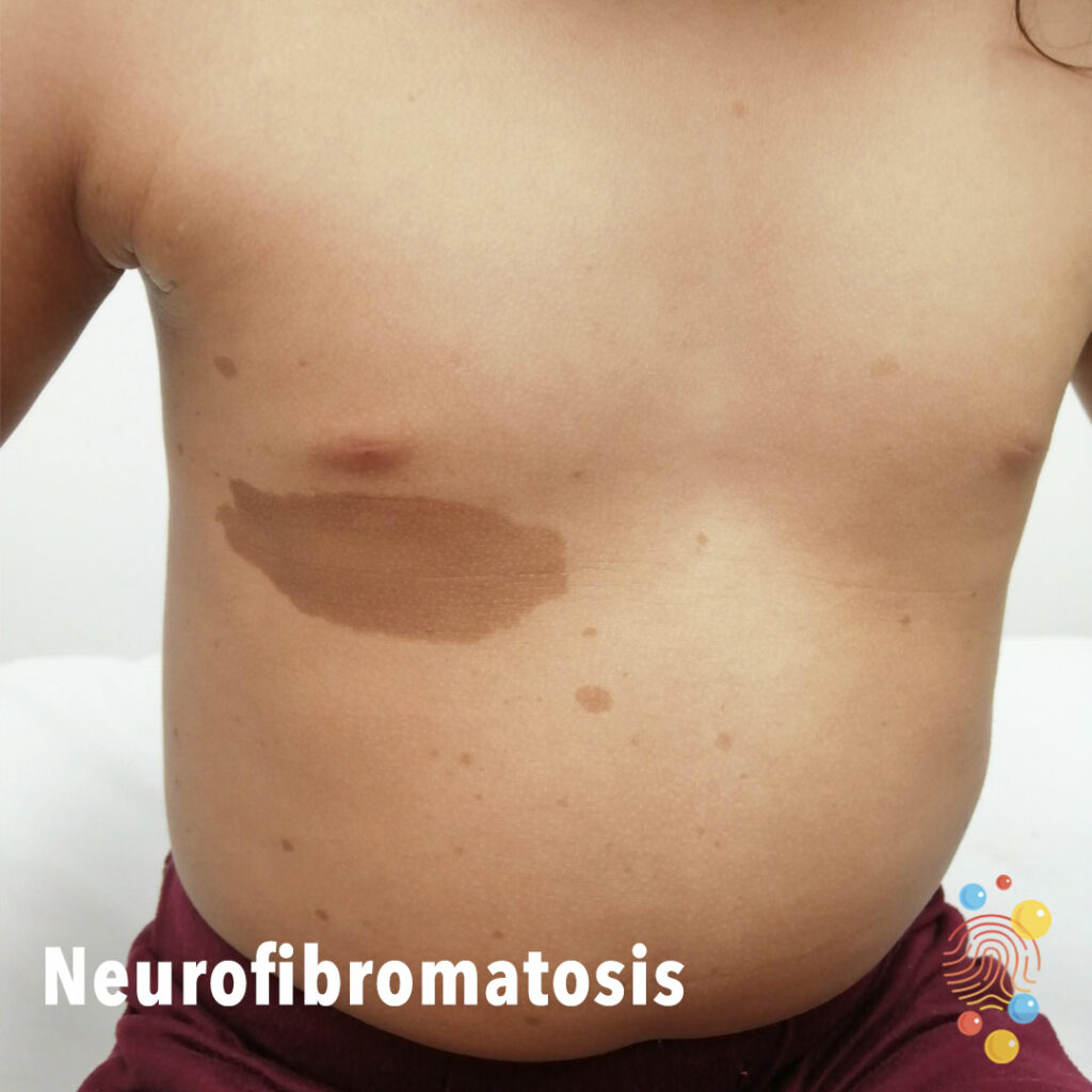
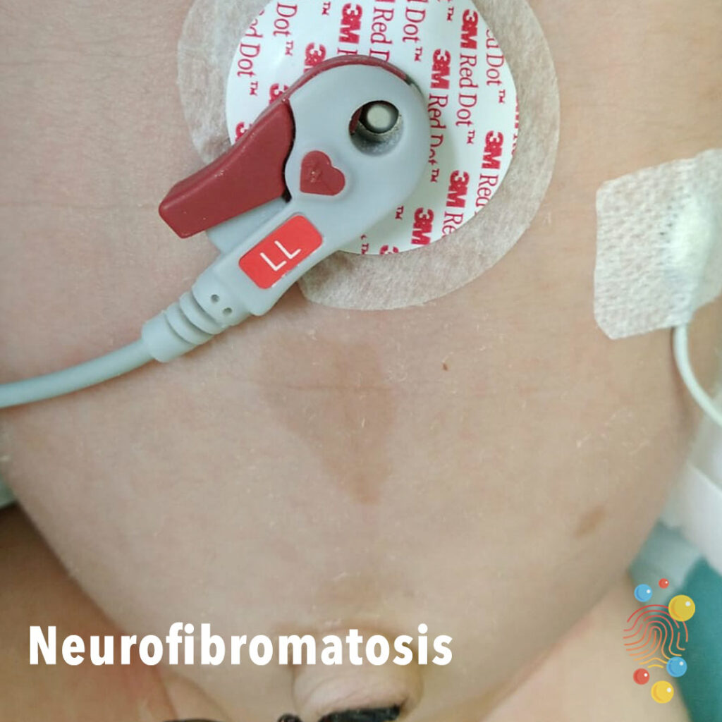
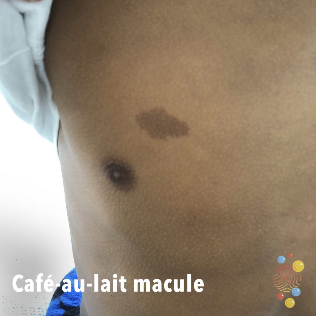
A 4-year-old girl with café-au-lait macula lesions on the chest, abdomen and extremities from birth. By maternal branch, all generations present the same type of café-au-lait mácula.

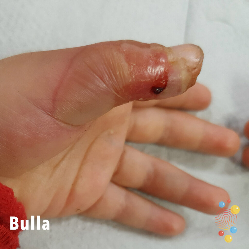
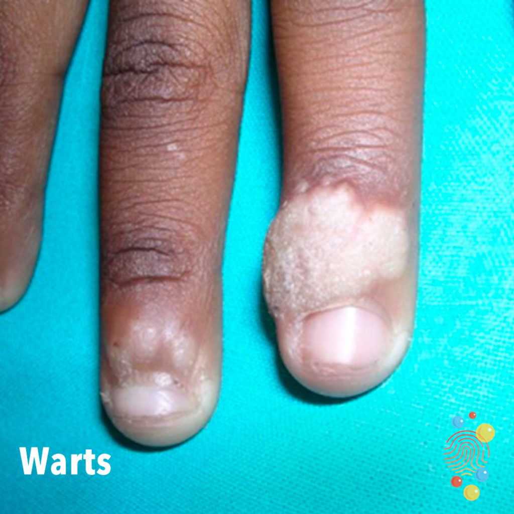
Pre de- roof: skin coloured bulla with verruciform lesion at the inferior pole. Post de-roof: deroofed bullae with centralised detached epidermis and underlying denuded skin. Fleshy verruciform lesion at inferior pole.
Learn more about warts
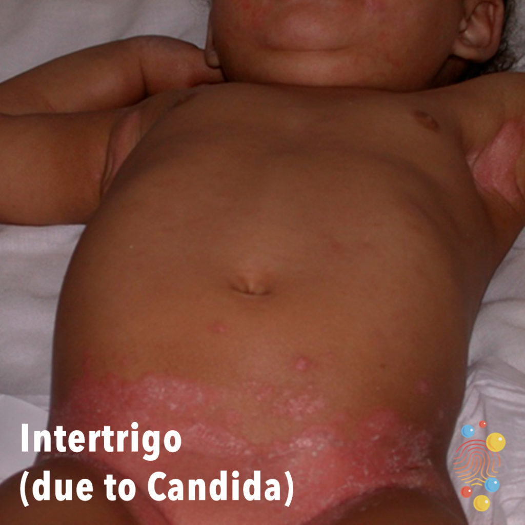
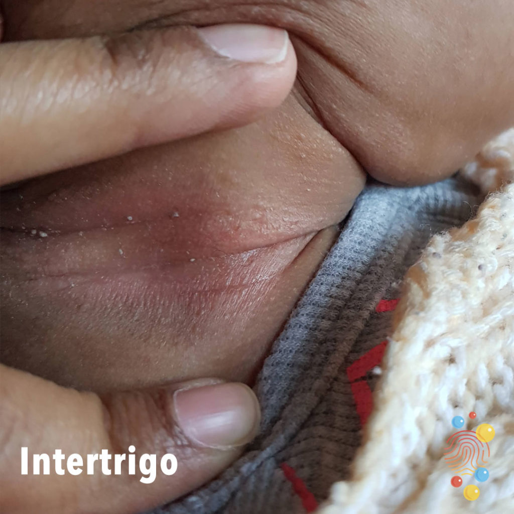
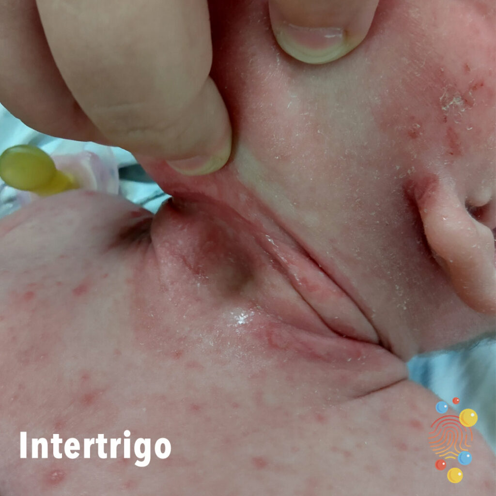
Confluent bright pink patches in skin folds of groin and axillae with scattered satellite areas.
Learn more about intertrigo
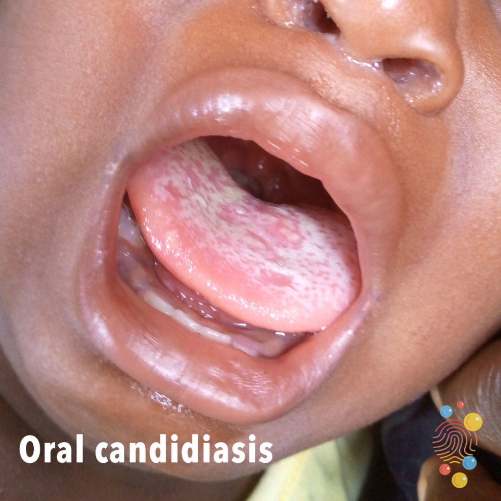
Milky discolouration of the tongue.
Learn more about neonatal thrush

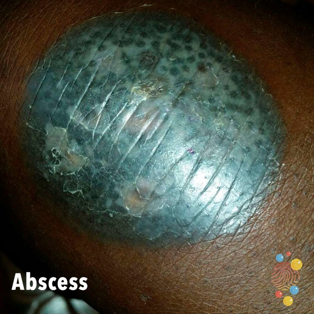
An abscess (tuberculosis colliquativa cutis). An ulcerated abscess with well-defined + raised borders.
Learn more about scrofulderma
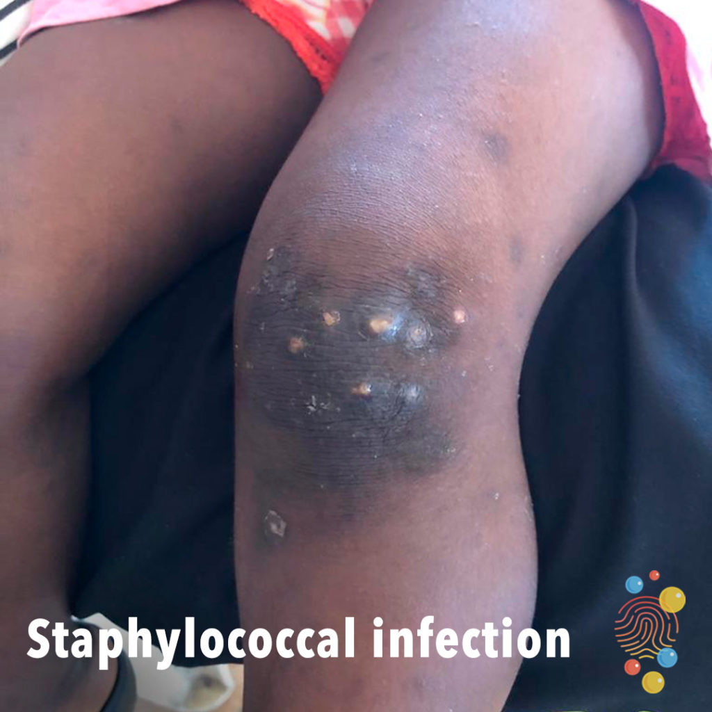
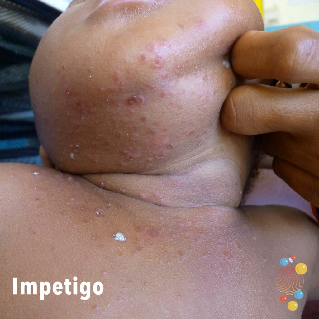
Pus-filled pustules + nodules with honey-colored crusts + ragged edges.
Learn more about staphylococcal infection
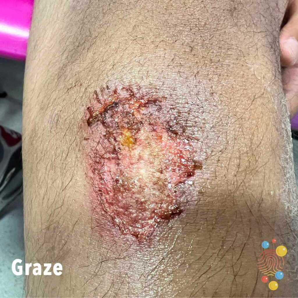
Grazed Knee – 13 year old boy

Erythematous papules arranged in an annular distribution with central clearing. One erythematous satellite papule superiorly
Learn more about eczema
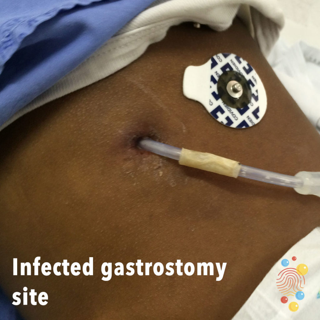
Erythema around gastrostomy site.
Learn more about gastrostomies
Blue pigmented patches over buttocks and sacrum
Learn more about dermal melanocytosis
Multiple erythematous nodules of varying sizes with overlying and peripheral scale.
Learn more about abscesses

Multiple clustered erosions with central ulceration on the back

Blue black macular patches over lumbosacral area.
Learn more about dermal melanocytosis
Scar overlying the medial malleolus of the left foot. Scattering of erythematous papules, xerosis of the skin (fine overlying scale)

Hypopigmented papules and plaques on posterior neck.
Learn more about eczema
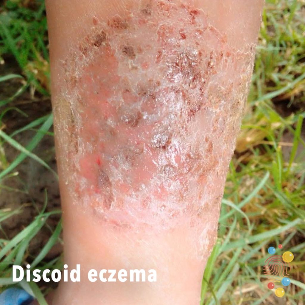
Discoid plaque with crusting, erosions and ooze.
Learn more about eczema
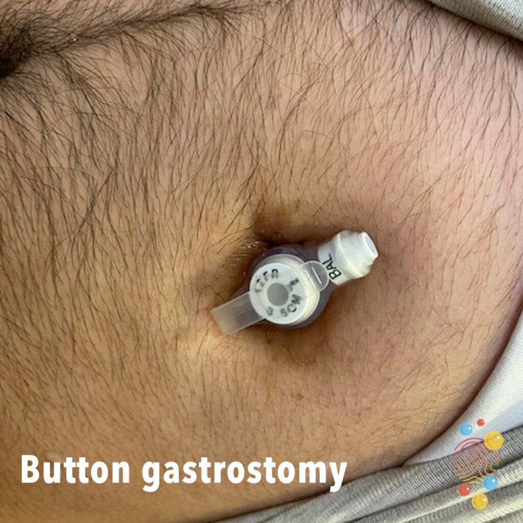
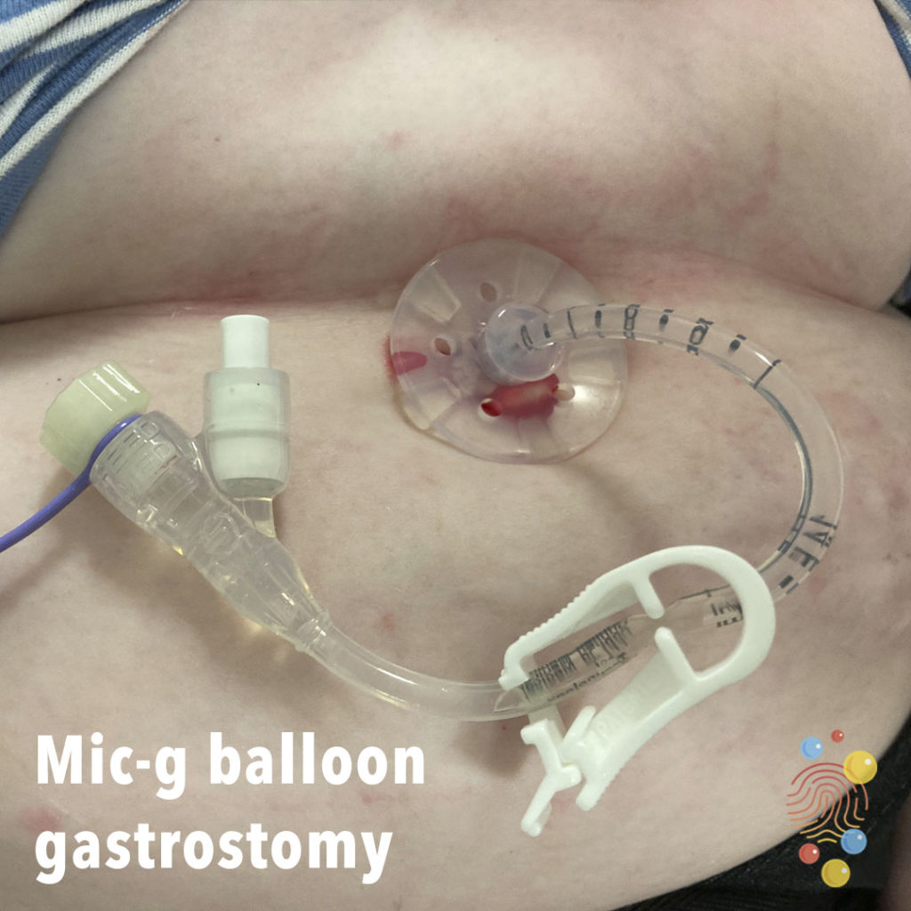
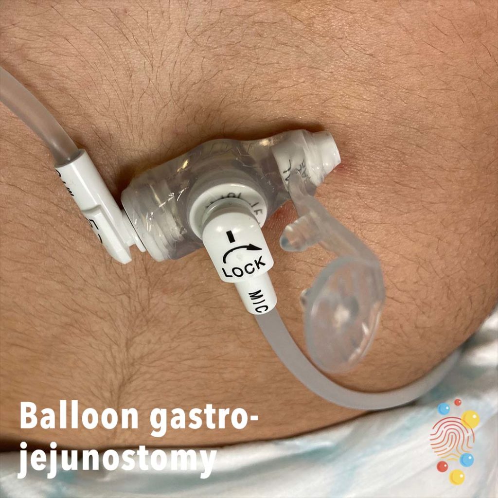
A button gastrostomy. The width (12Fr) and size (3cm) are written on the top of the tube. The side port (labelled ‘BAL’) is used for inflating and deflating the balloon.
Learn more about gastrostomies
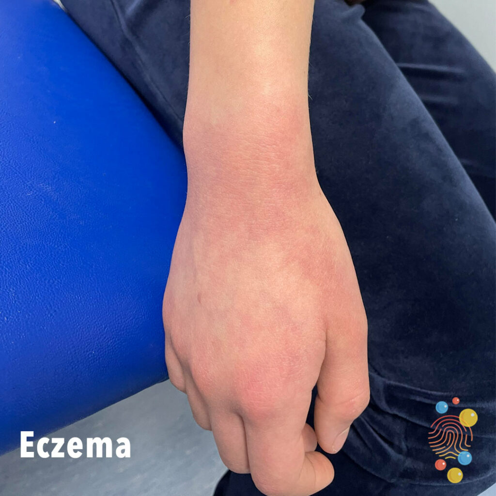
Mild erythema seen on wrist and dorsum of right hand.
Learn more about eczema


Punctate lesion with associated erythematous swelling on the left forearm. There is also a macular papular rash on the chest.
Learn more about BCGs
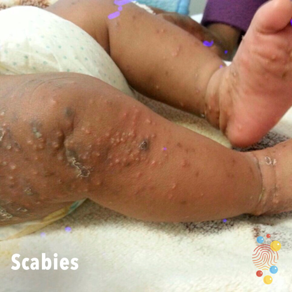
Excoriated papules on lower limbs including soles.
Learn more about scabies
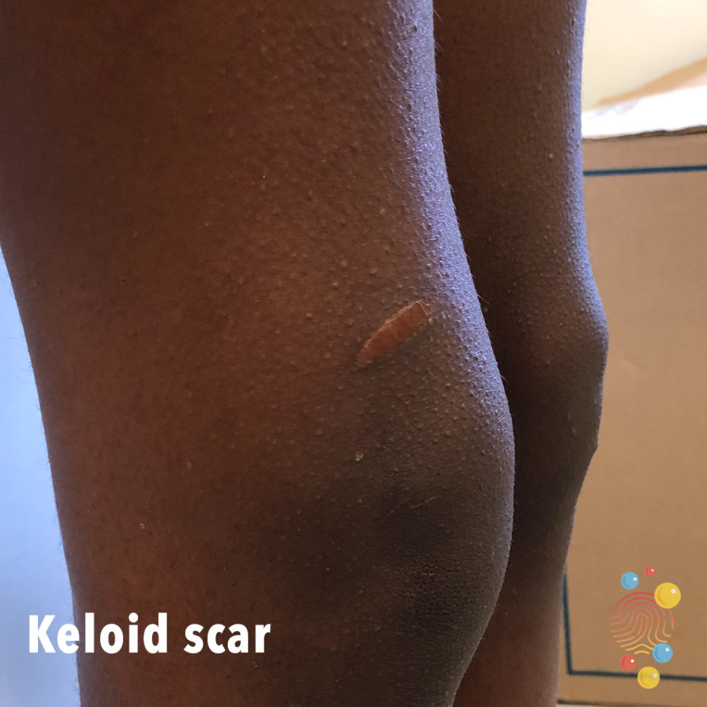
Exaggerated scarring response to injury.
Learn more about keloid scars.
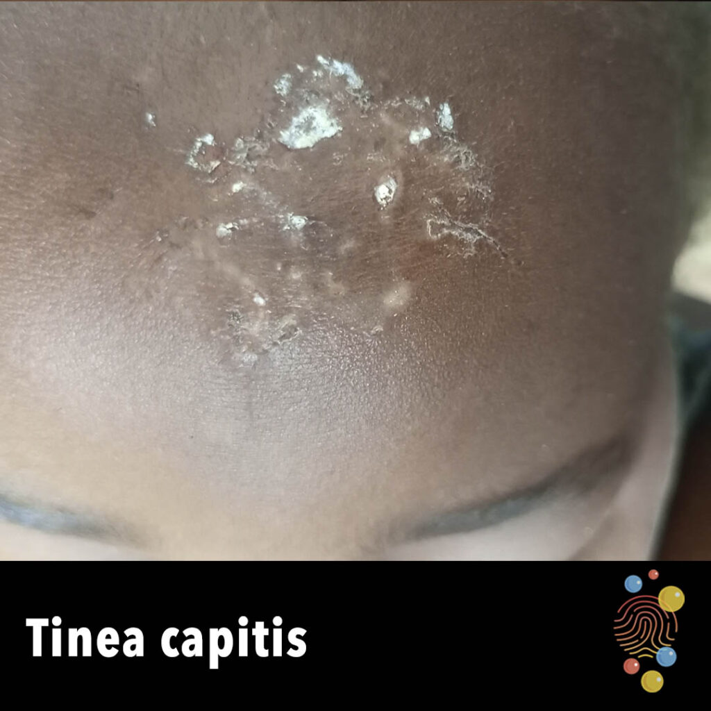
Erythema and scaling on the scalp.
Learn more about tinea capitis
Learn more about gastrostomies
Extensive wheals.
Learn more about urticaria
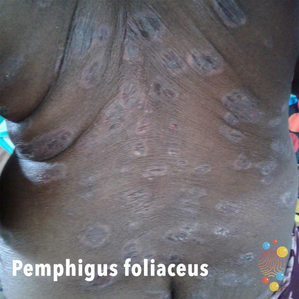
Multiple annular plaques with central erosion and hyperpigmentation and peripheral hypopigmentation.
Learn more about pemphigus
Unilateral erythematous rash well circumscribed around the eye.
Learn more about eczema

Stomatitis in child with bilateral pneumonia, urticaria rash and cardiovascular instability requiring >40ml/kg fluid + inotropes.
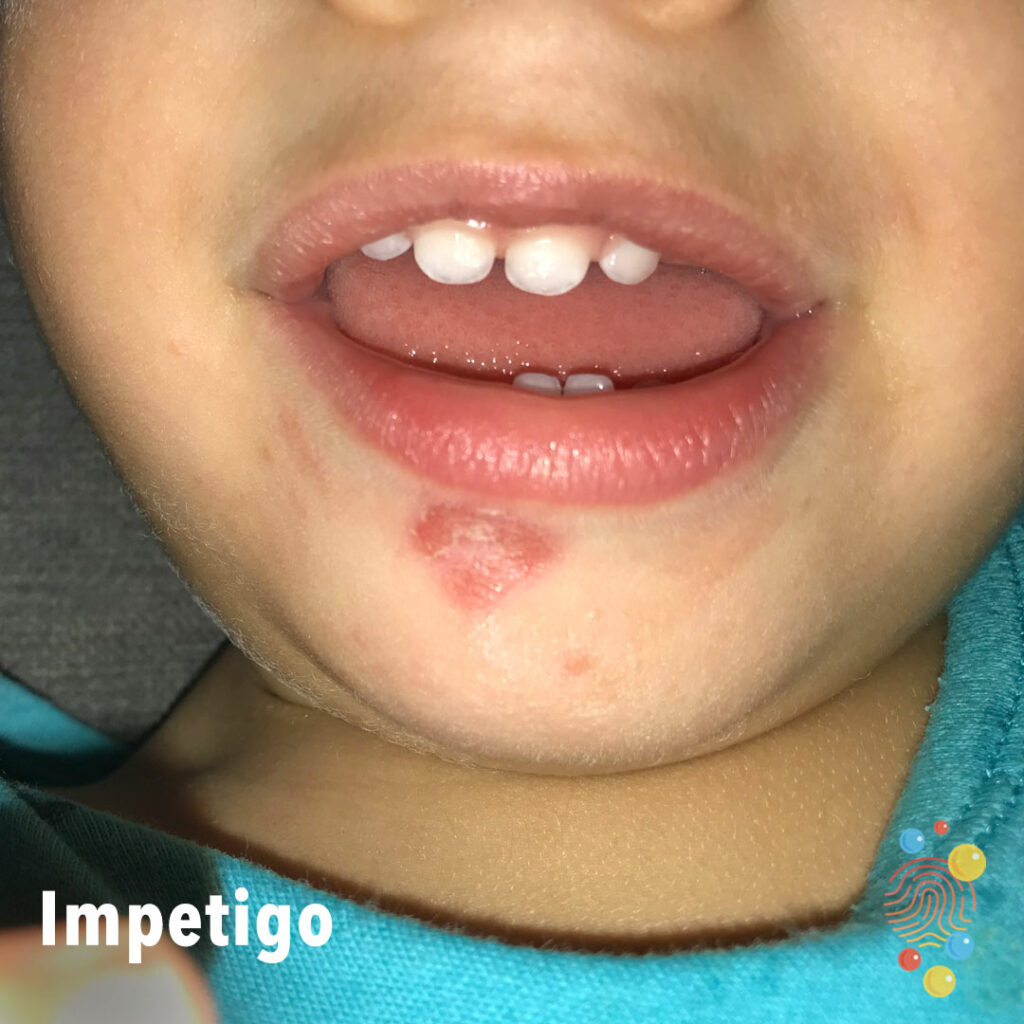
Superficial erosion over chin with crusting.
Learn more about bullous impetigo
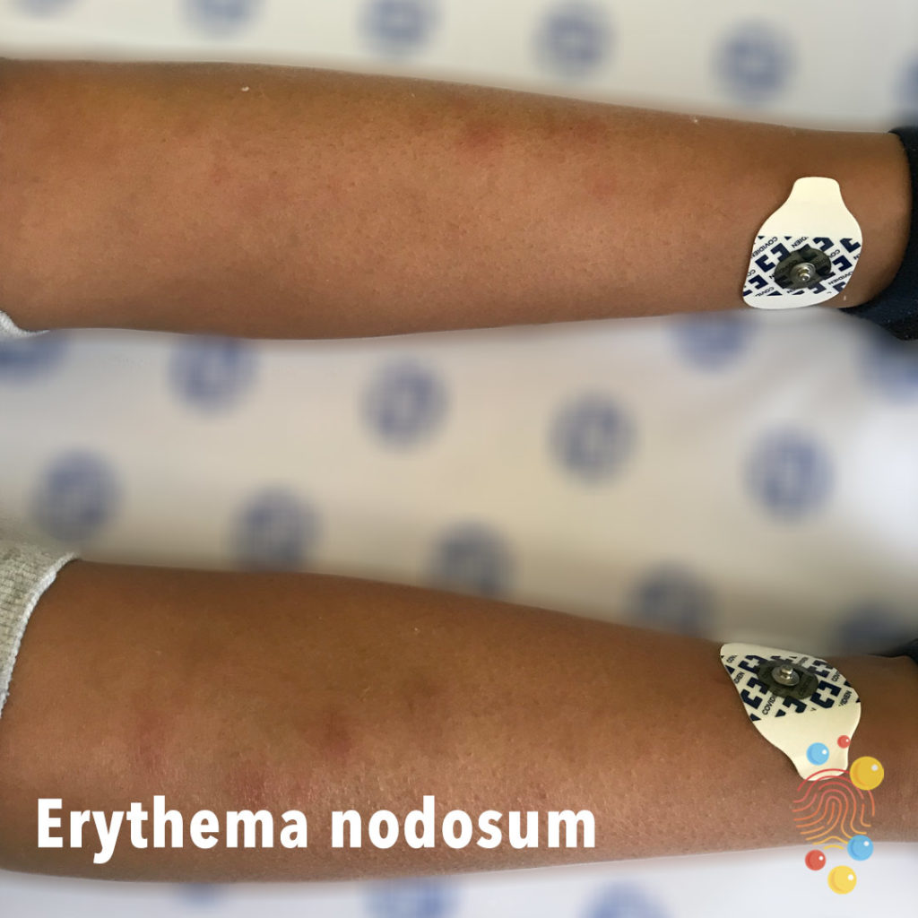
Bilateral tender erythematous lesions on the lower legs.
Learn more about erythema nodosum
Blue black macular patches on arms.
Learn more about dermal melanocytosis
Diffuse micropapules coalescing in areas with underlying erythema
Learn more about eczema
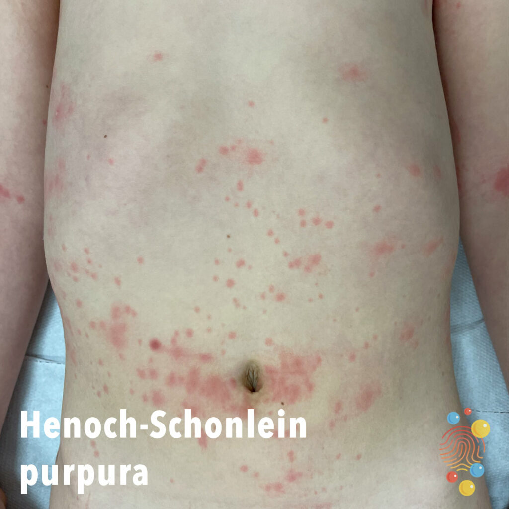
Erythematous papules with surrounding pallor on lower trunk.
Learn more about Henoch-Schonlein purpura
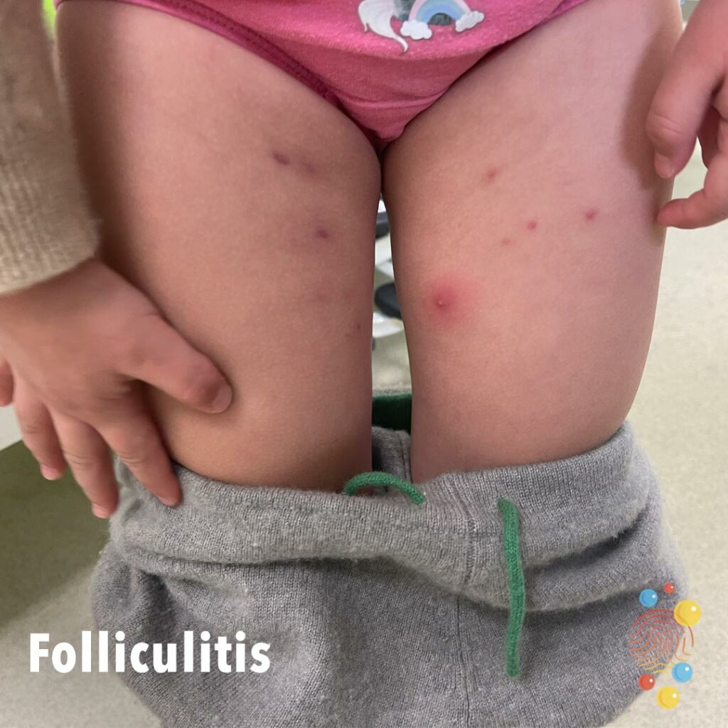
Erythematous papules inner thighs with a solitary pustule on left thigh in a caucasian child.
Learn more about folliculitis
Scarlet Fever
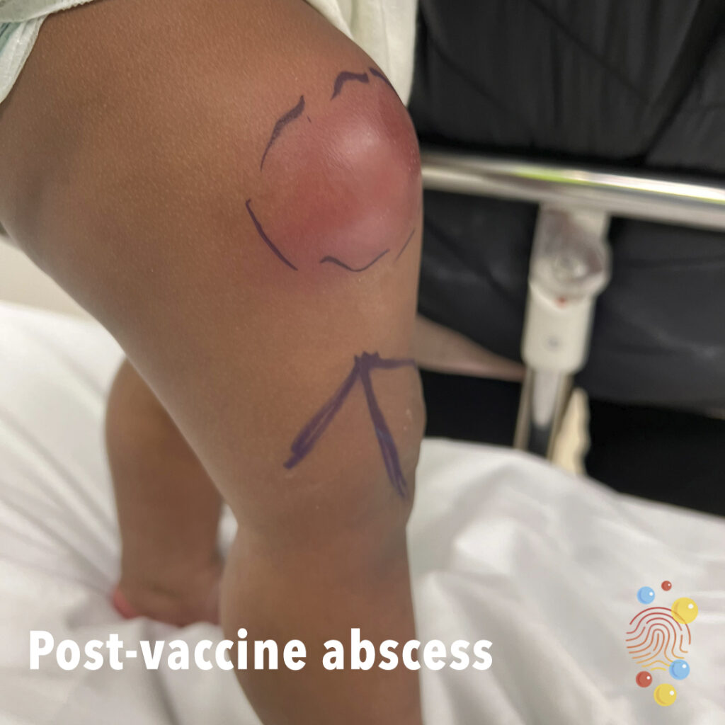
Thigh abscess post men c vaccine
Scarlet Fever

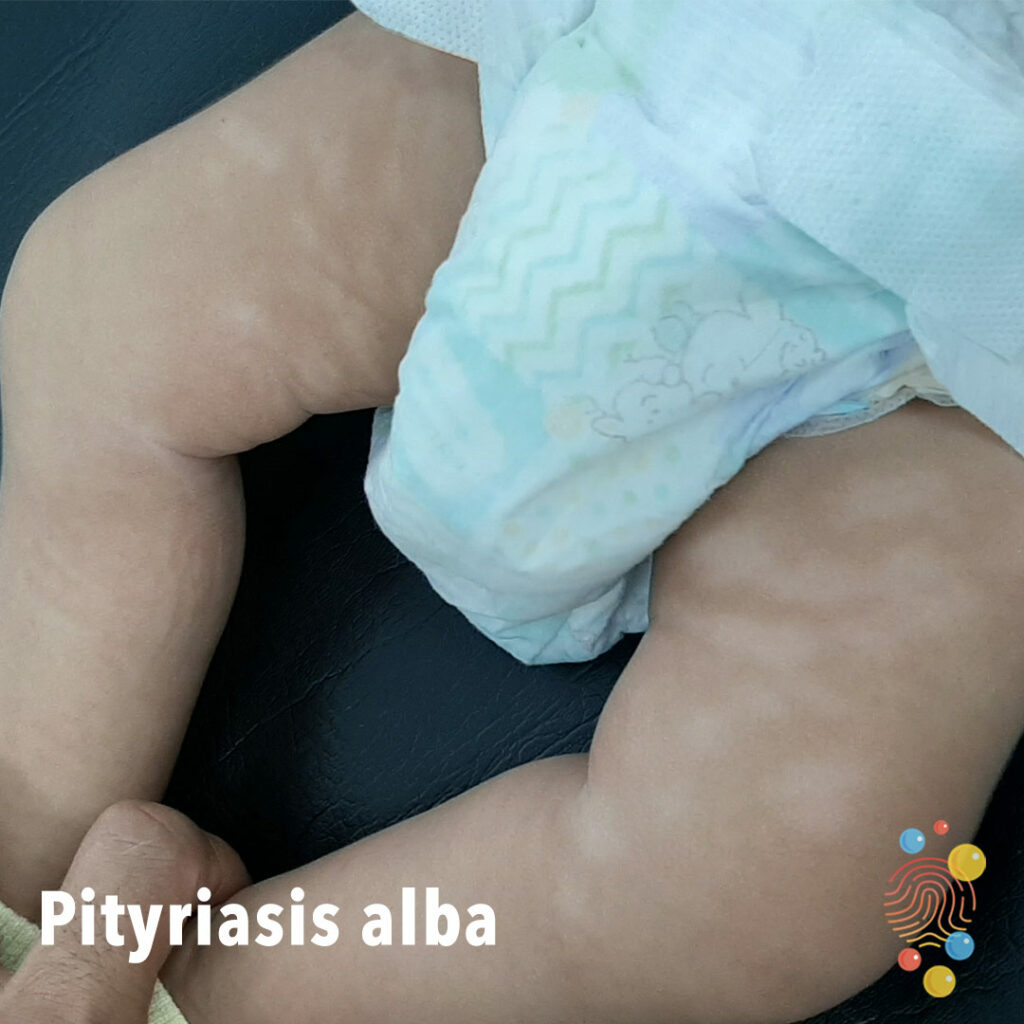
Hypopigmented patches on the anterior thighs.
Learn more about pityriasis alba

Inflammatory nodules with sinus tracts of axilla.
Learn more about hidradenitis suppurativa
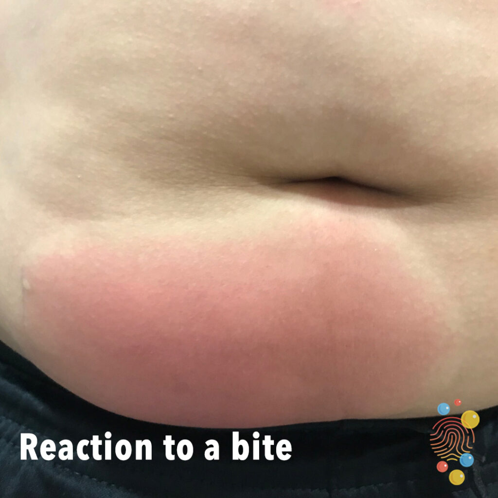
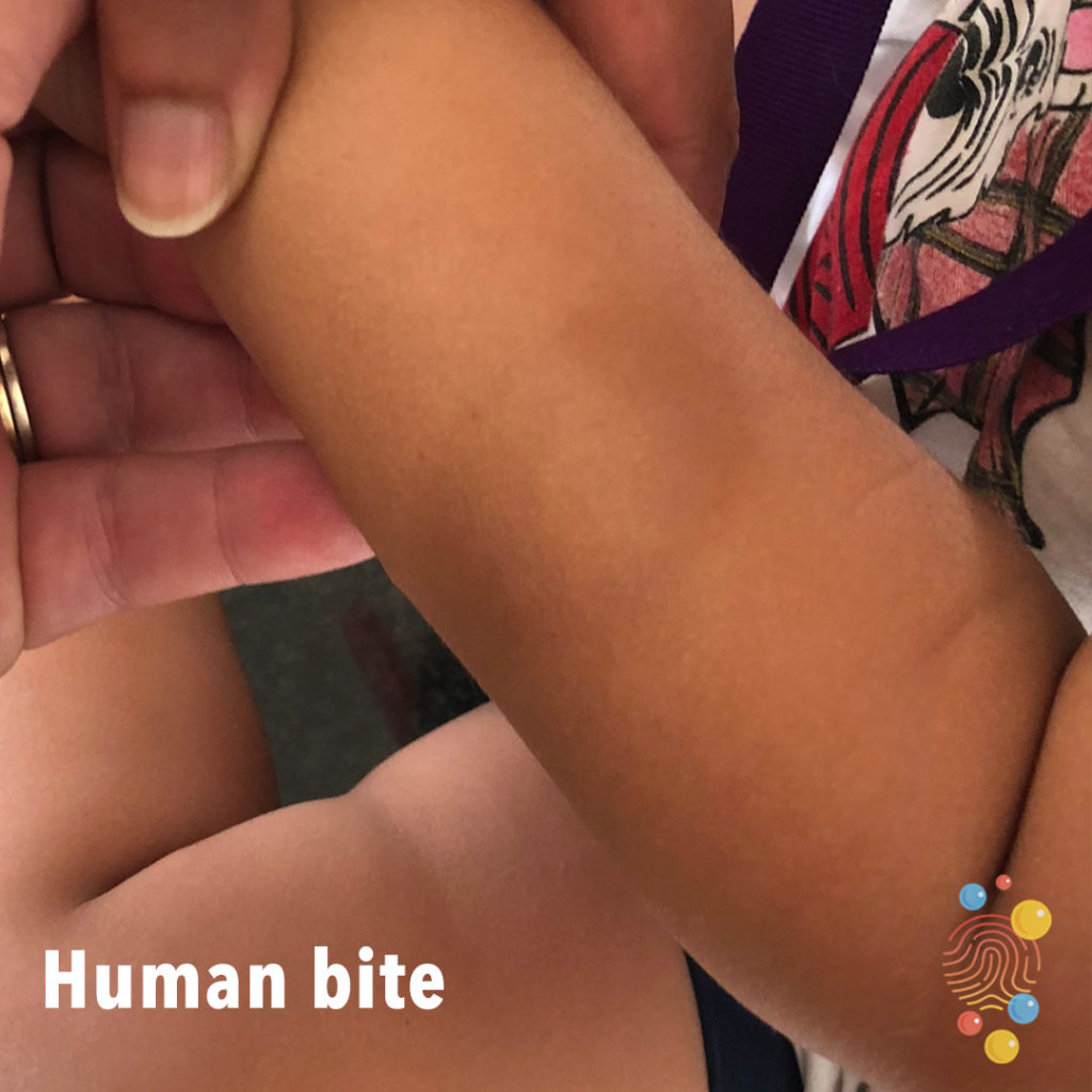
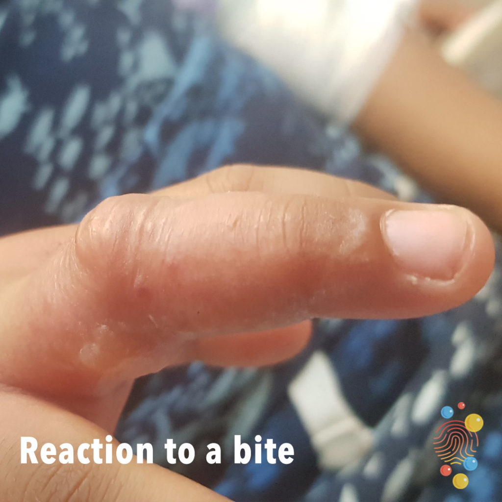
No epidermal change. Macular pale pink erythema with a white edge/skin coloured edge and appearance of sub epidermal swelling.
Learn more about bites.

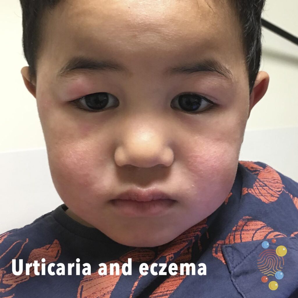
Periorbital diffuse swelling with mild erythema.
Learn more about periorbital cellulitis
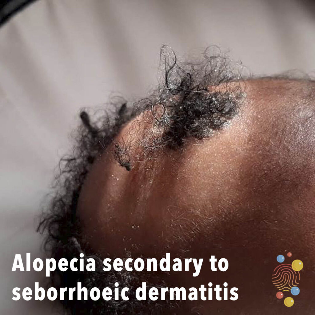
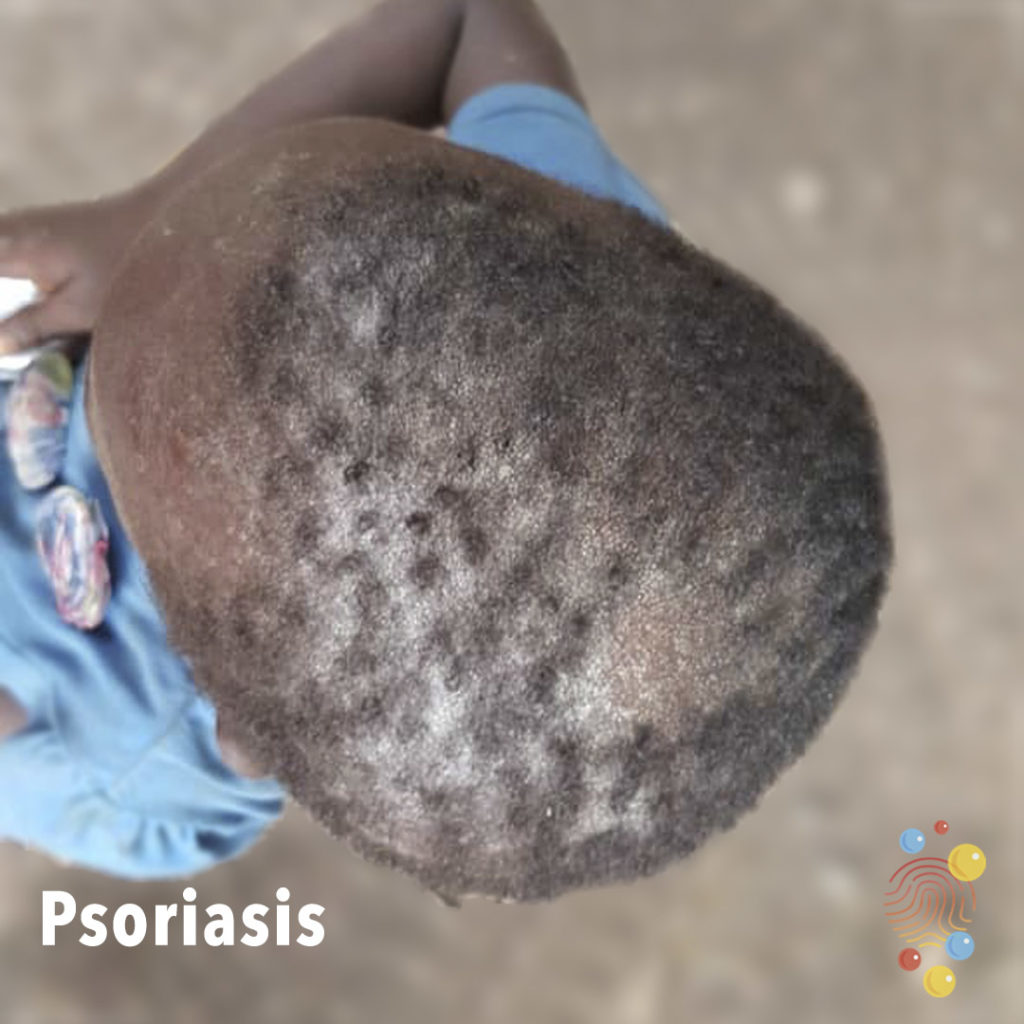
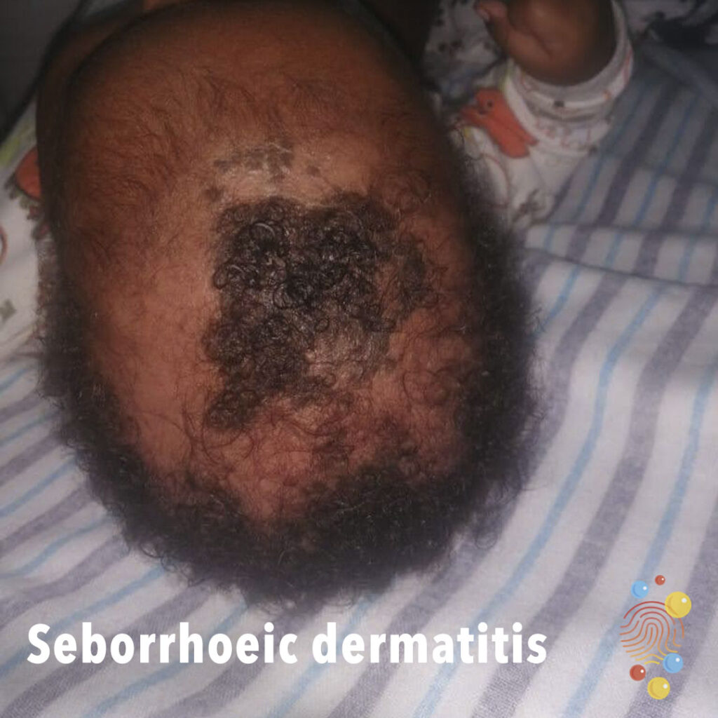
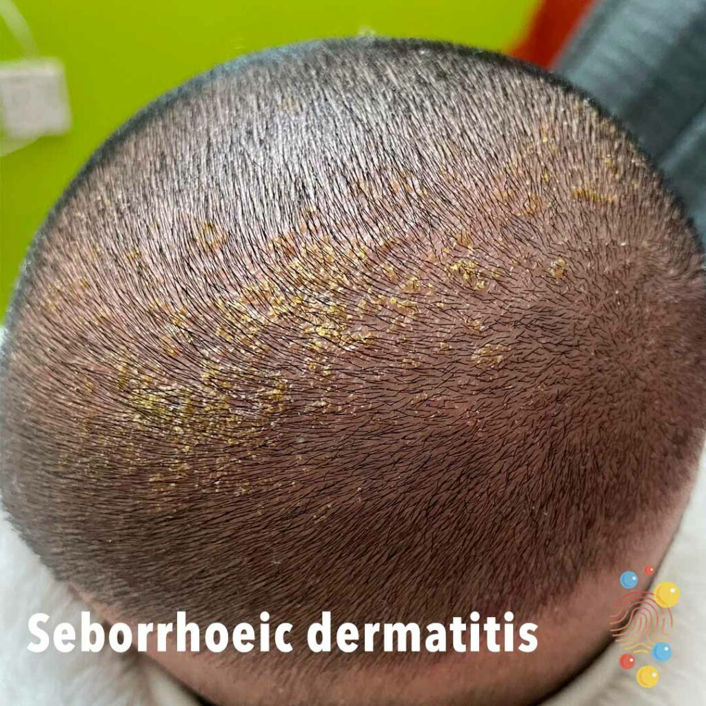
Multi-focal non-scarring alopecia with preservation of follicular ostia. Scaly, adherent plaque on the scalp.
Learn more about seborrhoeic dermatitis

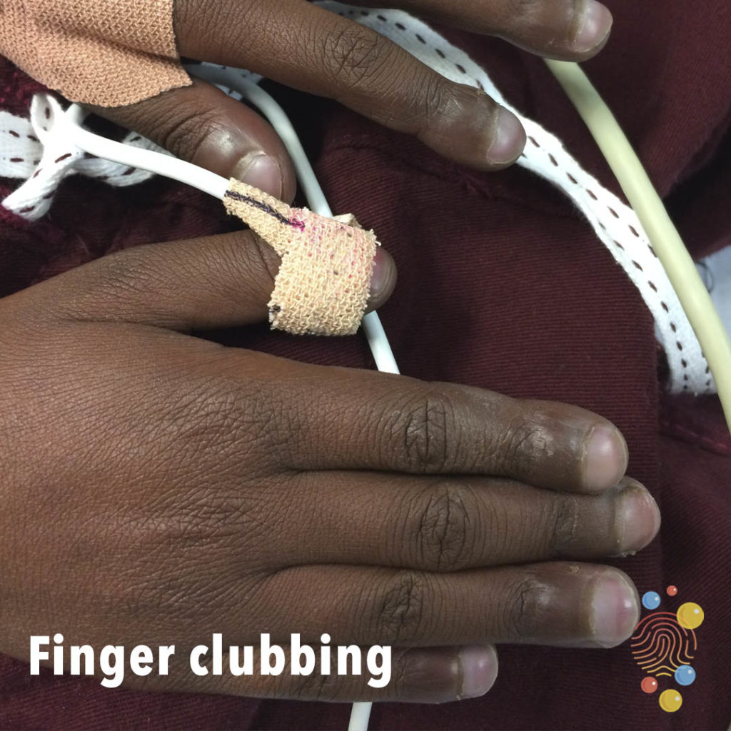
Clubbing or bulbous uniform swelling of the terminal phalanx involving all fingers with loss of normal angle between the nail and the nail bed.
Learn more about clubbing
Widespread homogeneous pink macules, patches and plaques with no obvious epidermal change.
Learn more about urticaria
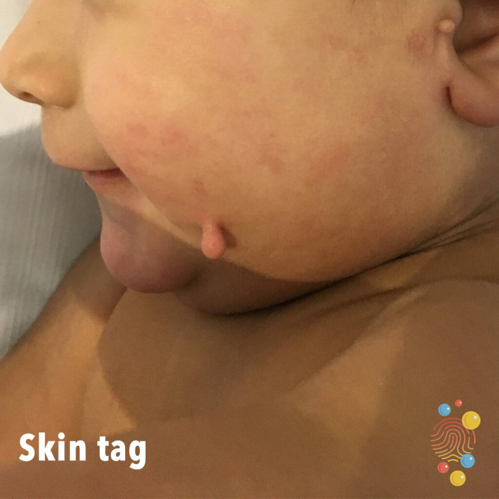
Learn more about skin tags
Extensive crusted erosions on the trunk and upper limb.
Learn more about eczema herpeticum
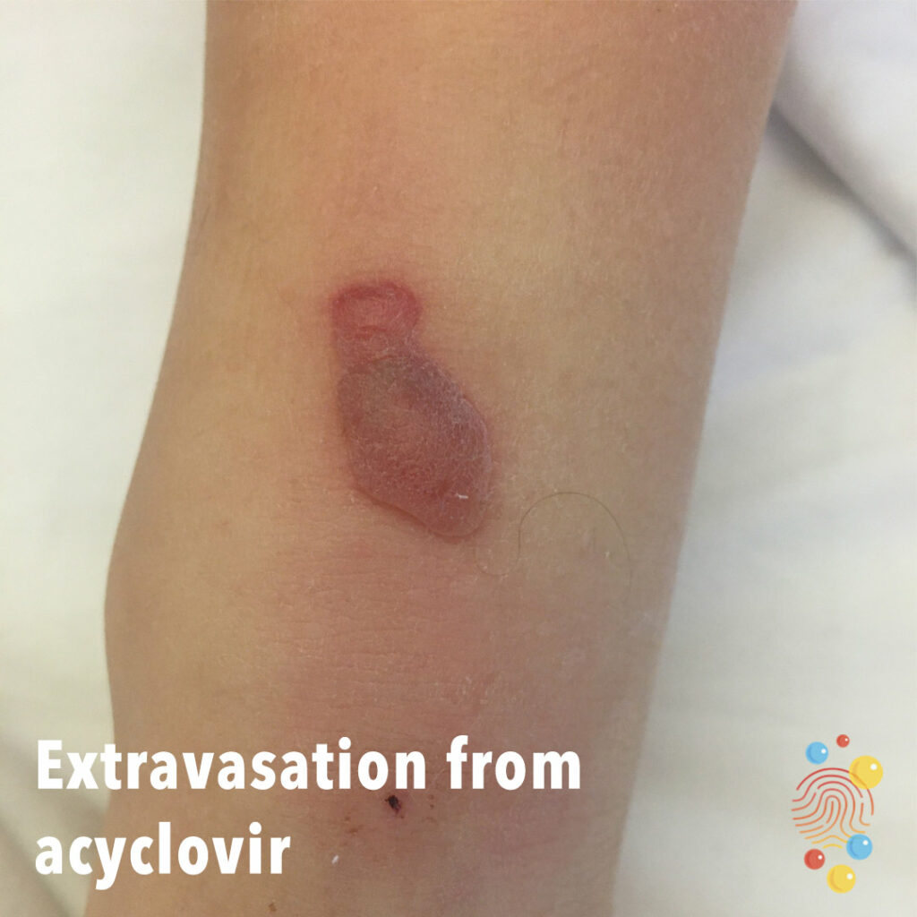
Puncture site noted inferiorly.
Learn more about extravasation
Multiple vesicles and bullae on the face and upper chest. Lesions in clusters with some slight evidence of golden crust.
Learn more about bullous impetigo
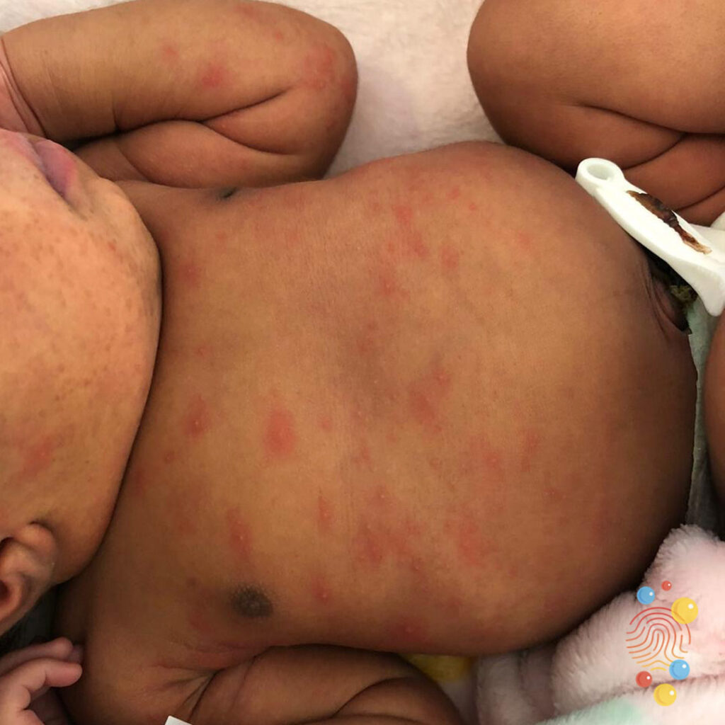
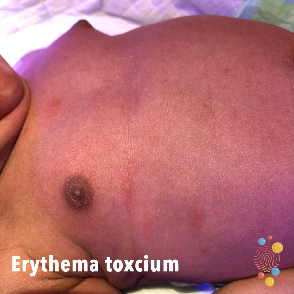
Multiple papules on an erythematous base on the trunk. Occasional pustules with macular base.
Learn more about erythema toxicum

Bitonsillar hypertrophy with white exudate.
Learn more about streptococcal pharyngitis
Yellowing of the skin and sclera.
Learn more about jaundice

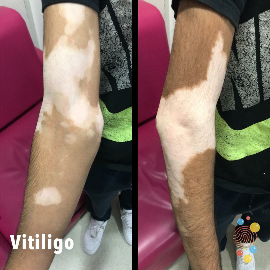
Patchy loss of skin colour with sharp margins bilaterally on arms and forearms.
Learn more about vitiligo
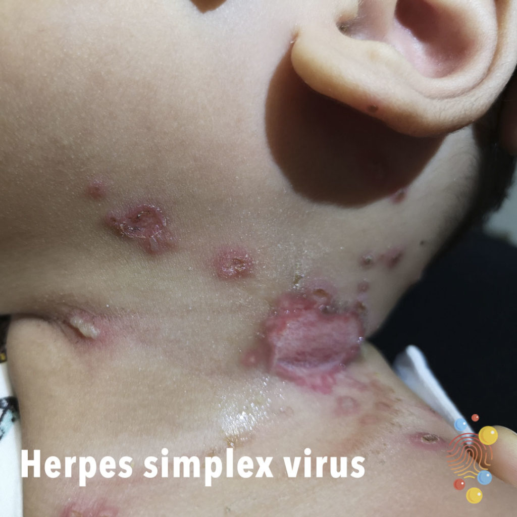
Punched out erosion affecting the left lateral neck fold with multiple discrete eroded areas. Lesions appear wet and a number feature deroofed blisters, with one cluster remaining medially. Largest lesion appears ulcerated.
Learn more about herpes simplex virus
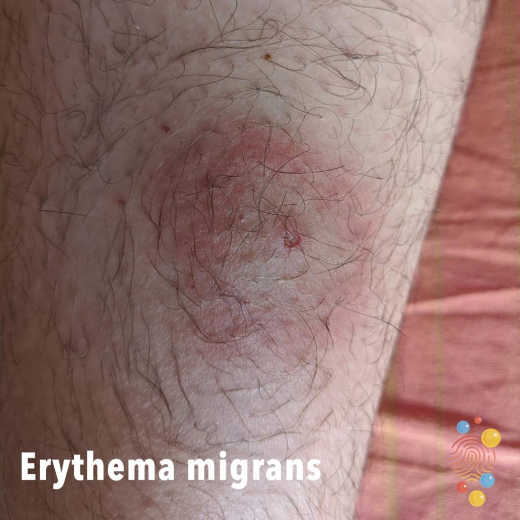
Annular erythematous eruption with central crusting and erosion.

Severe lichenified eczema with induration and impetiginisation
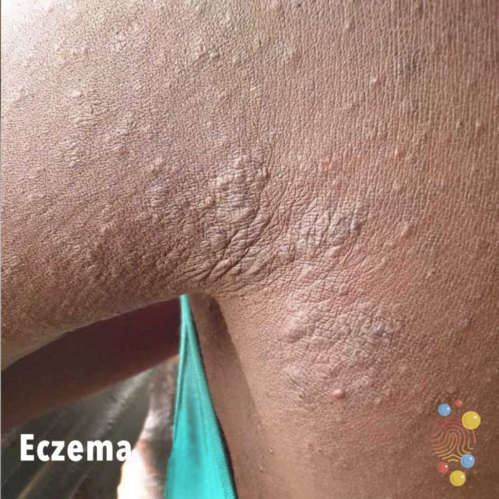
Papules + nodules.
Learn more about eczema

Small area of punctum/scab on scalp.
The lack of hair around the plaited braid suggest a degree of traction alopecia from braiding.
Learn more about epidermoid cysts
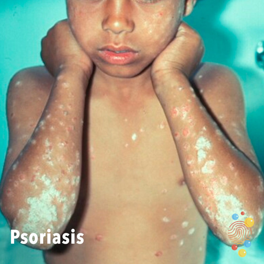
Skin desquamation and scaling at the periphery on background of erythema.
Learn more about psoriasis

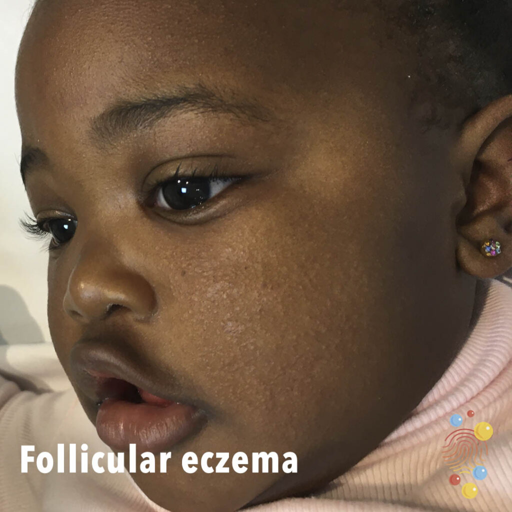
Lichenified hyperpigmented plaques on the abdomen with background follicular eczema.
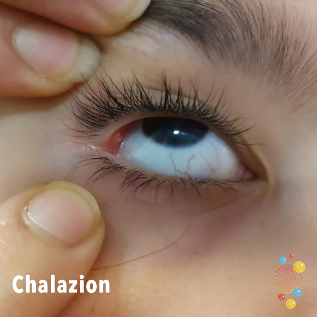
Chalazion

Extensive healing erosions with haemorrhagic crust and a collarette of scale
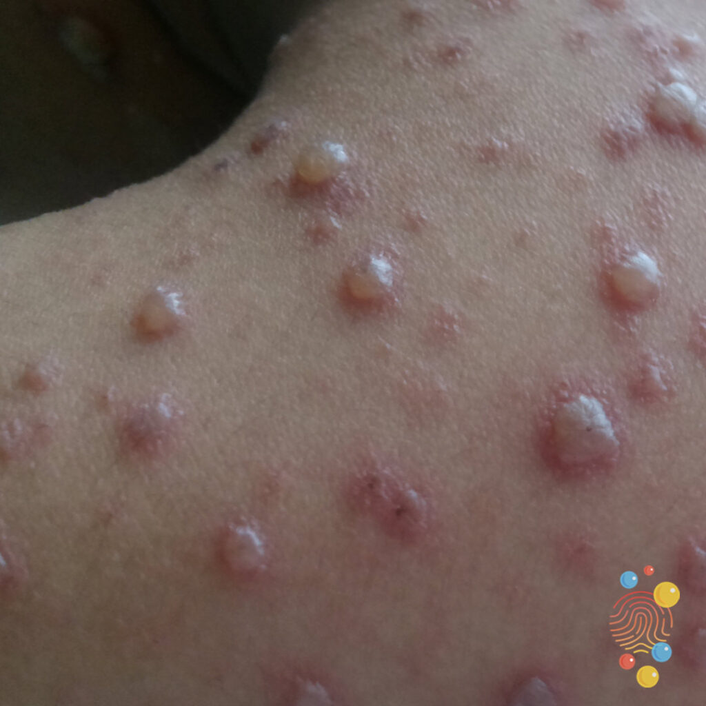
Multiple vesicles on an erythematous base.
Learn more about chicken pox

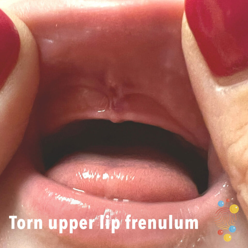
Torn upper lip frenulum
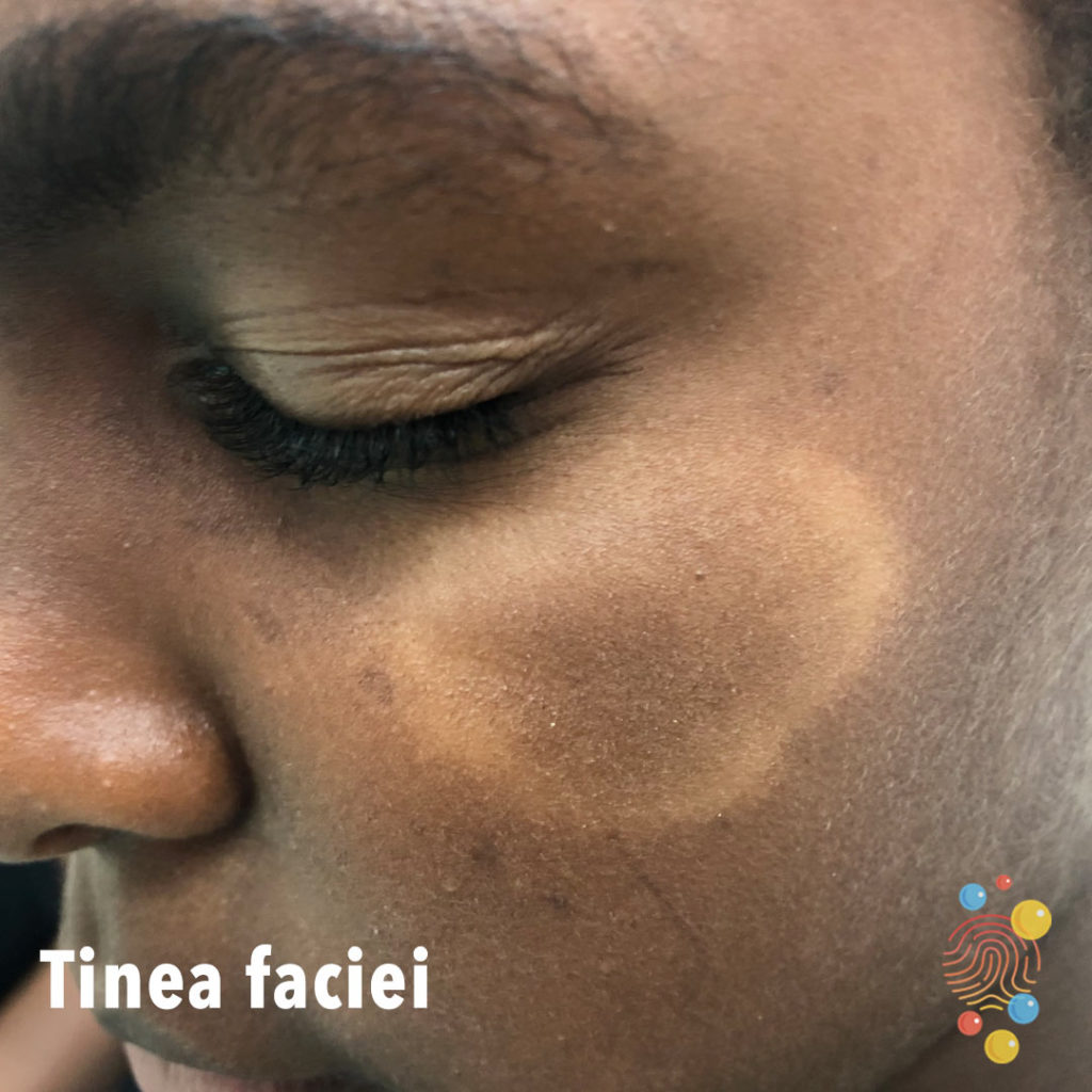
Annular hypopigmented patch with central scaling on a background of asteatotic skin.
Learn more about tinea faciei

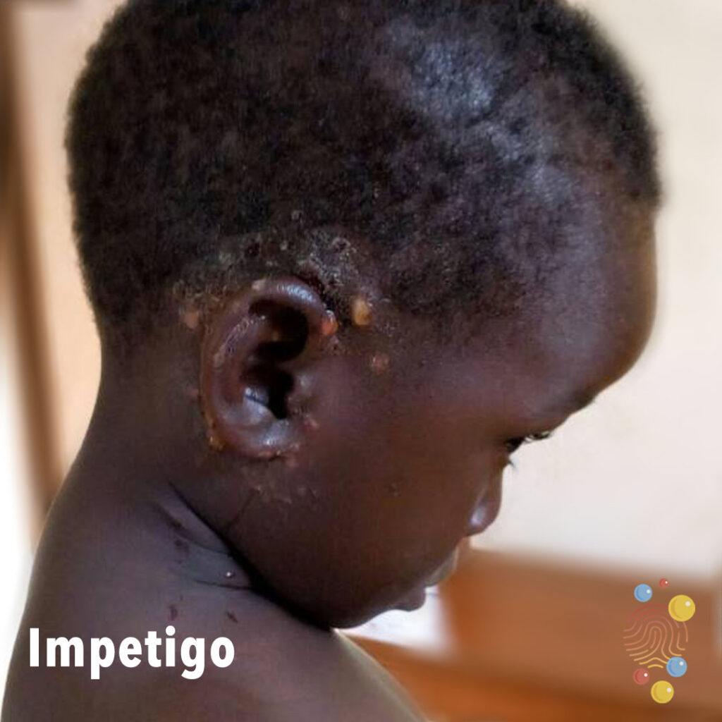
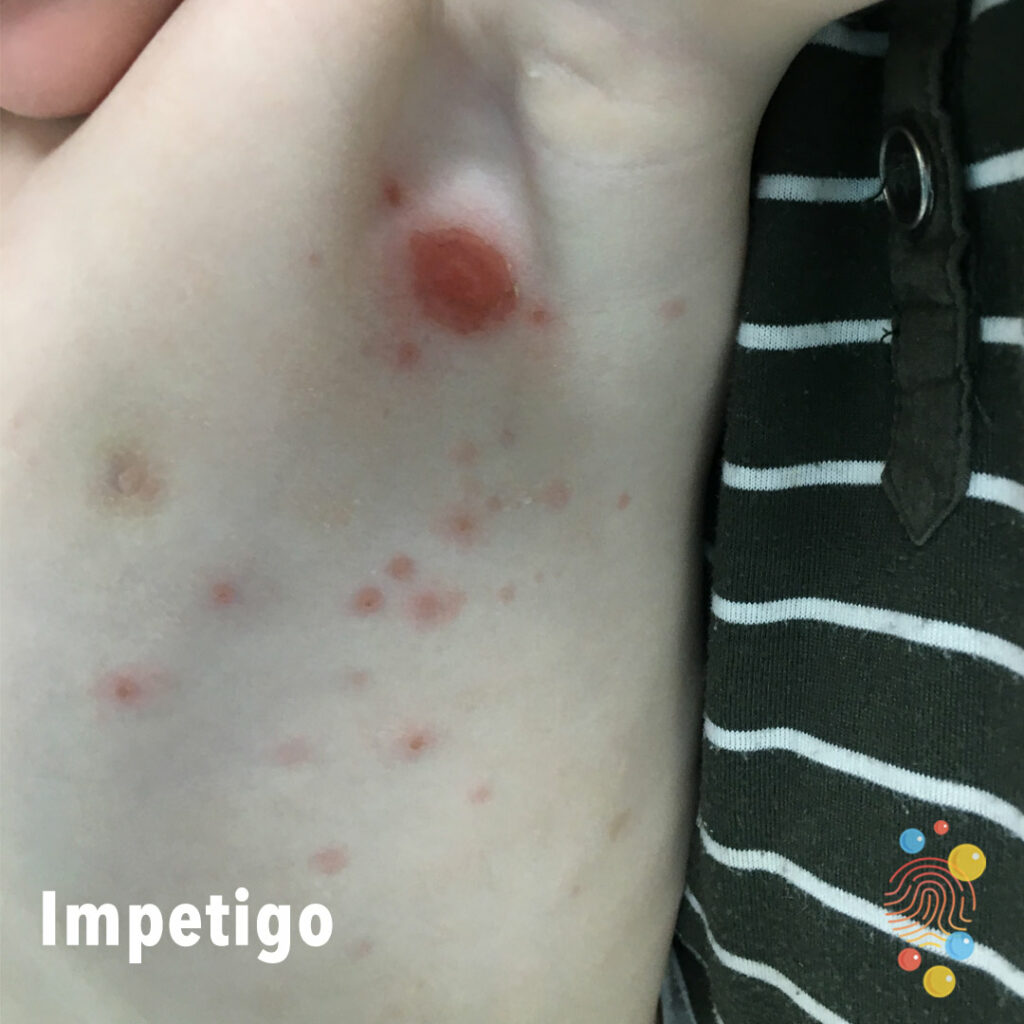
Impetigo
Abscess of the heel with surrounding redness and swelling with an incision wound to the heel.
Learn more about abscesses
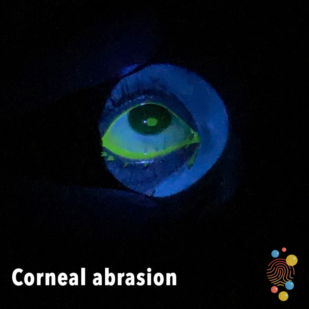
Learn more about corneal abrasions
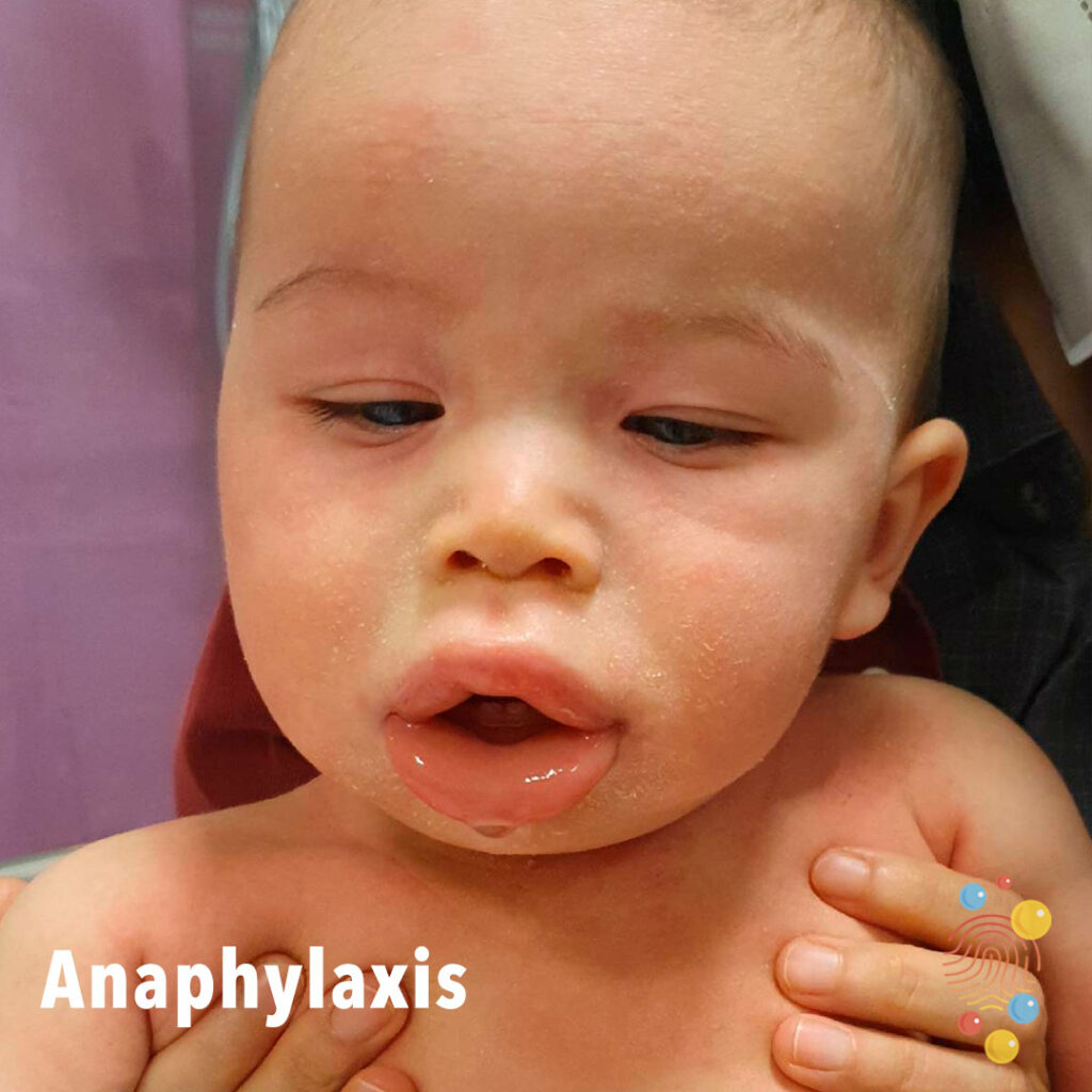
Swelling of the lip with associated drooling and rash.
Learn more about anaphylaxis
Patches of severely scaly skin.
Learn more about ichthyosis
Eczematous rash across the chest – erythema and lichenification.
Learn more about eczema
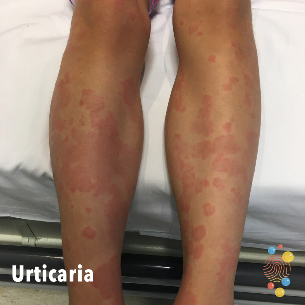
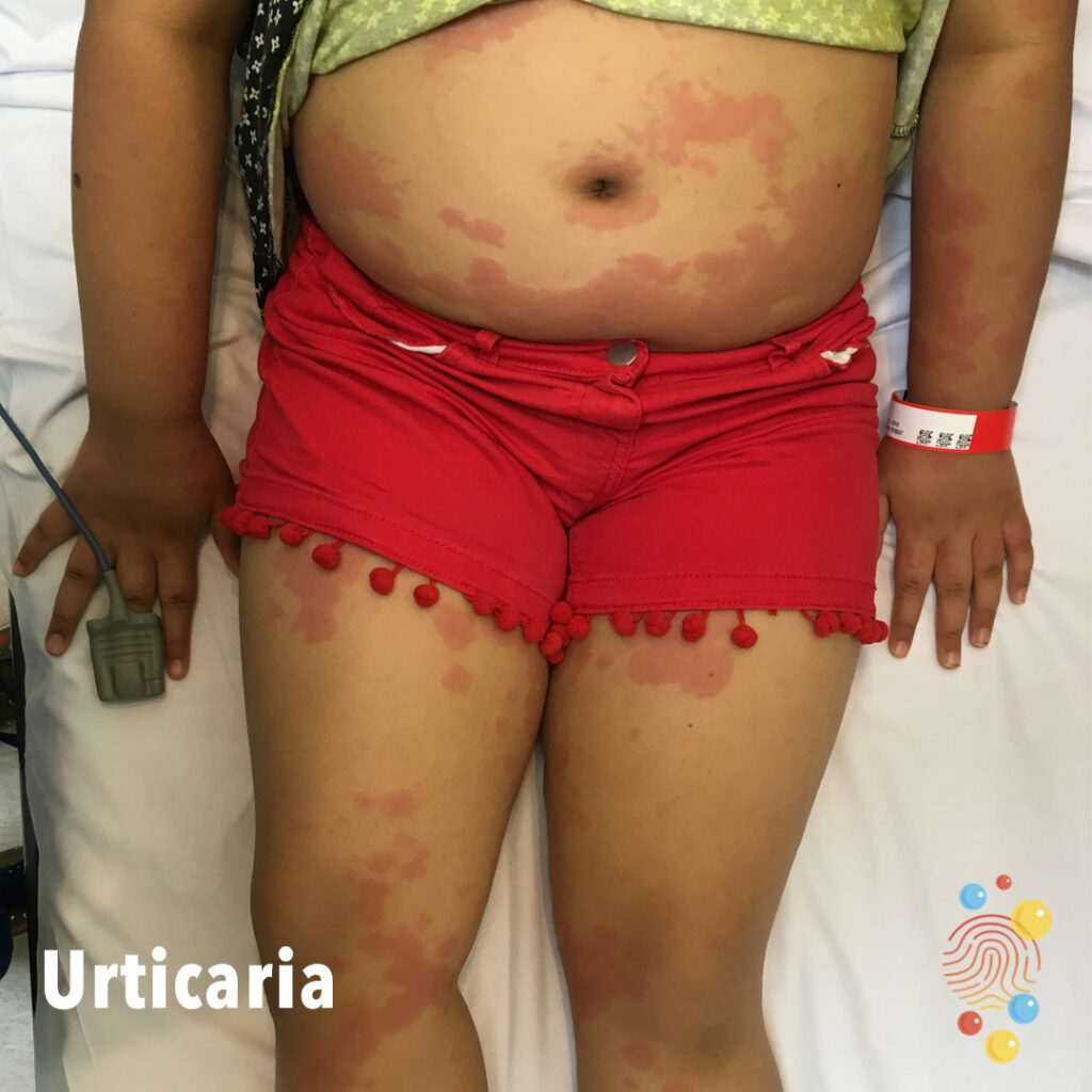
Erythematous edematous plaques “wheals” some of which are confluent on legs.
Learn more about urticaria
Umbilical erosion with multiple crusted erosions on the abdomen.
Learn more about impetigo
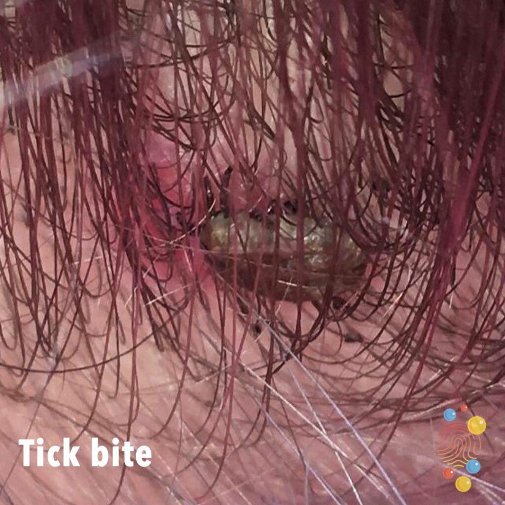
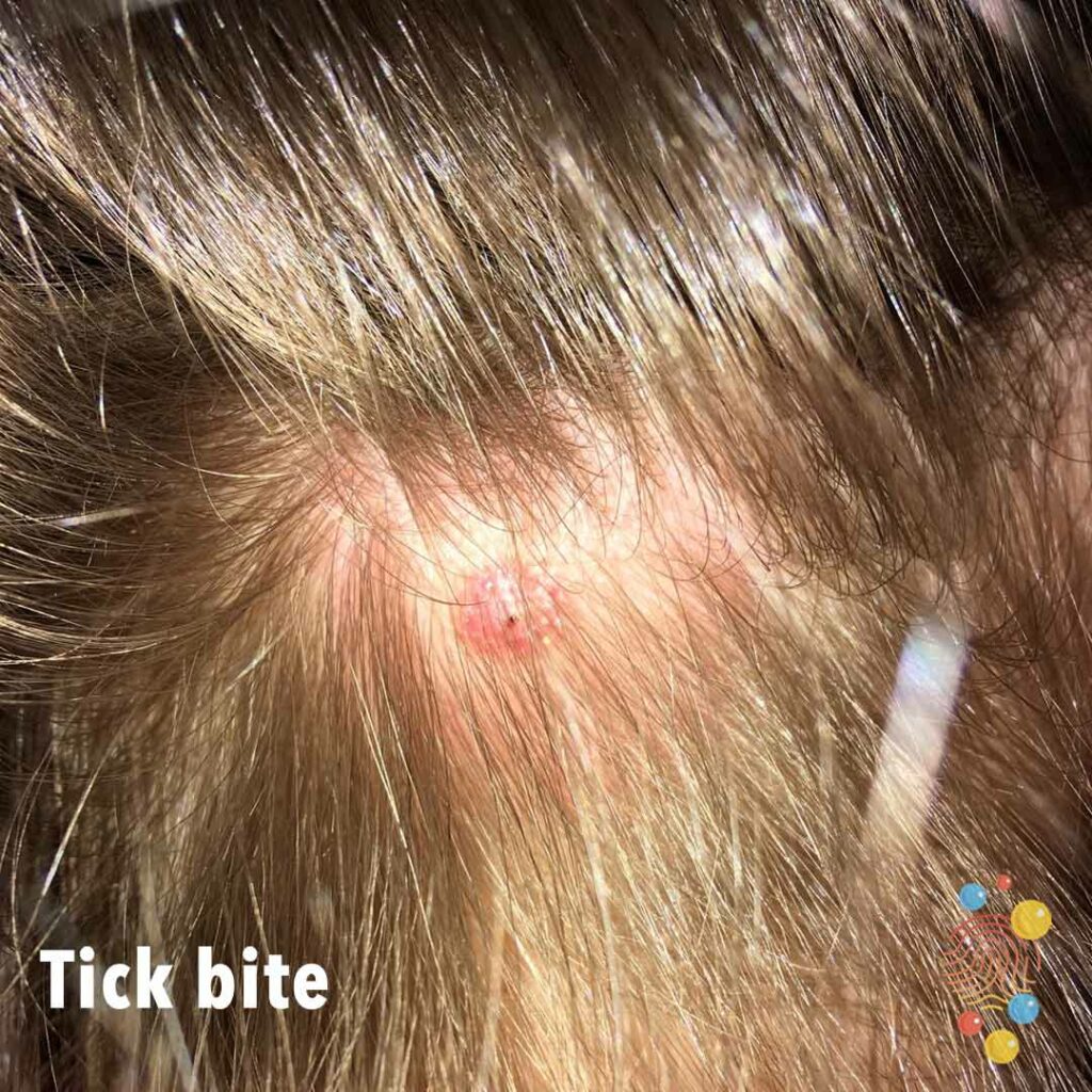
Tick is seen in this image amongst the hair.
Learn more about tick bites
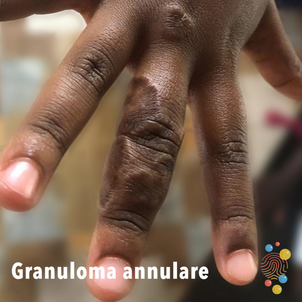
Raised reddish/purplish firm bumps arranged in multiple confluent rings localized on the third finger of the hand.
Learn more about granuloma annulare
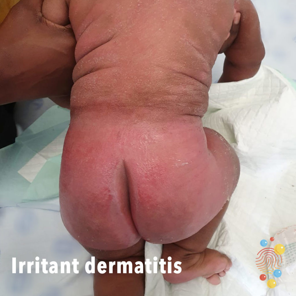
Widespread eczematous changes on back, bright erythema to buttocks, sparing of natal cleft.
Learn more about irritant dermatitis
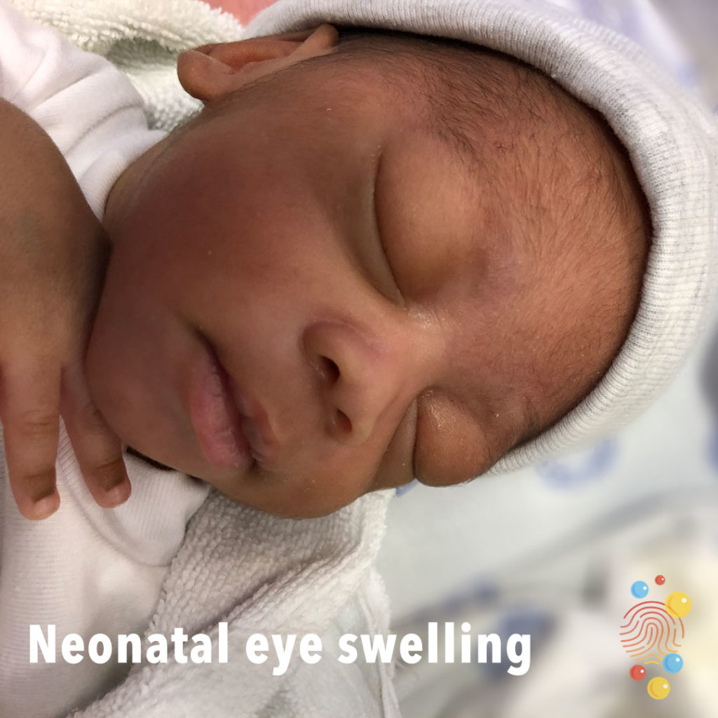
Bilateral eye swelling.
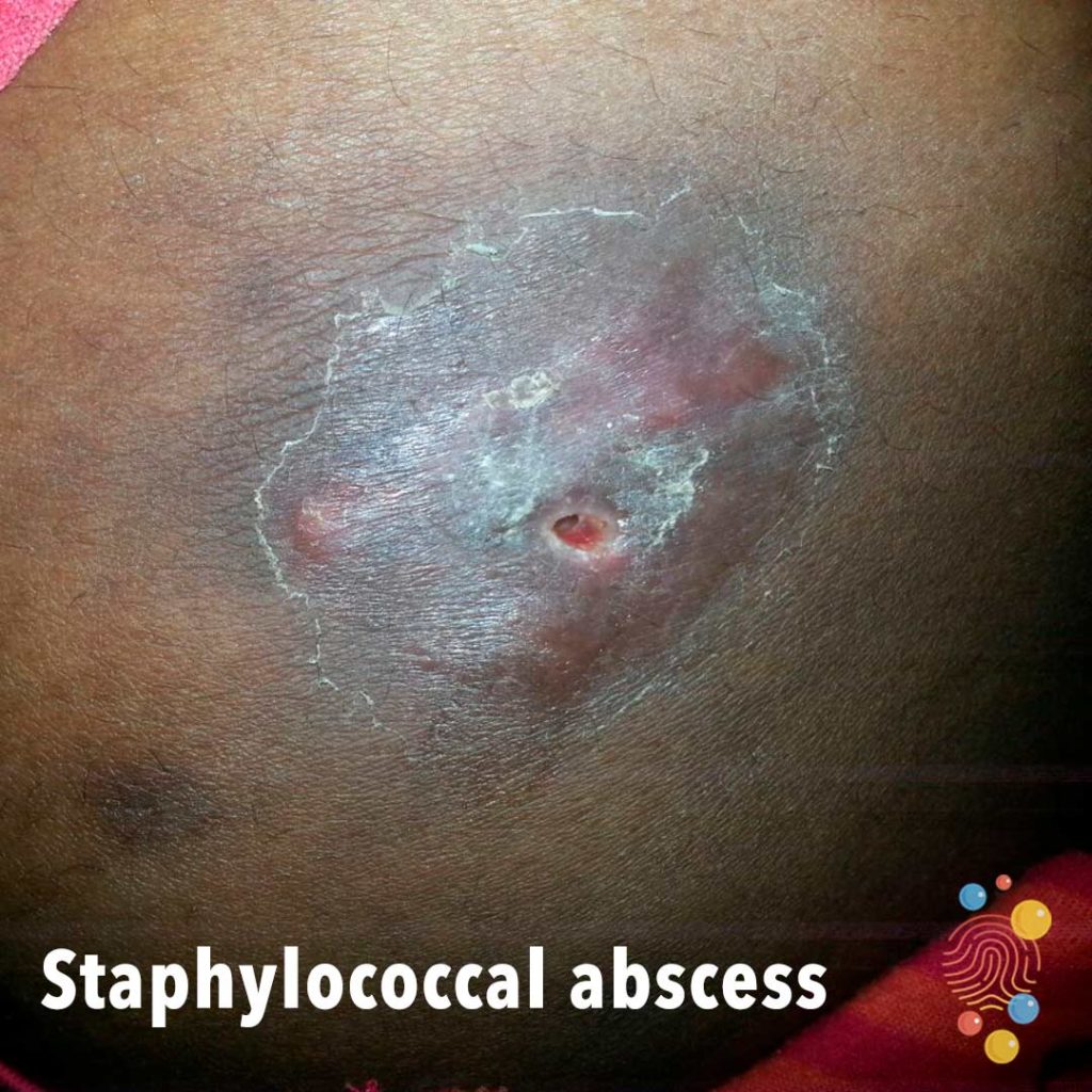
Indurated plaque with large central punctum and peripheral scale.
Learn more about staphylococcal abscesses
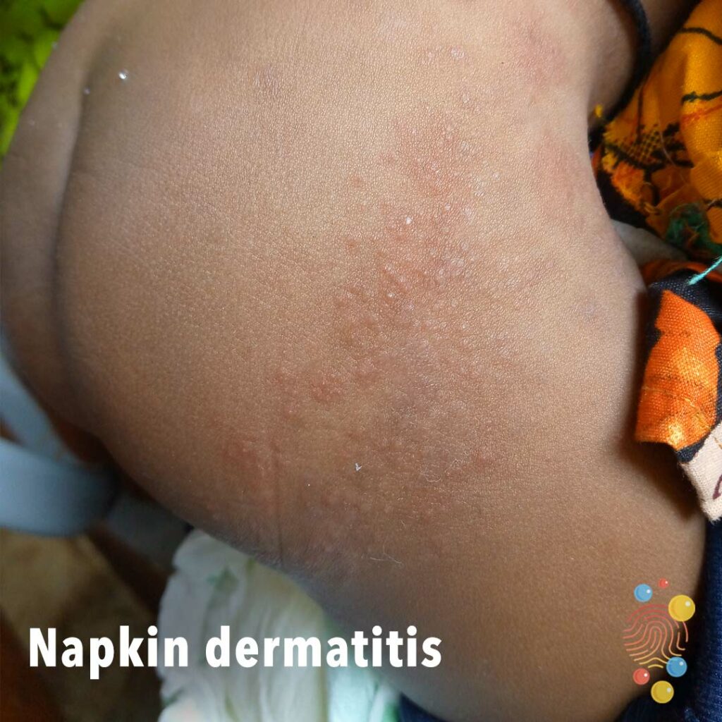
Ill-defined, erythematous, maculopapular rash, upper right thigh with post inflammatory hyperpigmentation.
Learn more about napkin dermatitis
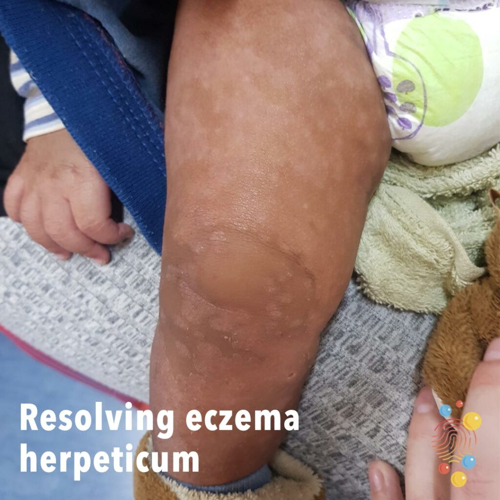
Postinflammatory hypopigmented macules.
Learn more about eczema herpeticum
Hyperpigmentation with scattered hypopigmentation and scale.
Learn more about pityriasis versicolor

Hyperpigmented plaques in concha, pinna, and periauricular skin.
Learn more about discoid lupus

Small pustule with underlying swelling.
Learn more about abscesses
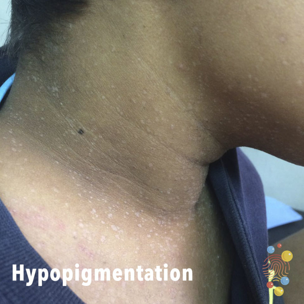
Confetti-like small hypopigmented macules affecting upper chest and neck.
Learn more about hypopigmentation
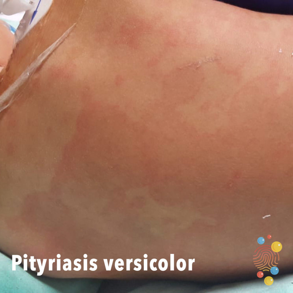
Round, scaly, confluent, erythematous patches.
Learn more about pityriasis versicolor
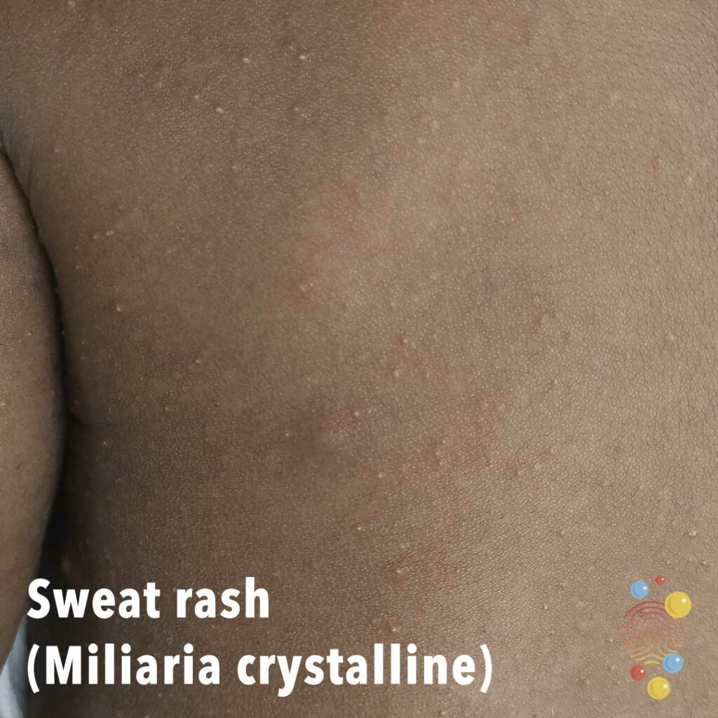
Multiple little tiny superficial blisters sweat-filled.
Learn more about miliaria

Erythema and crusting of the umbilical stump.
Learn more about omphalitis

Area of axillary lichenification and excoriation.
Learn more about eczema
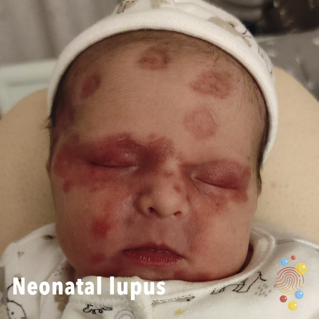
Discoid erythematous plaques affecting forehead and eyes, with a ‘raccoon-eye’ appearance, in a neonate with a mother with anti-SSA (Ro) antibodies.
Well-circumscribed blue/green fluctuant mass.
Learn more about abscesses
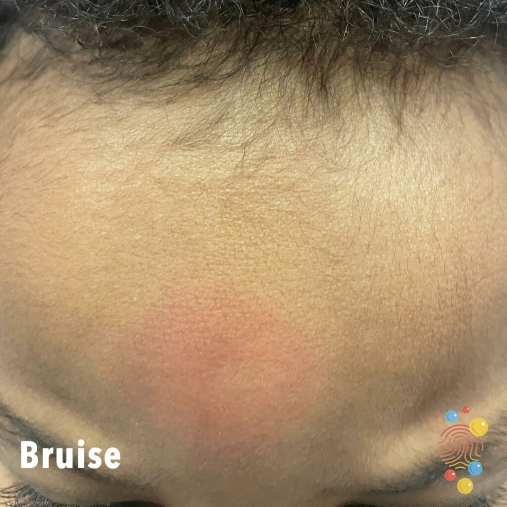
Central forehead bruise.
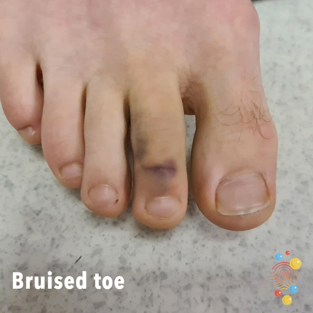
Bruised Toe
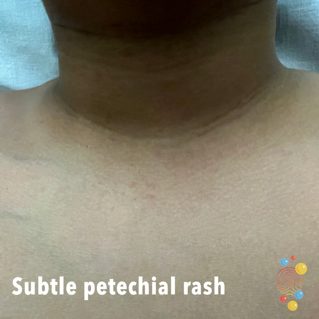
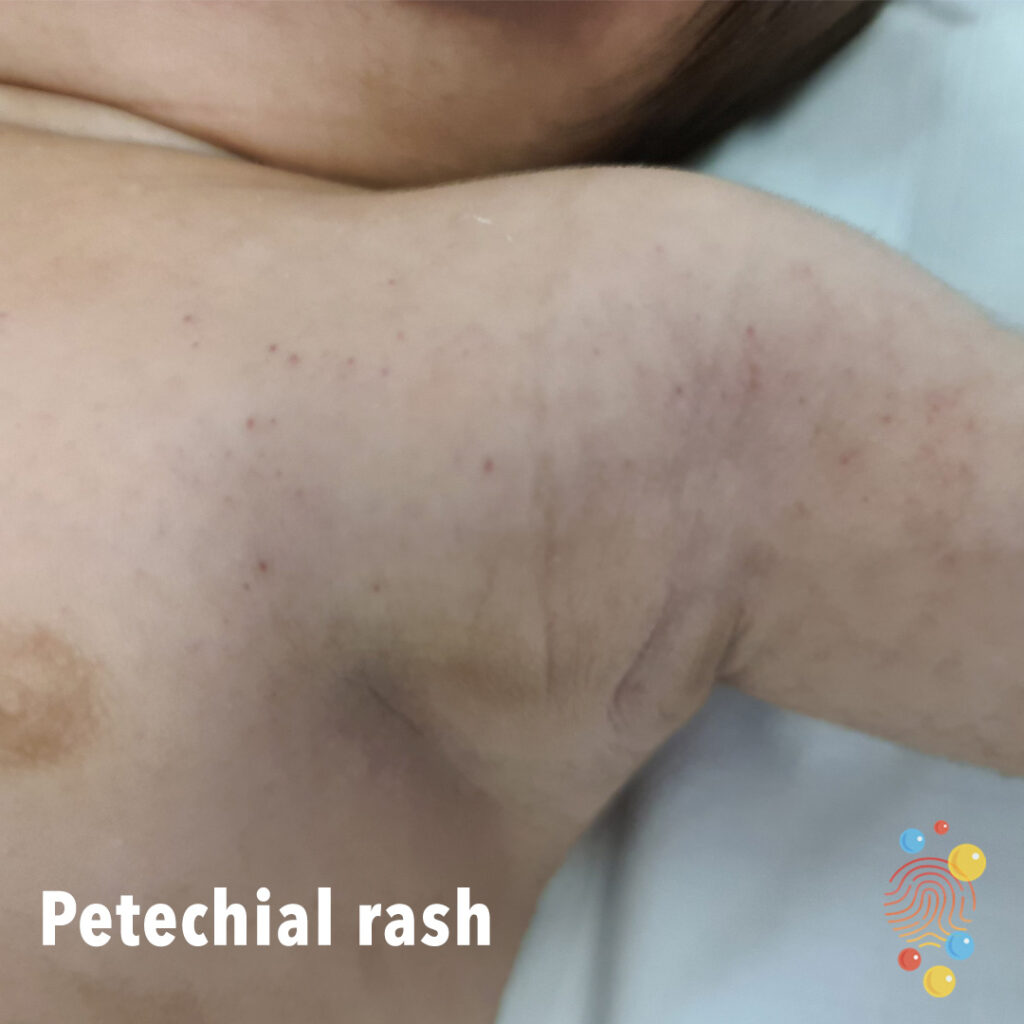
Subtle Petechial Rash
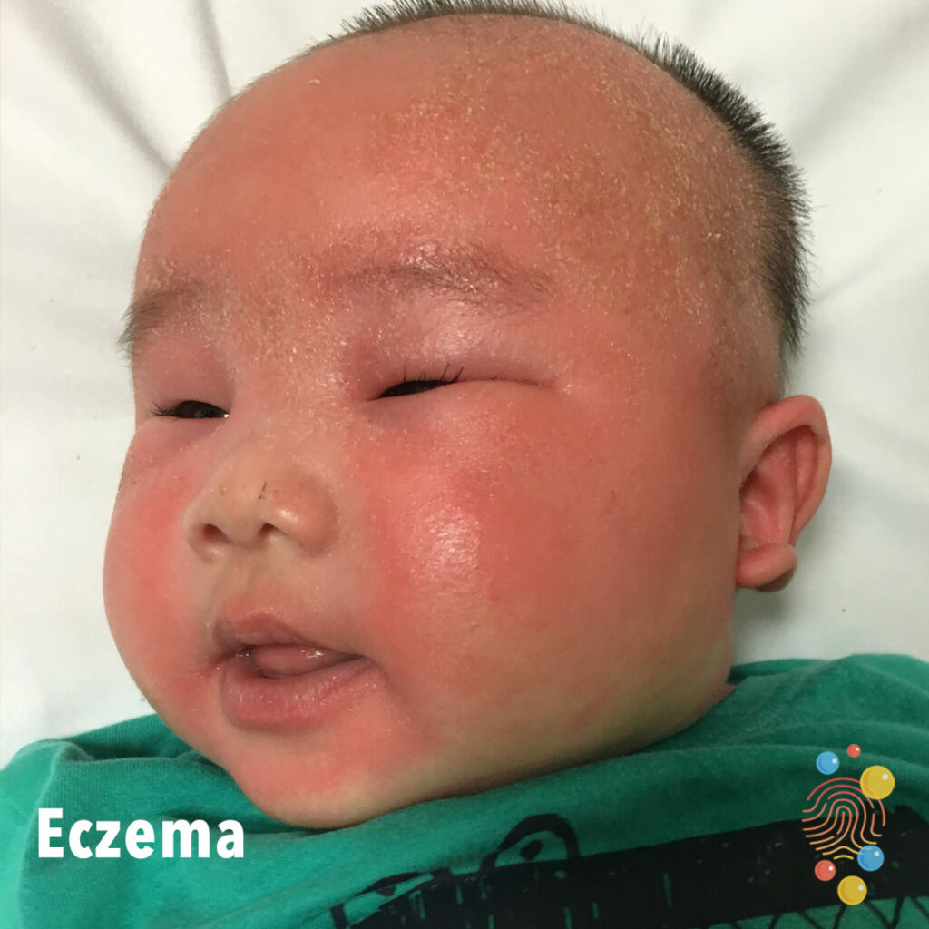
Hyperkeratosis over left forehead and glabella.
Learn more about eczema
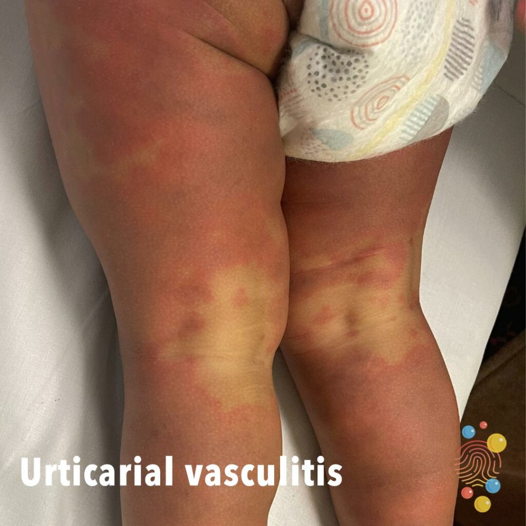
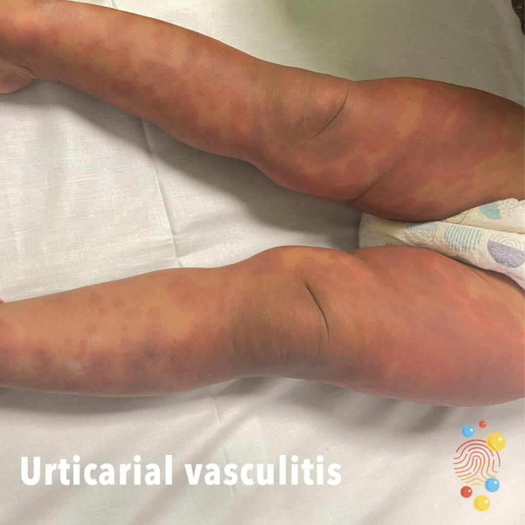
Urticarial Vasculitis
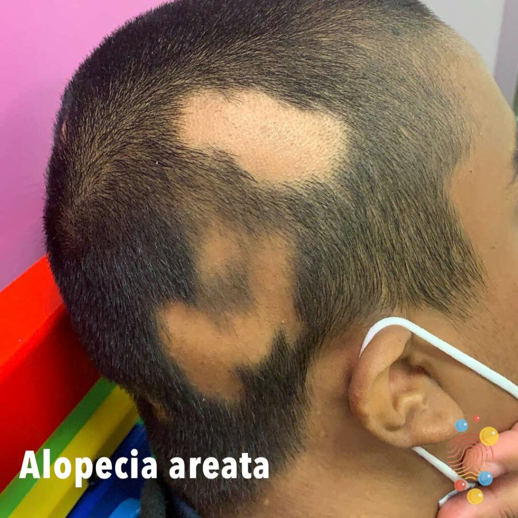
Multiple, discrete, annular patches of hair loss over right side (parietal area) of scalp.
Learn more about alopecia areata
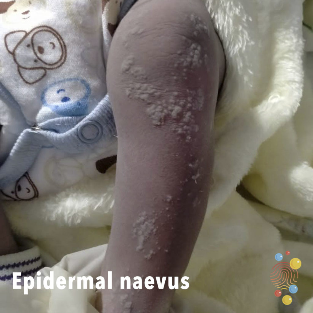
Blashkoid distribution of hyperkeratotic papules and plaques along arms.
Learn more about epidermal naevus

Erythematous tender nodules on shins with bruised appearance.
Learn more about erythema nodosum
Pustules on an erythematous base.
Learn more about erythema toxicum
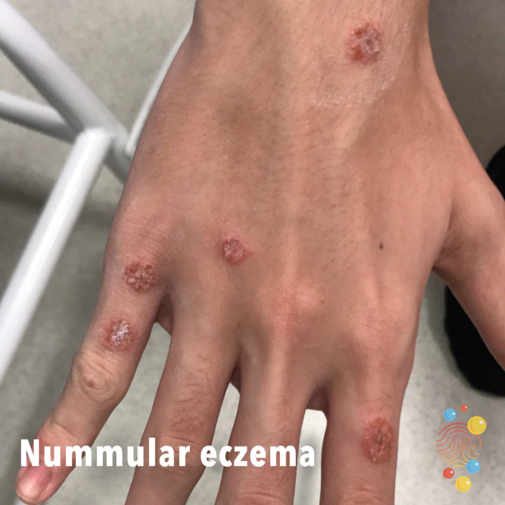
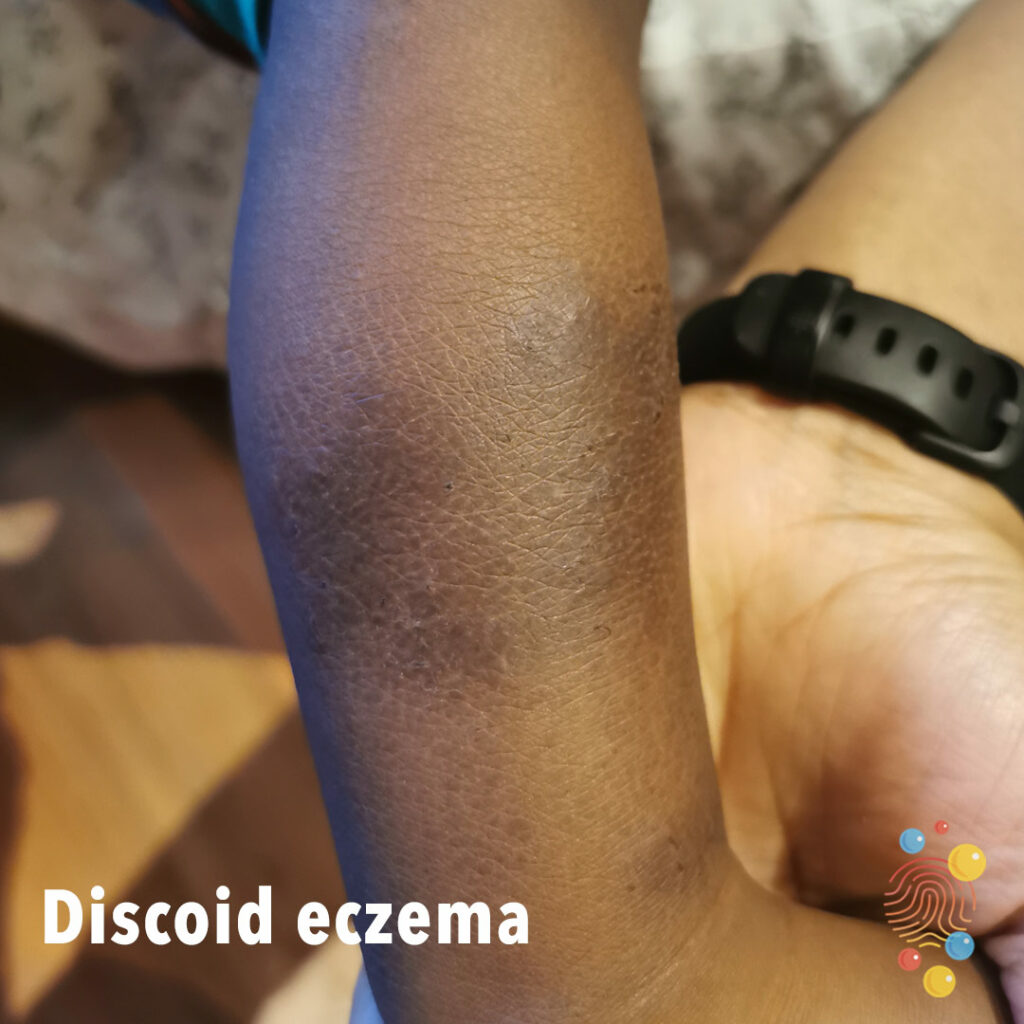
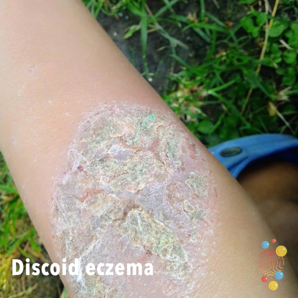
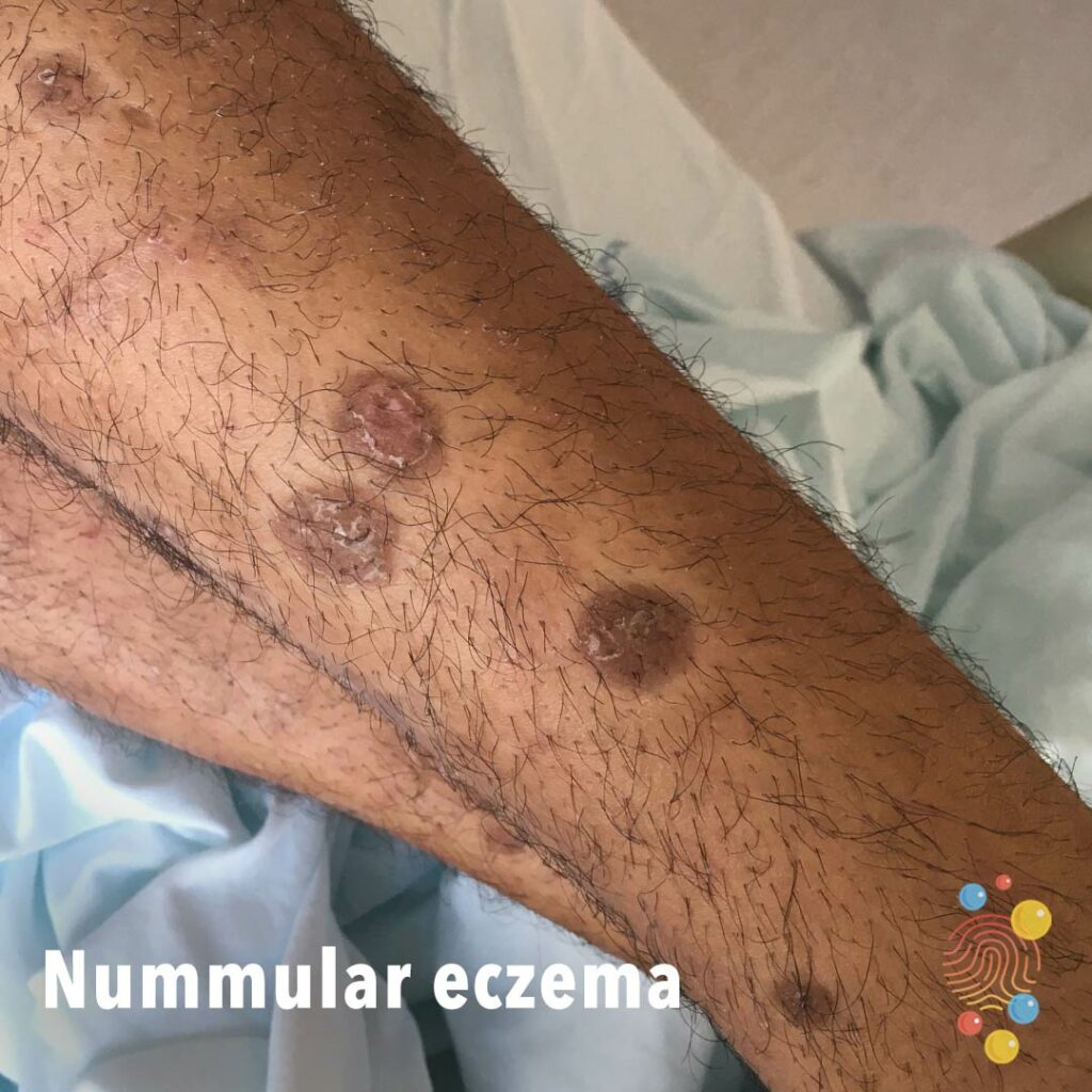
Multiple coin-shaped eczematous areas on the dorsal hands.
Learn more about eczema
Erythematous skin with maceration and superficial erosions on the posterior neck in the skin folds.
Learn more about intertrigo
Eroded superficial blisters with some crusting.
Learn more about bullous impetigo
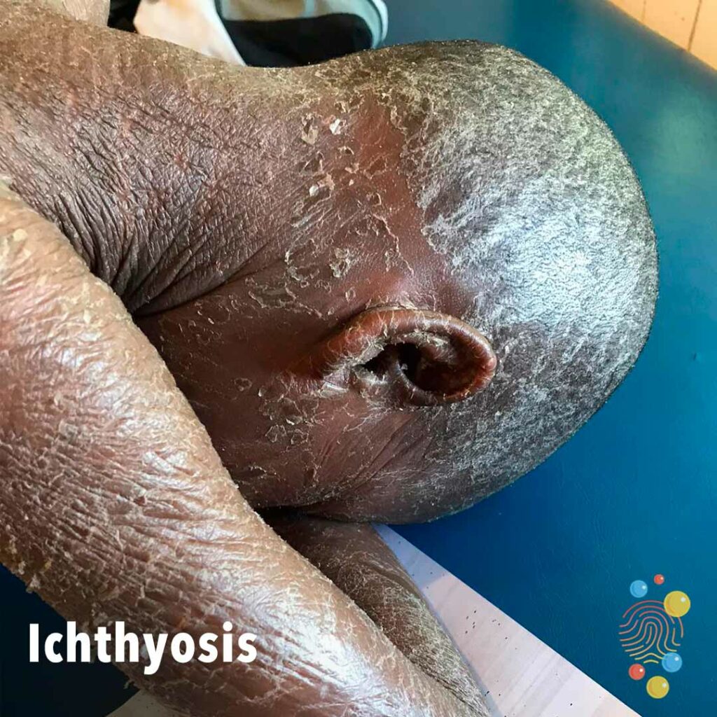
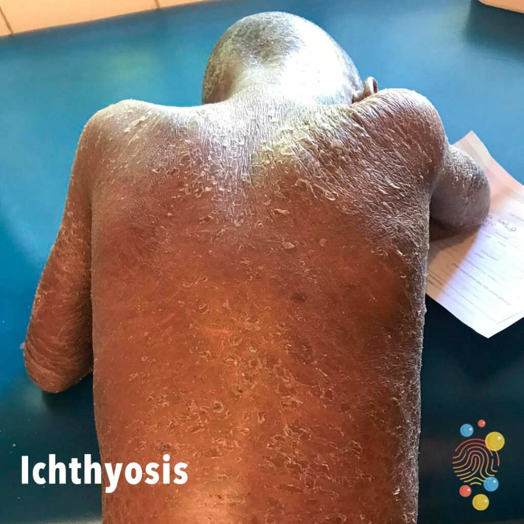
Widespread ichthyosis with severe lichenification and xerosis.
Learn more about ichthyosis
Severe erythema, lichenification, and bleeding of the lower limbs.

Multiple urticated bruises, some of which have a targetoid appearance

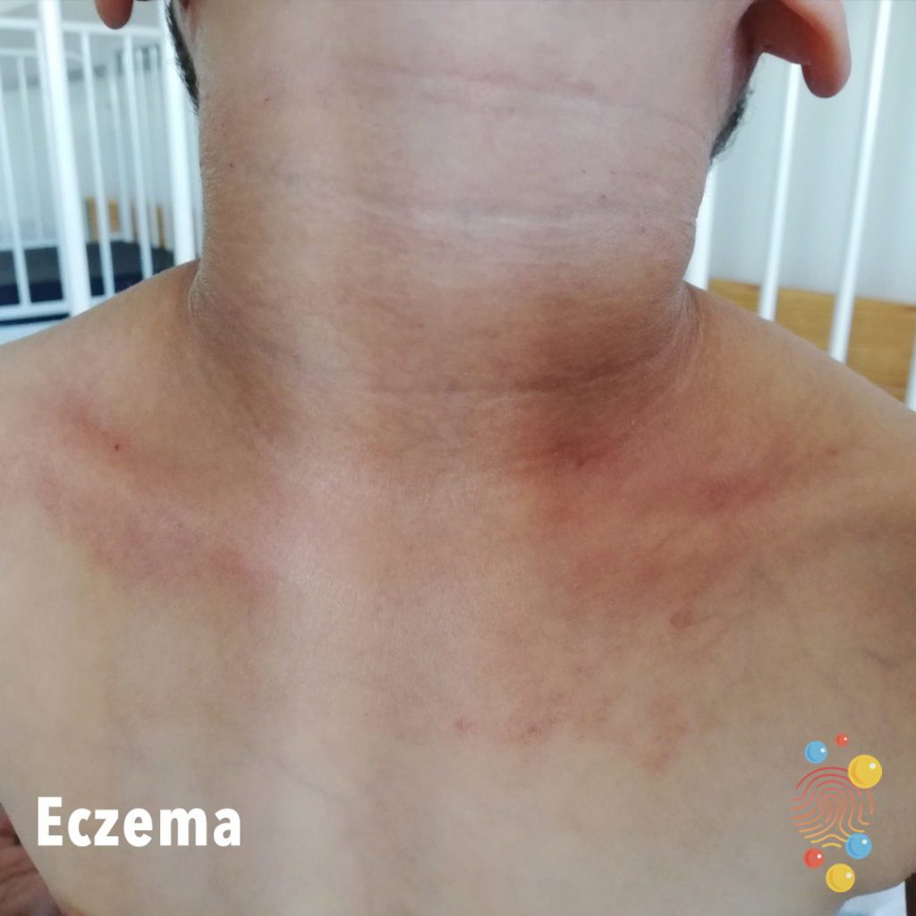
Erythema, lichenification, and hyperpigmentation of anterior neck folds.
Learn more about eczema
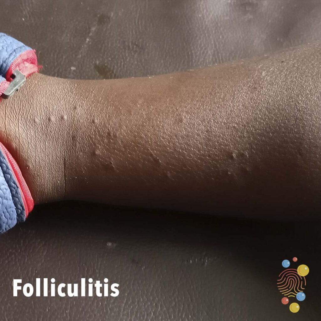
Skin coloured papules centred around hair follicles.
Learn more about folliculitis

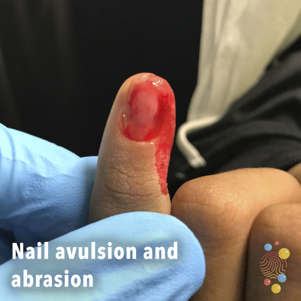
Nailbed injury pre and post repair.
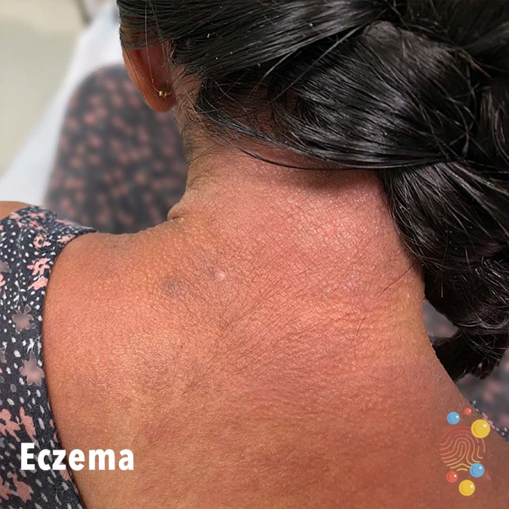
Xerosis, lichenification, and erythema of the posterior neck.
Learn more about eczema
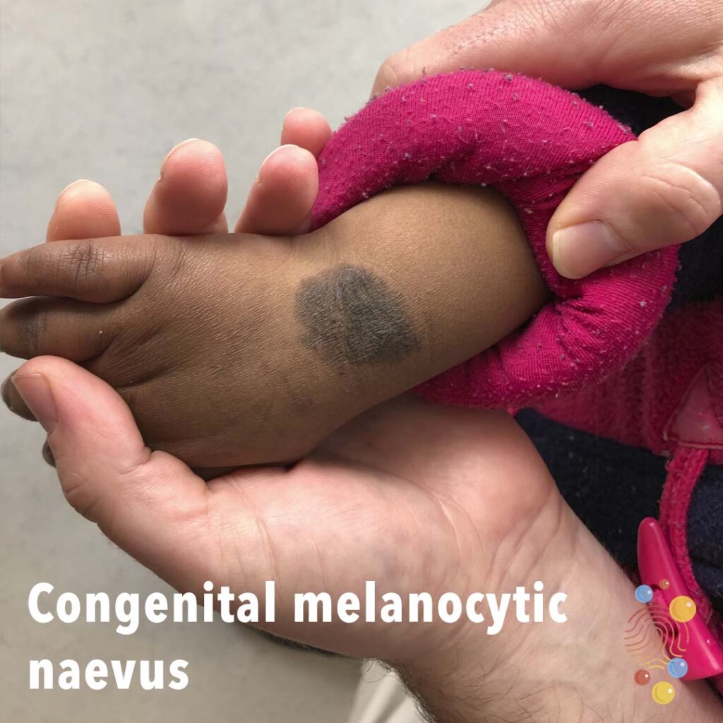
Dark brown pigmented lesion of dorsal wrist.
Learn more about congenital melancytic naevi

Symmetric swelling of lower limbs associated with hyperkeratosis, plantar keratoderma, and dystrophic toenails
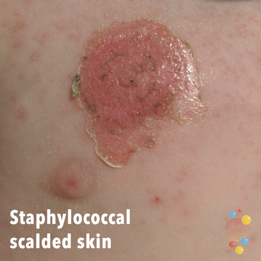
Erythematous rash with surrounding blister and peeling of the skin.
Learn more about staphylococcal scalded skin
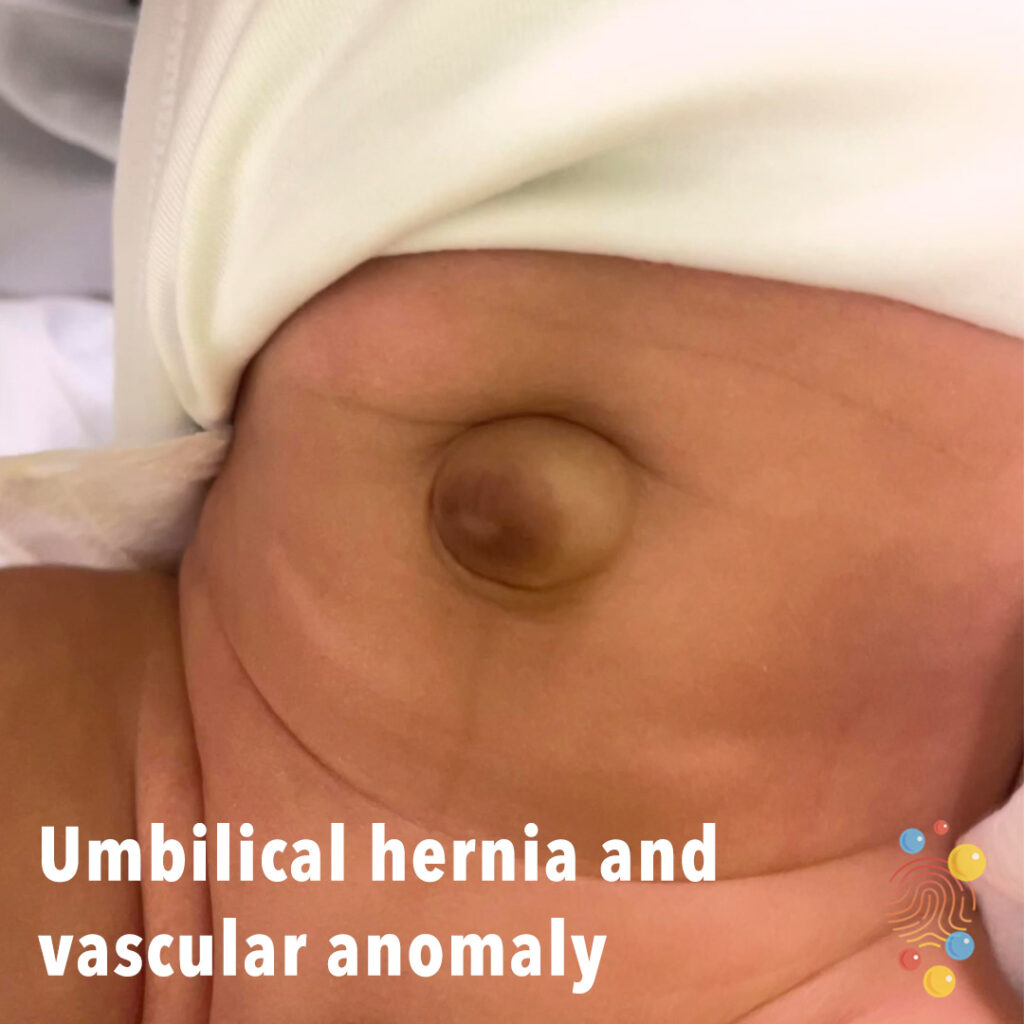
Bulge at the site of the umbilicus with a dark-coloured discolouration of the skin.
Learn more about umbilical hernias
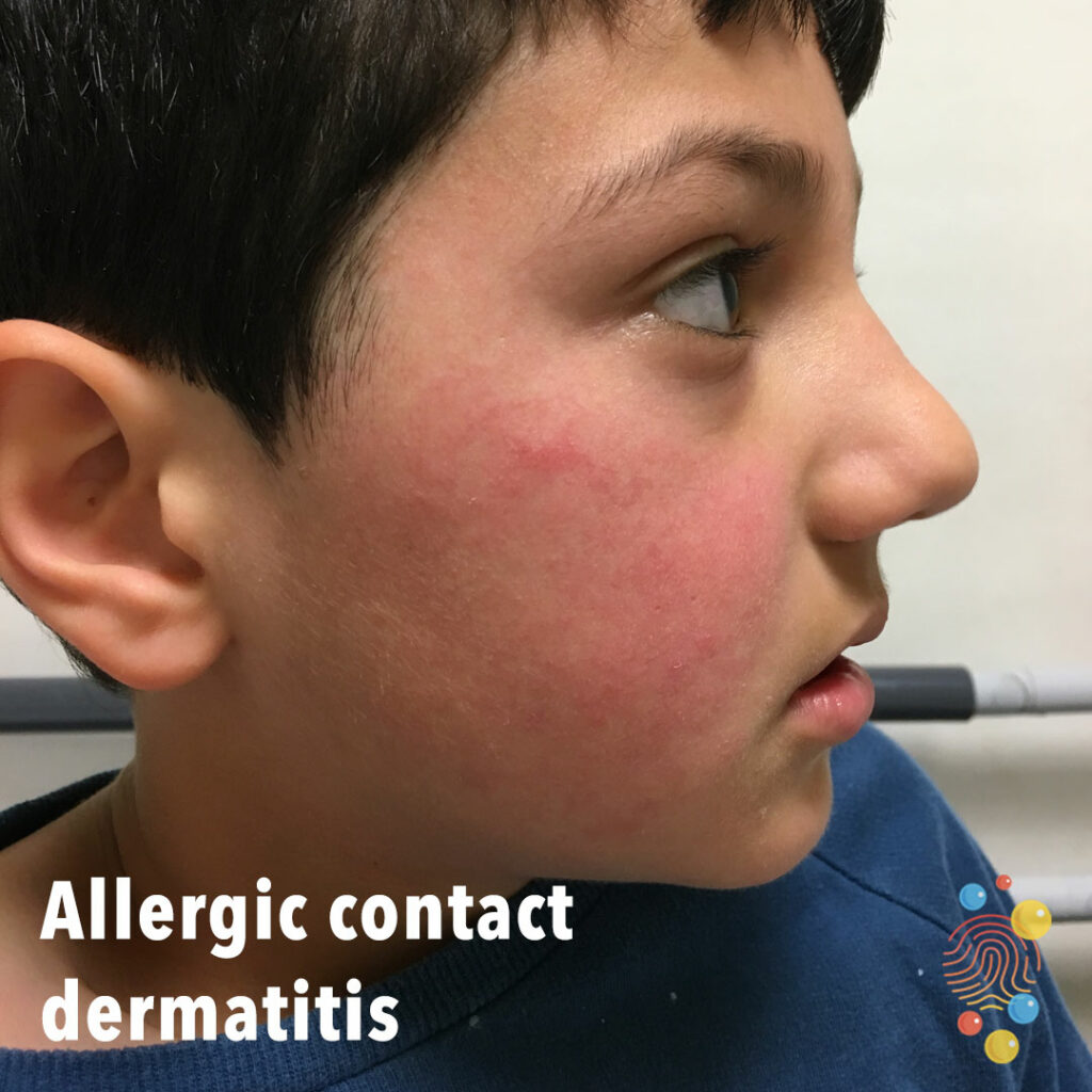
Facial erythema.
Learn more about eczema

Multiple urticated bruises, some of which have a targetoid appearance
Intertrigo
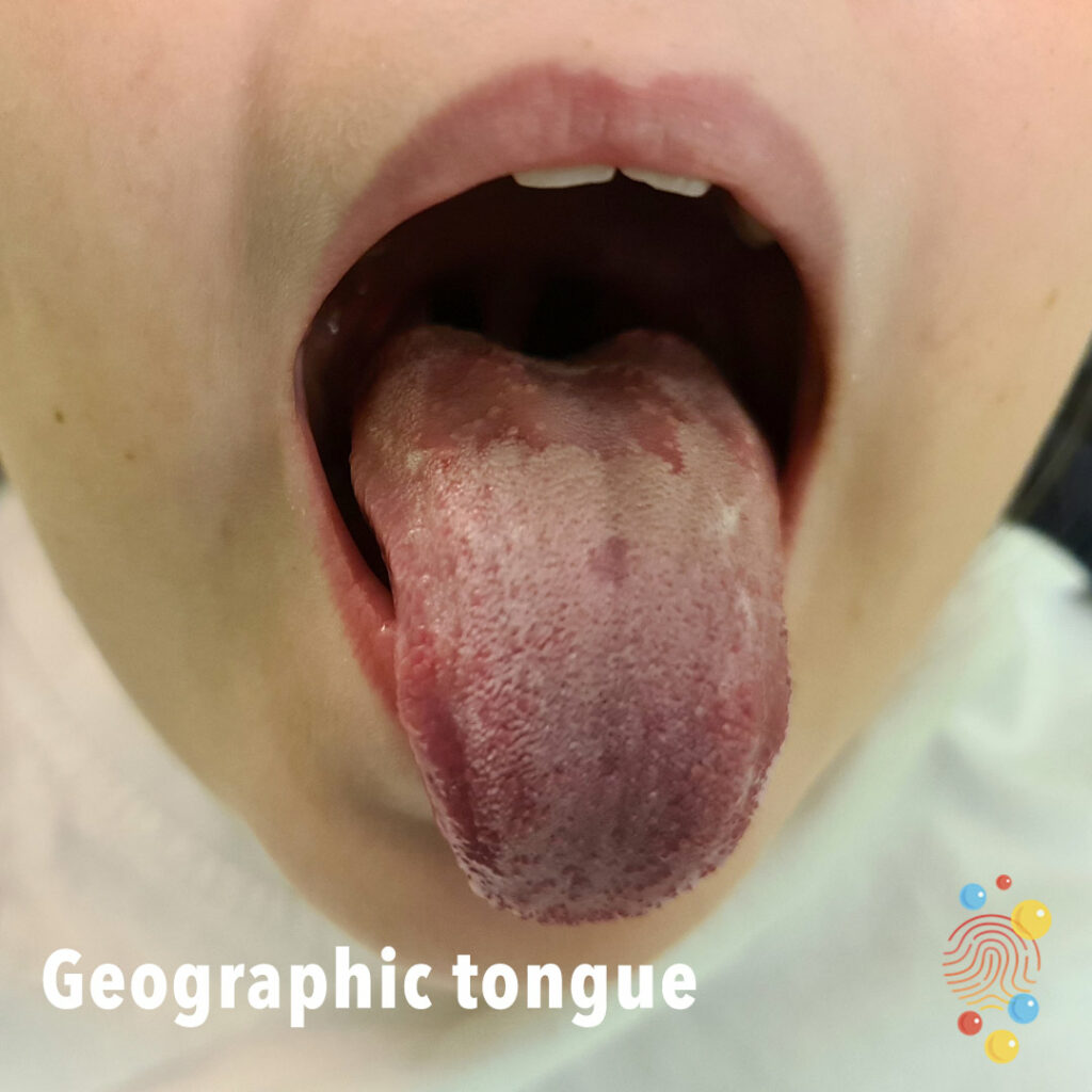
Coated tongue with areas of loss of normal rugosity revealing smooth flat areas.
Learn more about geographic tongue
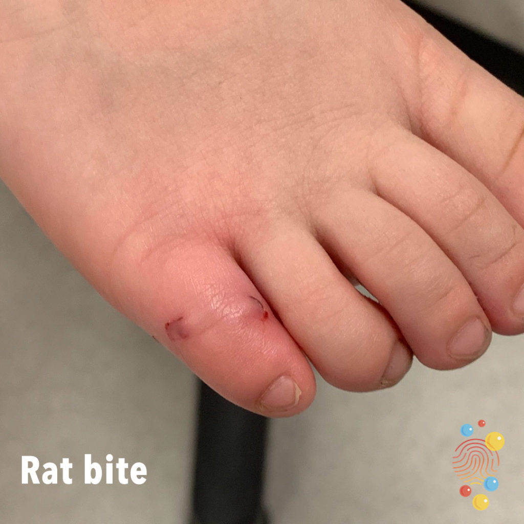
Bite mark on fifth toe.
Learn more about bites
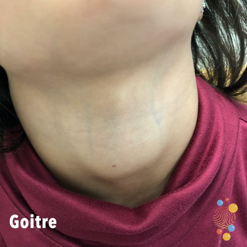
Subcutaneous swelling of anterior neck.
Learn more about goitres
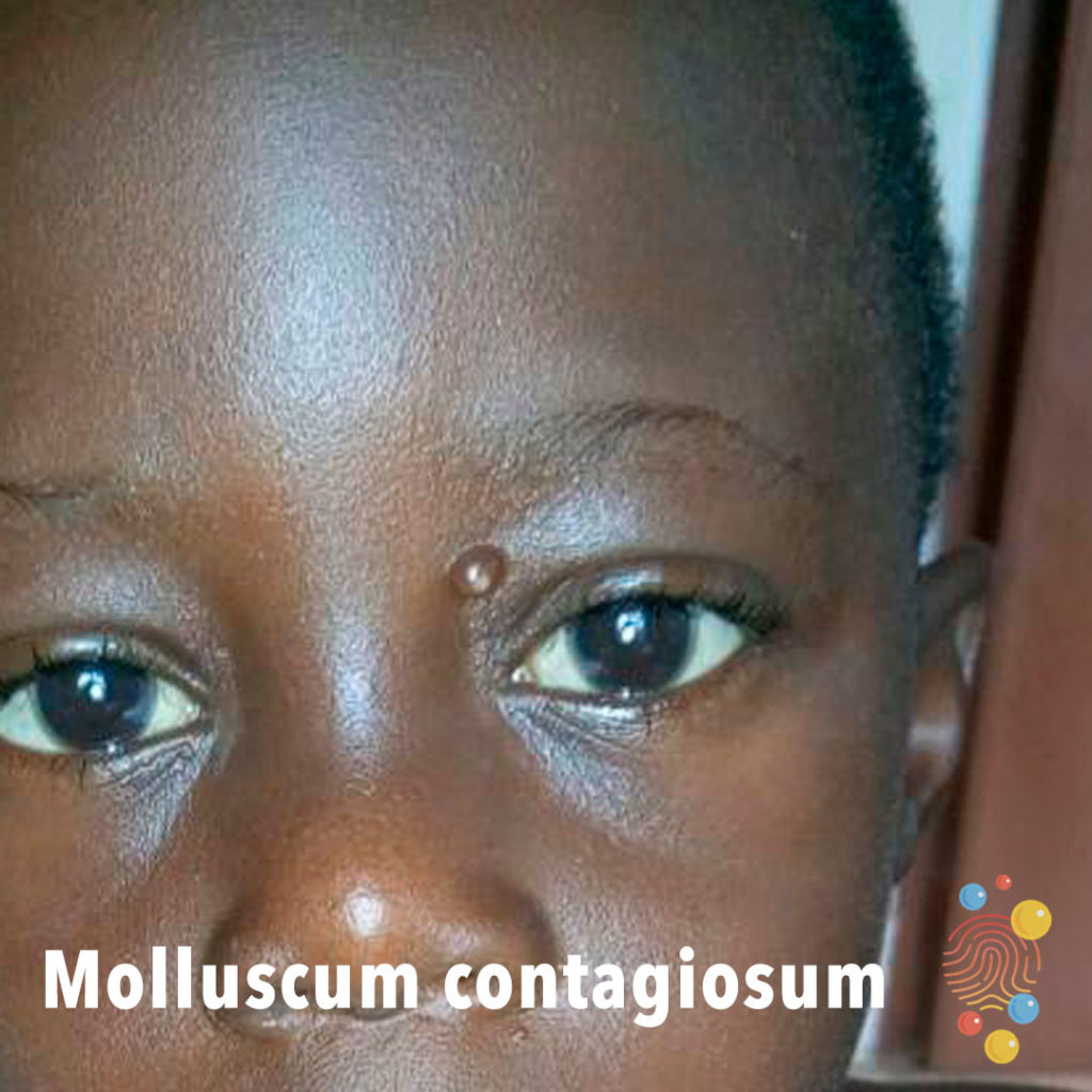
Pearl-like papules with a dimple in the centre.
Learn more about molluscum contagiosum
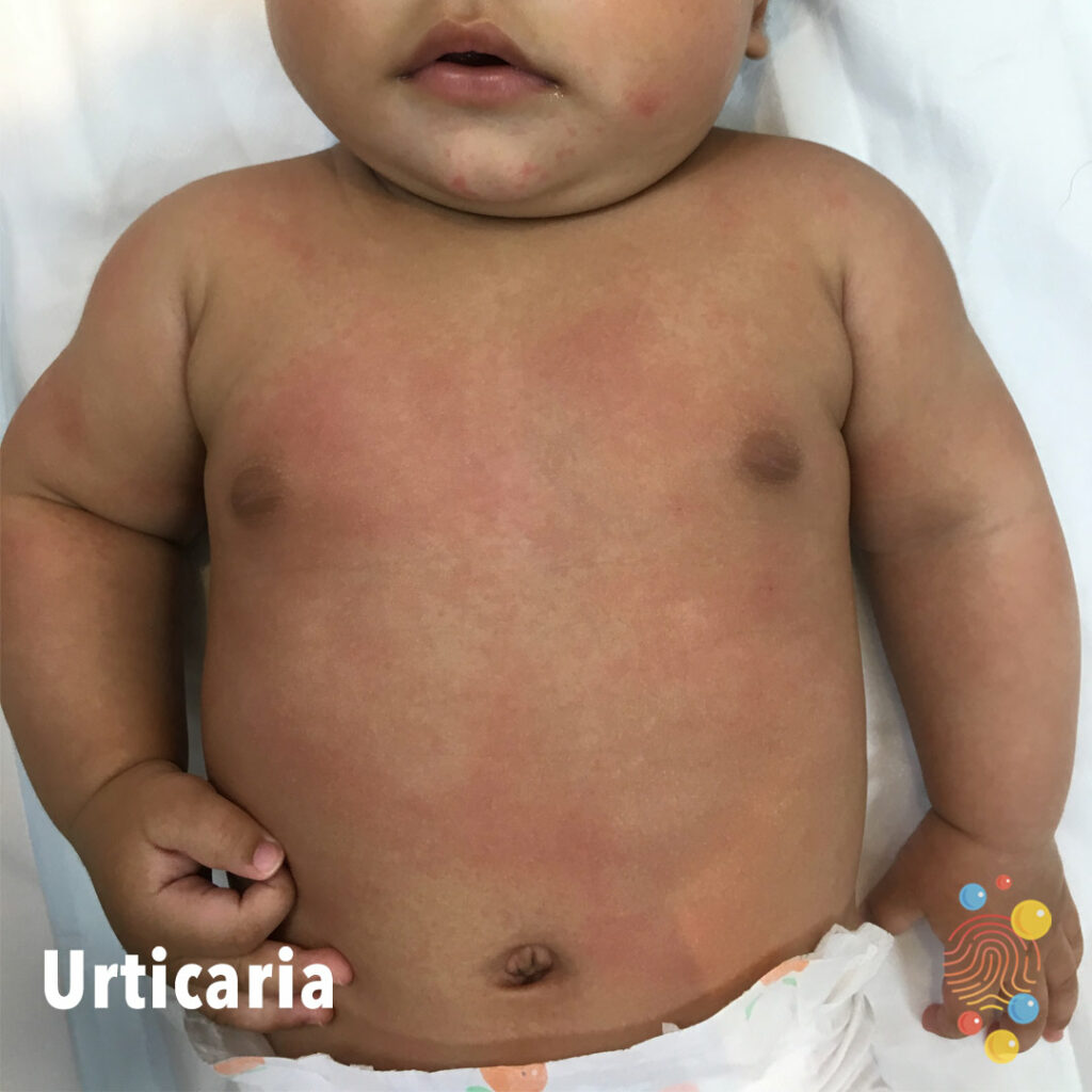
Blotchy macular erythema on trunk. Could also be consistent with viral exanthem.
Learn more about urticaria

Eruption of dark red macules, vesicles, and erosions distributed across areas previously affected by atopic dermatitis, with relative sparing of the trunk
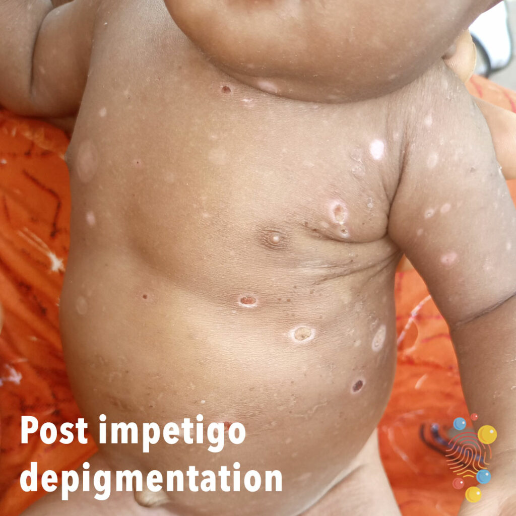
Multiple areas of oval depigmentation, some with scabs still present and some chronic changes.
Learn more about impetigo
Patch of hypopigmented skin of the left lower cheek.
Learn more about pityriasis alba

Widespread white scaling of the face on a background of erythema, some periorbital swelling and Dennie Morgan folds with nasal tip sparing. More marked scaling in the neck region.
Learn more about eczema
Multiple excoriated pustules with scaly ragged edge.
Learn more about bullous impetigo
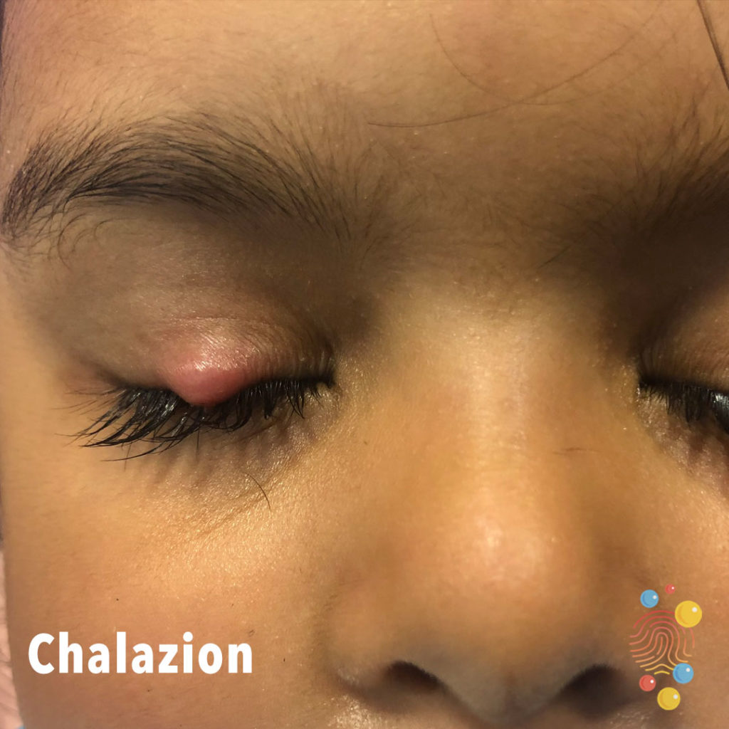
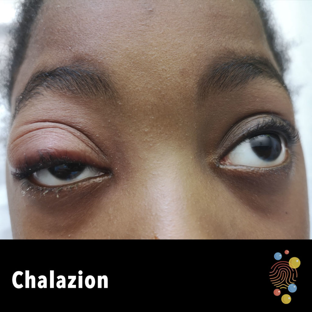
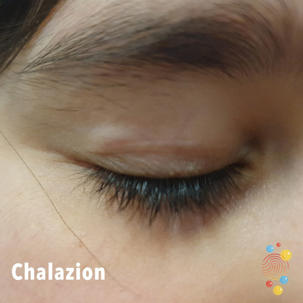
Erythematous lump on the upper eyelid.
Learn more about chalazion
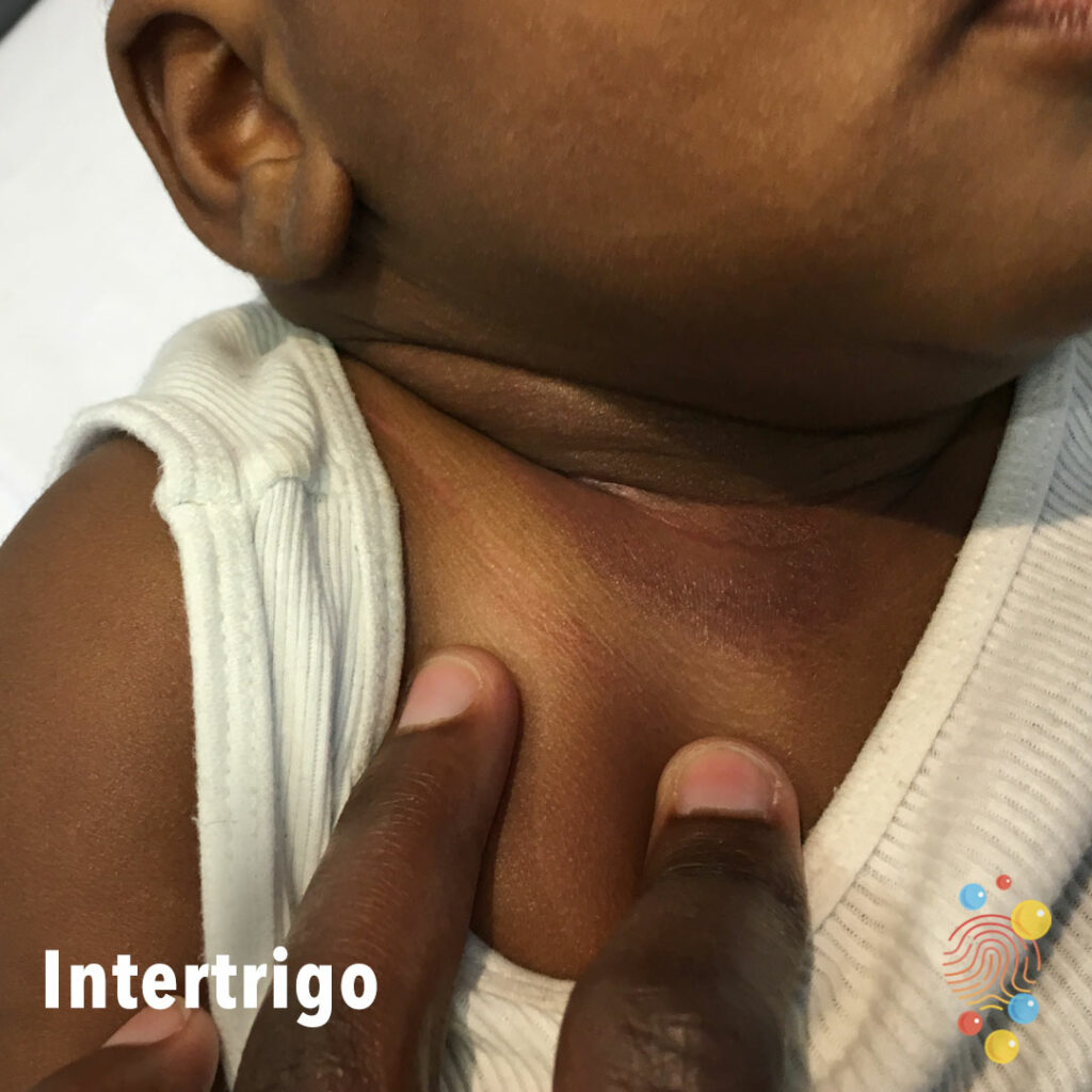
Moist erythema of the anterior neck fold.
Learn more about intertrigo
Central break in the skin in two lesions with surrounding erythema and impression of sub epidermal swelling, fading into normal skin at edge.
Learn more about bites
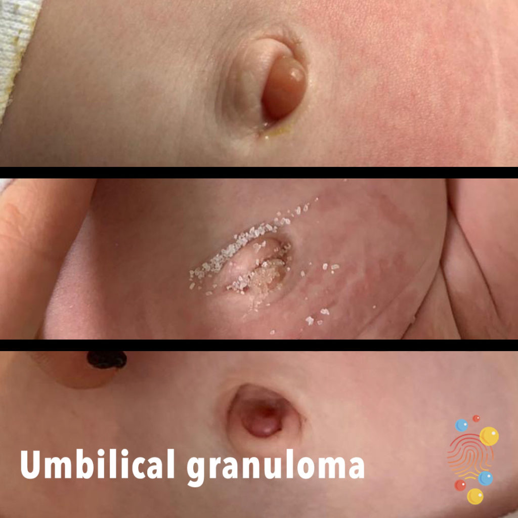
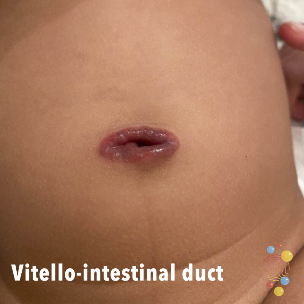
Progression of umbilical granuloma with salt treatment.
Learn more about umbilical granulomata
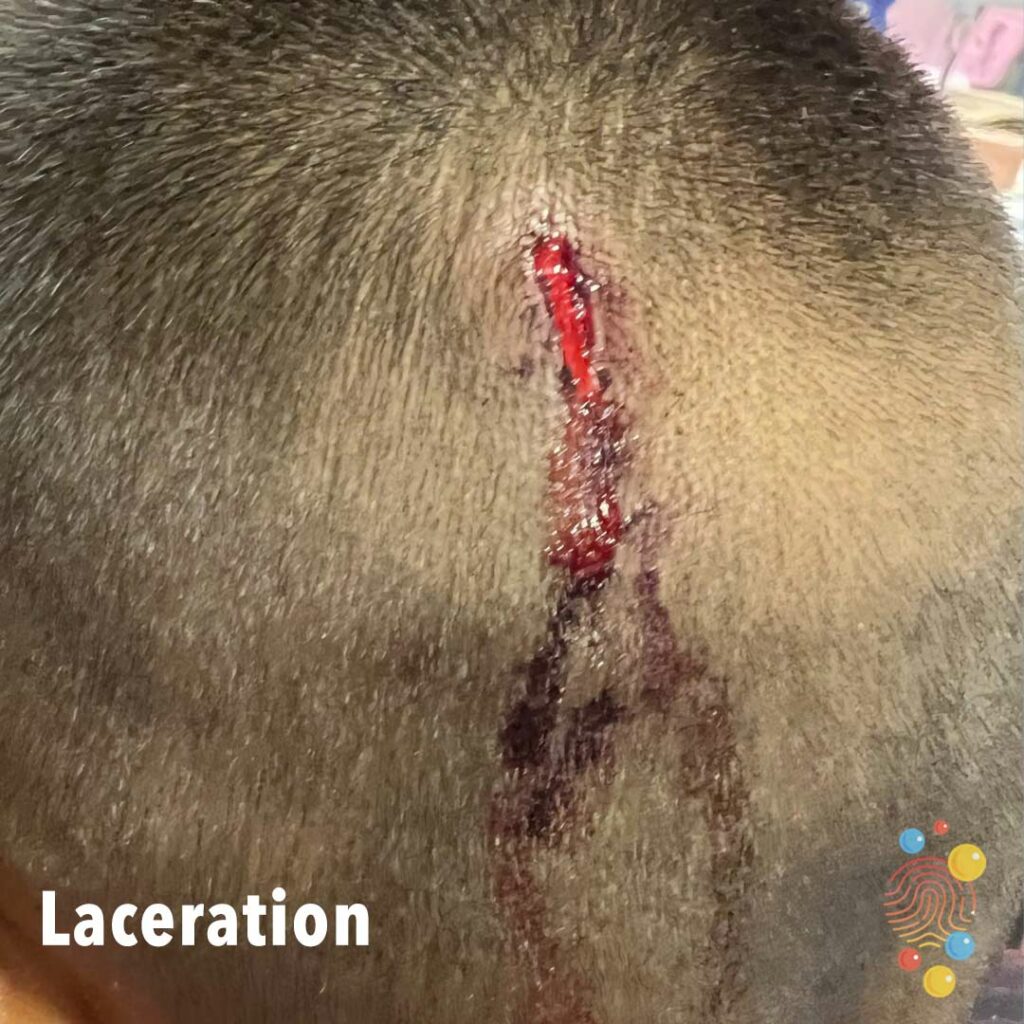
Head Laceration


Multiple monomorphic pearly white papules with central umbilication.
Learn more about molluscum contagiosum
Exacerbation of eczema with likely herpetic lesions

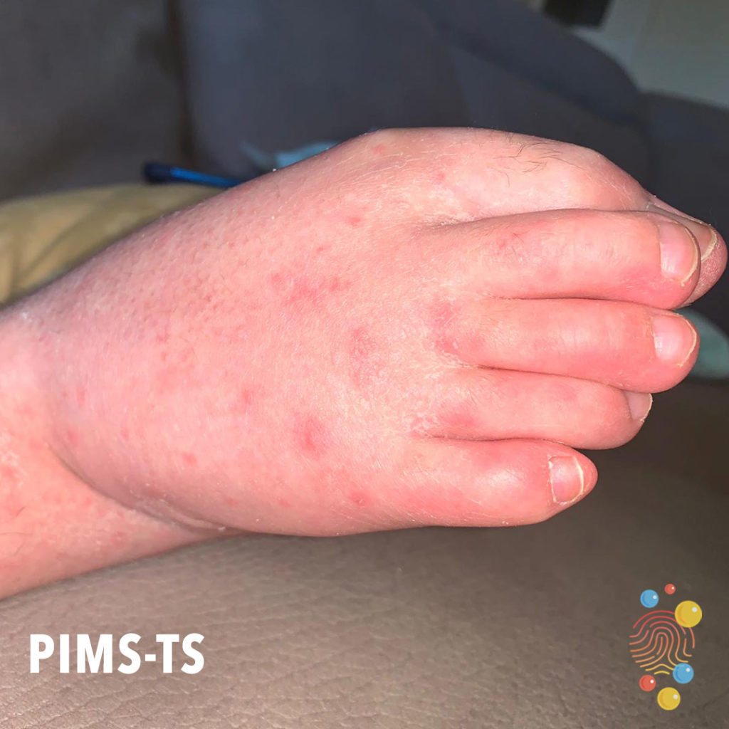
Erythematous papules on dorsa of both feet extending to the ankles.
Learn more about hand, foot and mouth
Urticarial Vasculitis
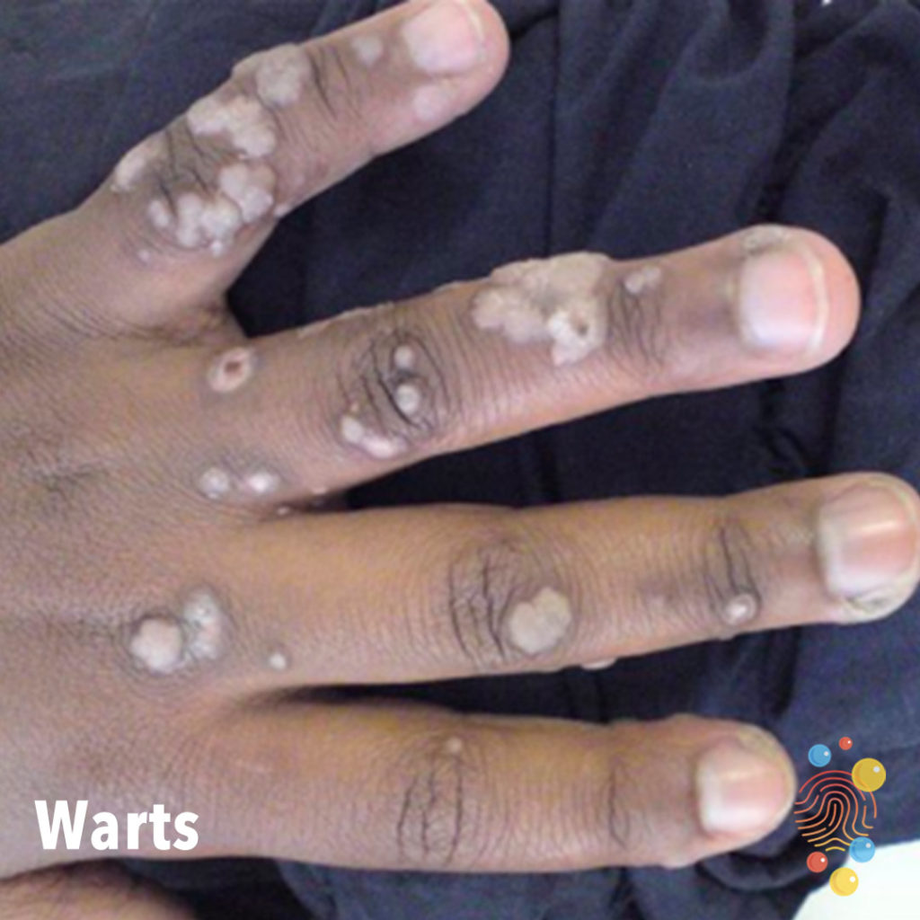
Rough verrucous flat-topped lesions on the dorsal fingers.
Learn more about warts
Crust on an erythematous base.
Learn more about chicken pox

Lichenified erythematous/violaceous plaque
Learn more about eczema

Swelling of left eyelid.
Learn more about periorbital oedemas
Dermal melanocytosis over the lower lumbar region. Scattered erythematous papules over back.
Learn more about dermal melanocytosis
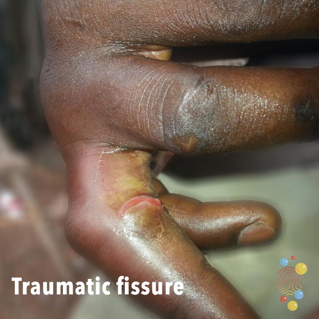
Pink superficial cracks between the linear fissures probably caused by injury or trauma.
Learn more about traumatic fissures
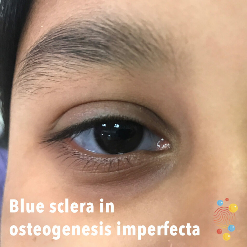
Blue sclera is associated with osteogenesis imperfecta.
Learn more about blue sclerae
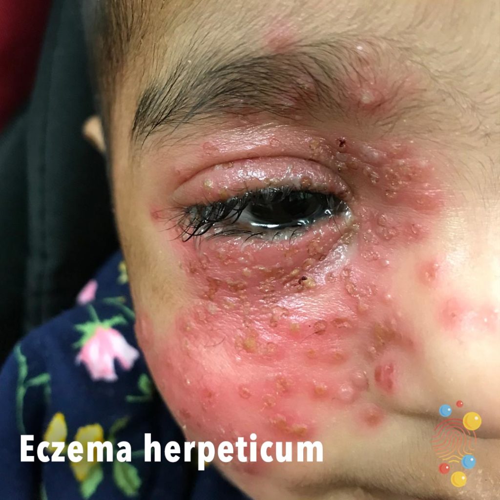
Multiple vesicles with erythema affecting the right cheek and periorbital area with purulent weeping from the right eye.
Learn more about eczema herpeticum

Chelitis of lips. The lesions on the chin are flat hypopigmented macules likely post-inflammatory.
Learn more about Kawasaki disease

Vesiculopustular eruption of lips with crust and ulceration.

Swelling to right side of face.
Learn more about abscesses
Patches of lichenified skin in the antecubital fossa with post-inflammatory hyperpigmentation.
Learn more about eczema
Blue discolouration affecting the leg.
Learn more about dermal melanocytosis

Multiple scattered crateriform nodules on trunk with surrounding erythema.
Learn more about leukaemia cutis
Scarlet Fever
Redness and swelling of upper eyelid with associated ptosis.
Learn more about chalazion

Multiple crusty erosions on the nose and chin with associated vesicles/blisters on the chin.
Learn more about bullous impetigo
Post-inflammatory hyperpigmentation.
Learn more about erythema nodosum
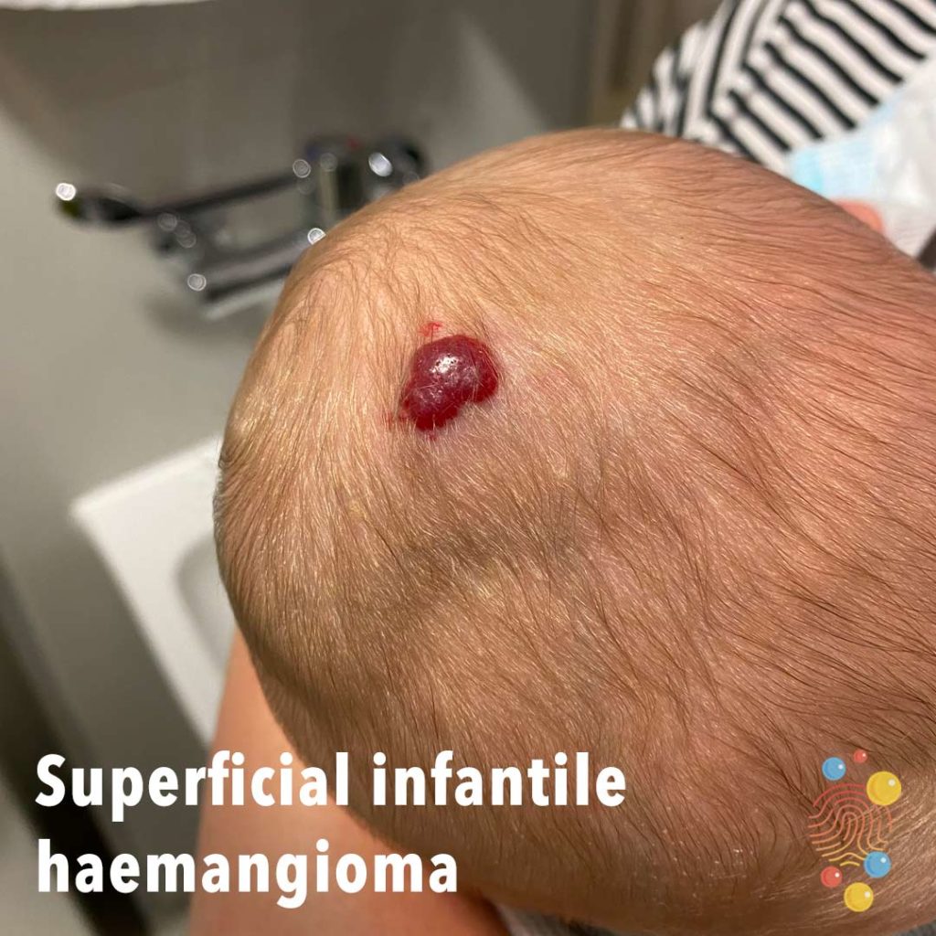
Raised bright red strawberry lesion with associated swelling of the scalp.
Learn more about haemangiomas
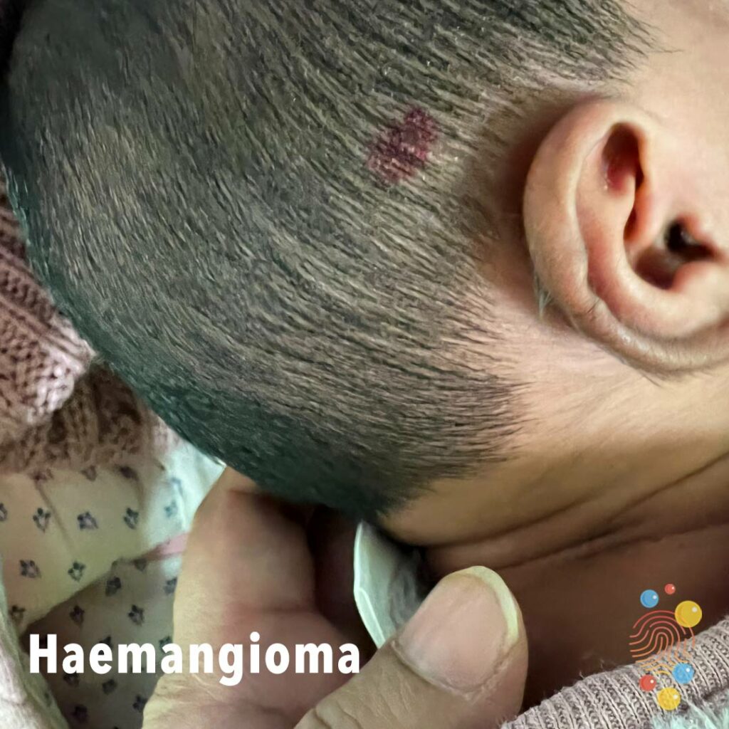
Haemangioma to scalp
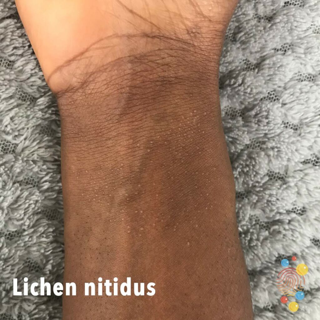
Multiple, discrete 1-2mm, hypopigmented papules over the flexor aspect of the wrist.
Learn more about lichen nitidus

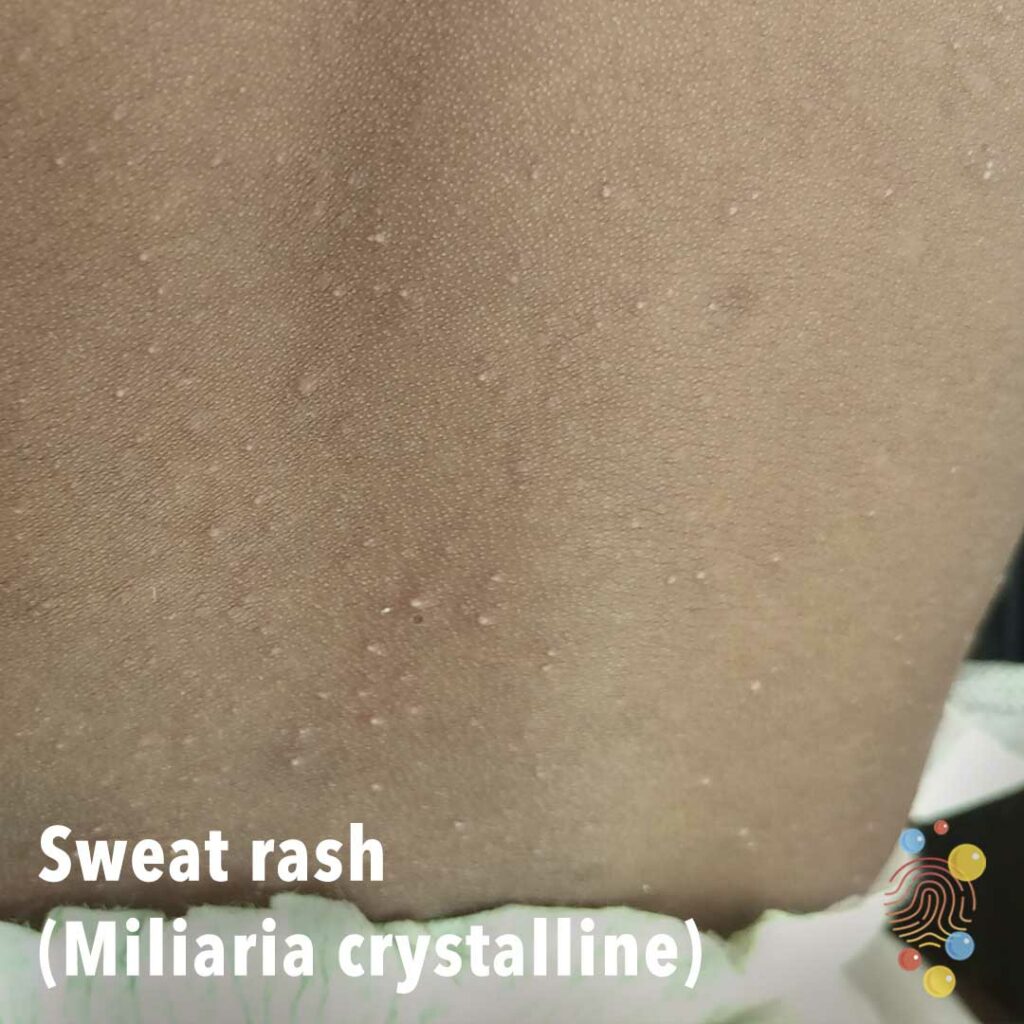
Multiple little tiny superficial sweat-filled blisters.
Learn more about miliaria
Multiple flaccid bullae with erosions on upper limb.
Inflammed lesion with central punctum and pustular eruption surrounding this. Background erythema.
Learn more about bites
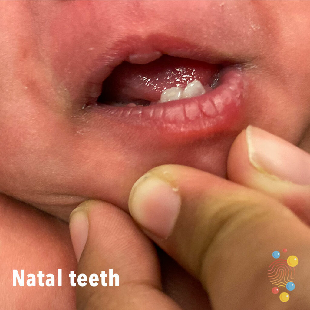
Mature natal teeth nearly or fully developed.
Learn more about natal teeth
Infected round ulcer with well-defined borders.
Learn more about ecthymas
Normal Bruising Pattern

Mouth injury with impacted tooth.

Multiple papulopustules with early punched-out erosions.
Learn more about eczema herpeticum

Grouped vesicles over knee.
Learn more about bites
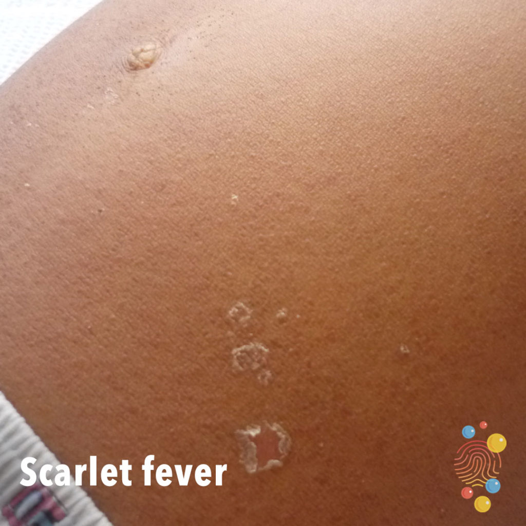
Sandpaper-like popular eruption with desquamation.
Learn more about scarlet fever

Hyperpigmented patch with raised border and evidence of scar from previous surgery.
Learn more about haemangiomas
Scaly hypopigmented patches on posterior neck.
Learn more about pityriasis versicolor
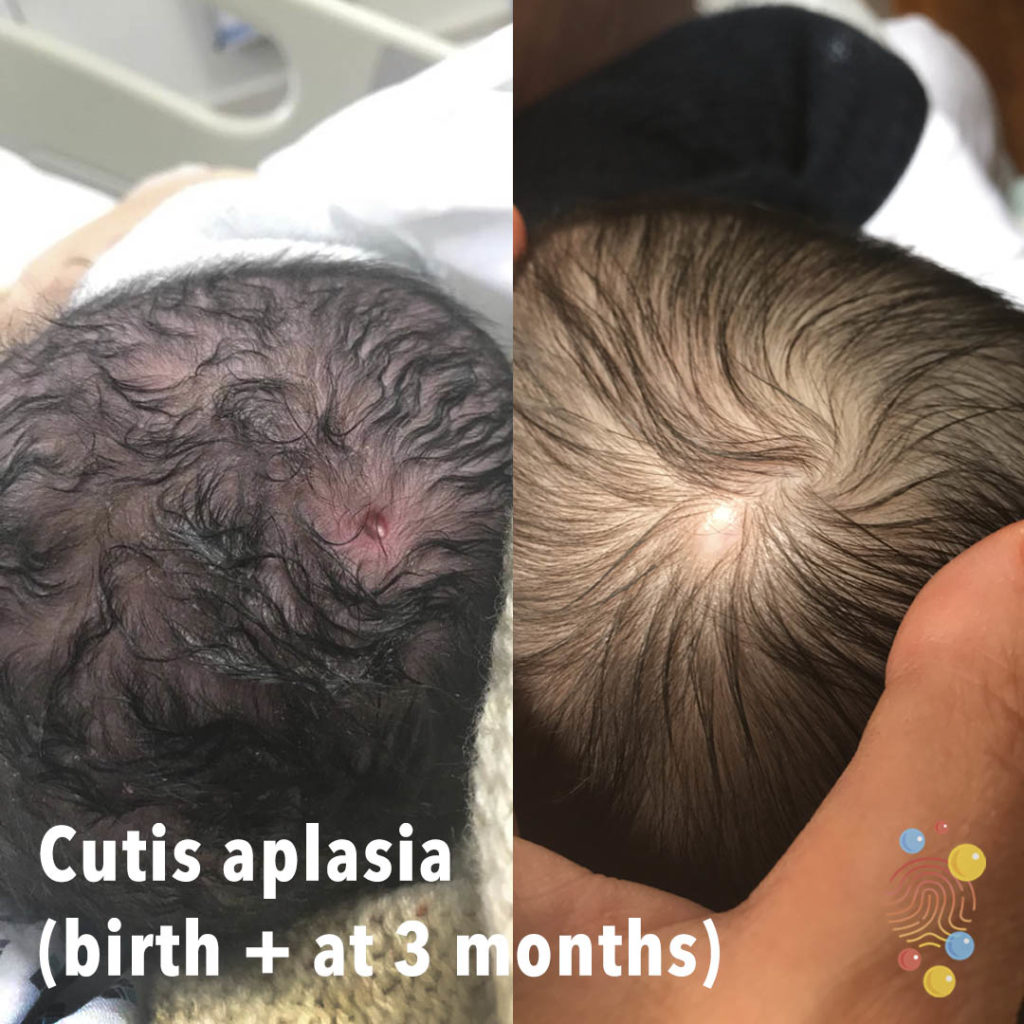
Oval membranised nodule lateral to midline of scalp at occipital hair whorl. Look for a ‘hair collar sign’. Heals with scarring.
Learn more about cutis aplasia
Chalazion
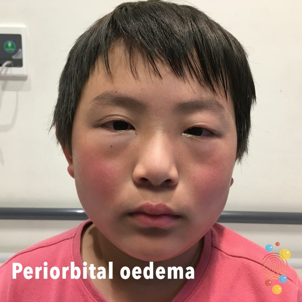
Swelling of the left upper eyelid and erythema of the cheeks bilaterally.
Learn more about periorbital oedema
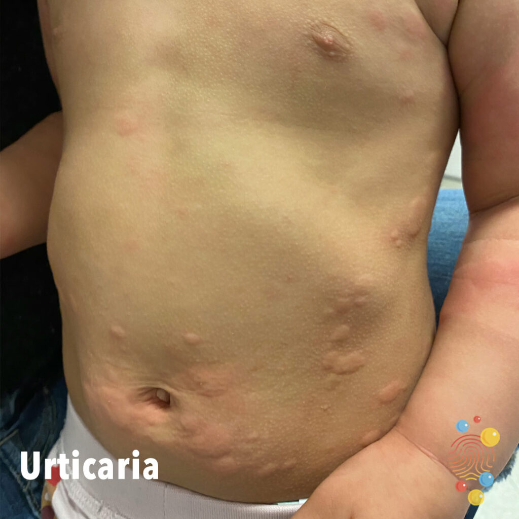
Multiple discrete /coalescing wheals over torso and abdomen.
Learn more about urticaria

Injected conjunctiva with some erythema.
Learn more about conjunctivitis
Pustular lesions on the scalp.
Learn more about folliculitis

Islands of repigmentation from patches of vitiligo on anterior shins.
Learn more about vitiligo

Extensive healing erosions with haemorrhagic crust and a collarette of scale

Eruption of dark red macules, vesicles, and erosions distributed across areas previously affected by atopic dermatitis, with relative sparing of the trunk
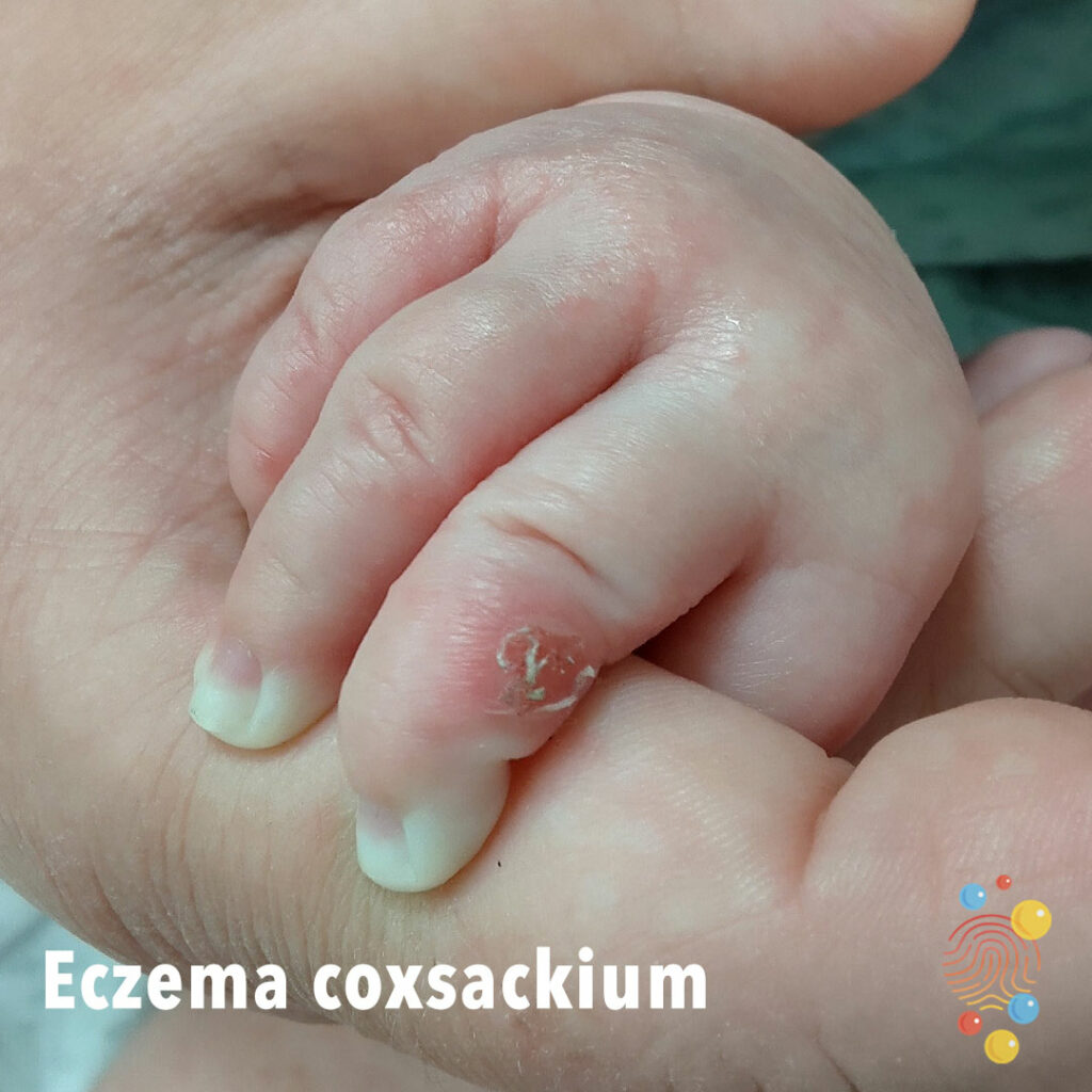
Erythematous lesions with blisters, erosions, + crusting on dorsal hands and perioral area.
Learn more about eczema coxsackium

Swelling and redness of the eyelid and surrounding soft tissues.
Learn more about cellulitis
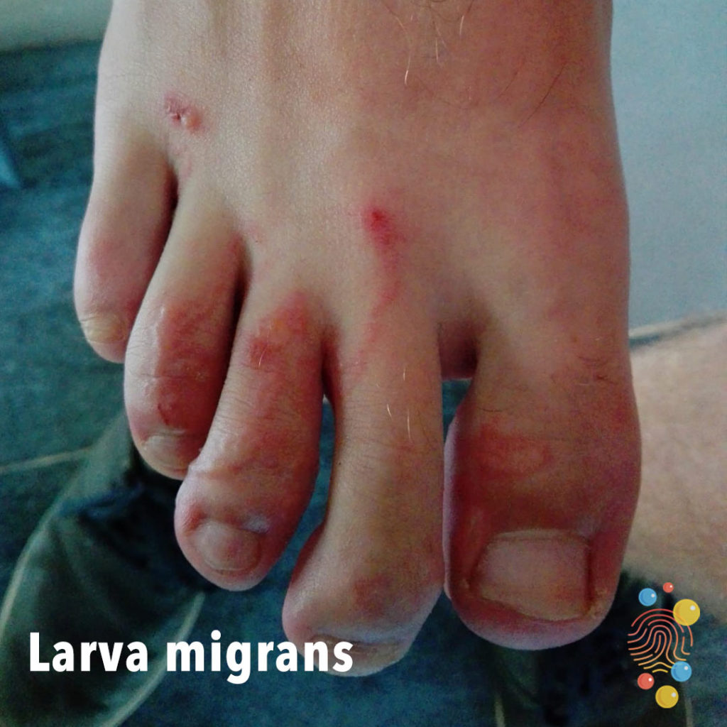
Multiple erythematous linear/actuate lines on toes and dorsal foot.
Learn more about larva migrans
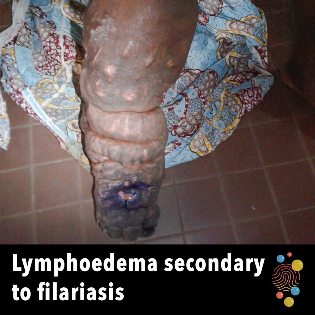
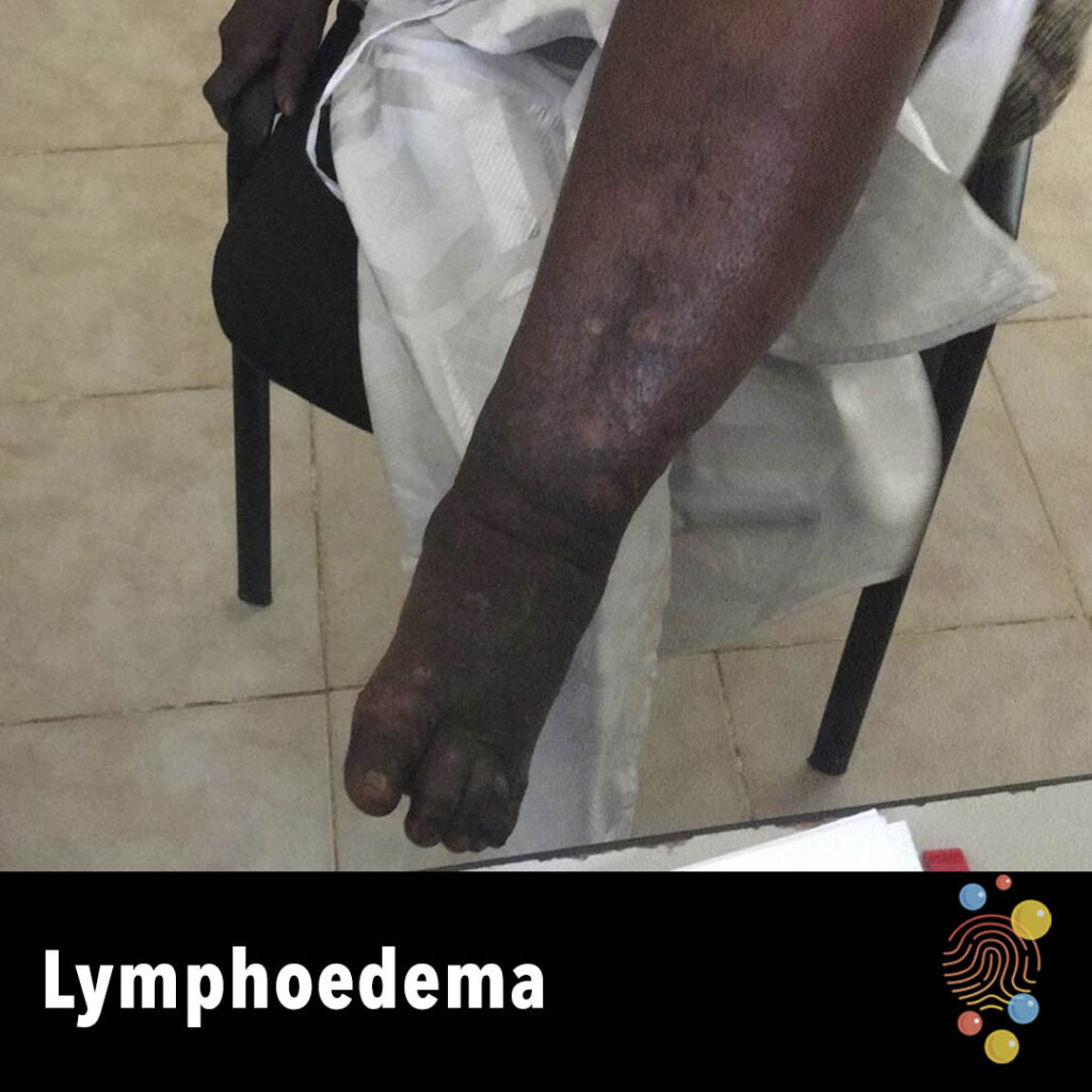
Lymphoedema with thickening of the skin (elephantiasis) and associated ulceration and hyperpigmentation.
Learn more about lymphoedema
Bruise to shin
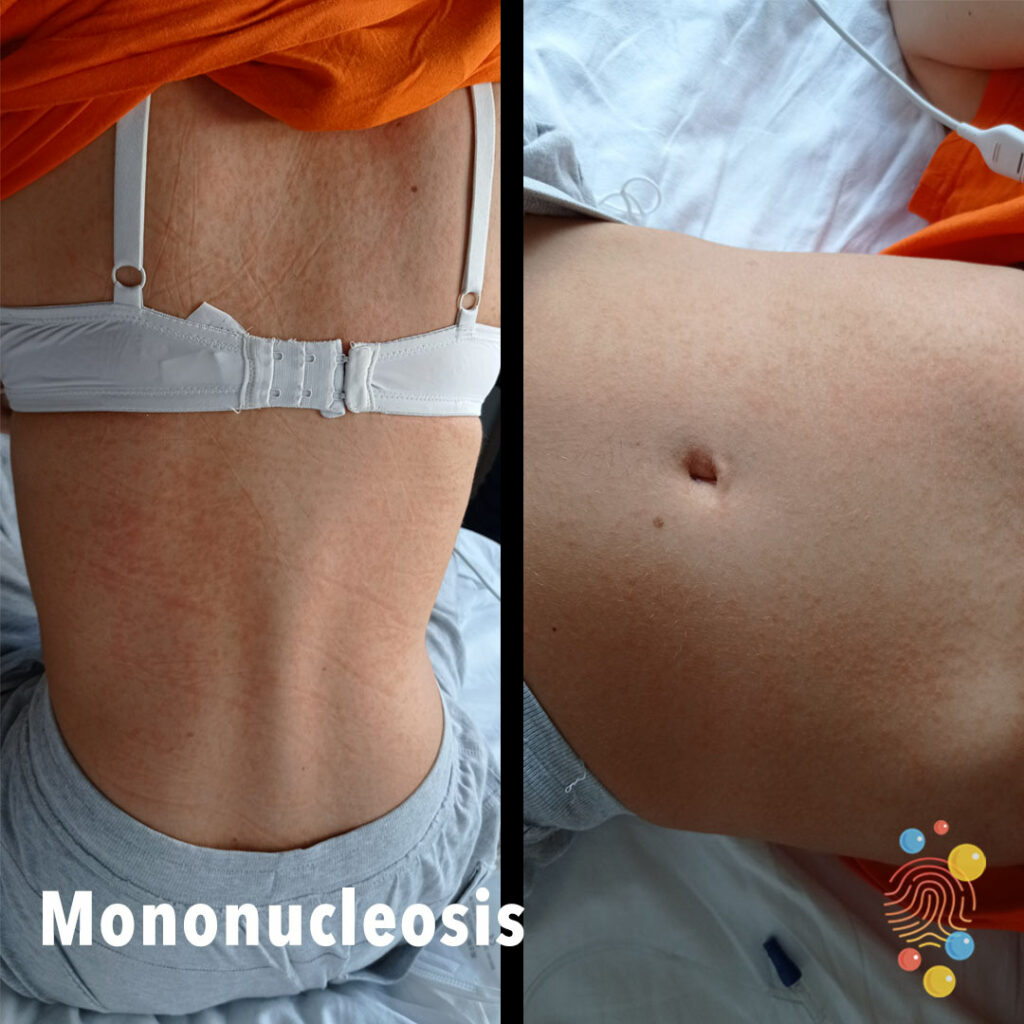
Widespread erythematous maculopapular rash.
Learn more about infectious mononucleosis

Papular eruption with erythematous base.
Learn more about viral exanthem
Moist confluently erythematous inflamed area with pustules.
Learn more about eczema
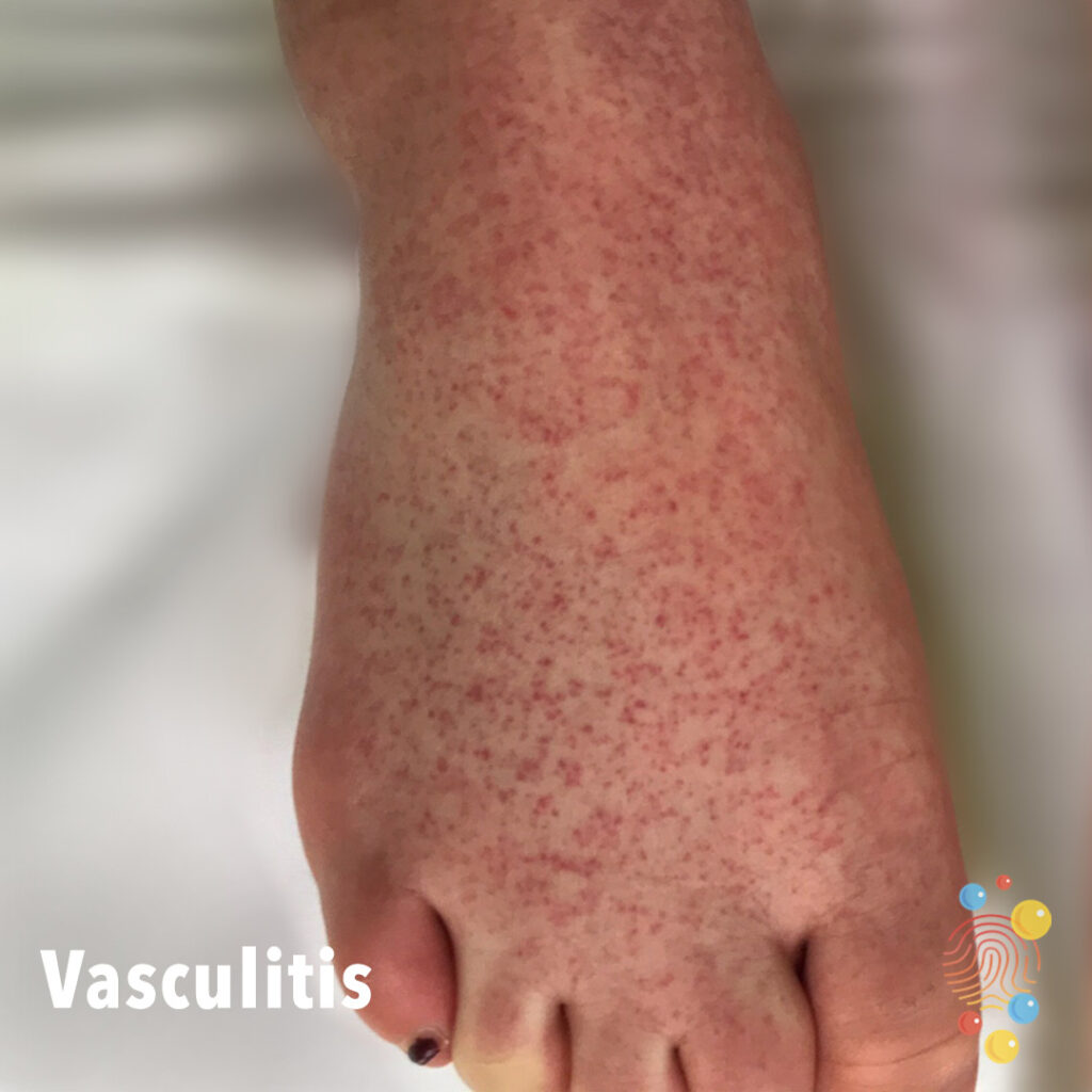
Multiple red petechiae across the dorsum of the foot.
Learn more about vasculitis

Blister following Mantoux.
Learn more about the Mantoux test
Discoid plaque with crusting, erosions and ooze.
Learn more about eczema
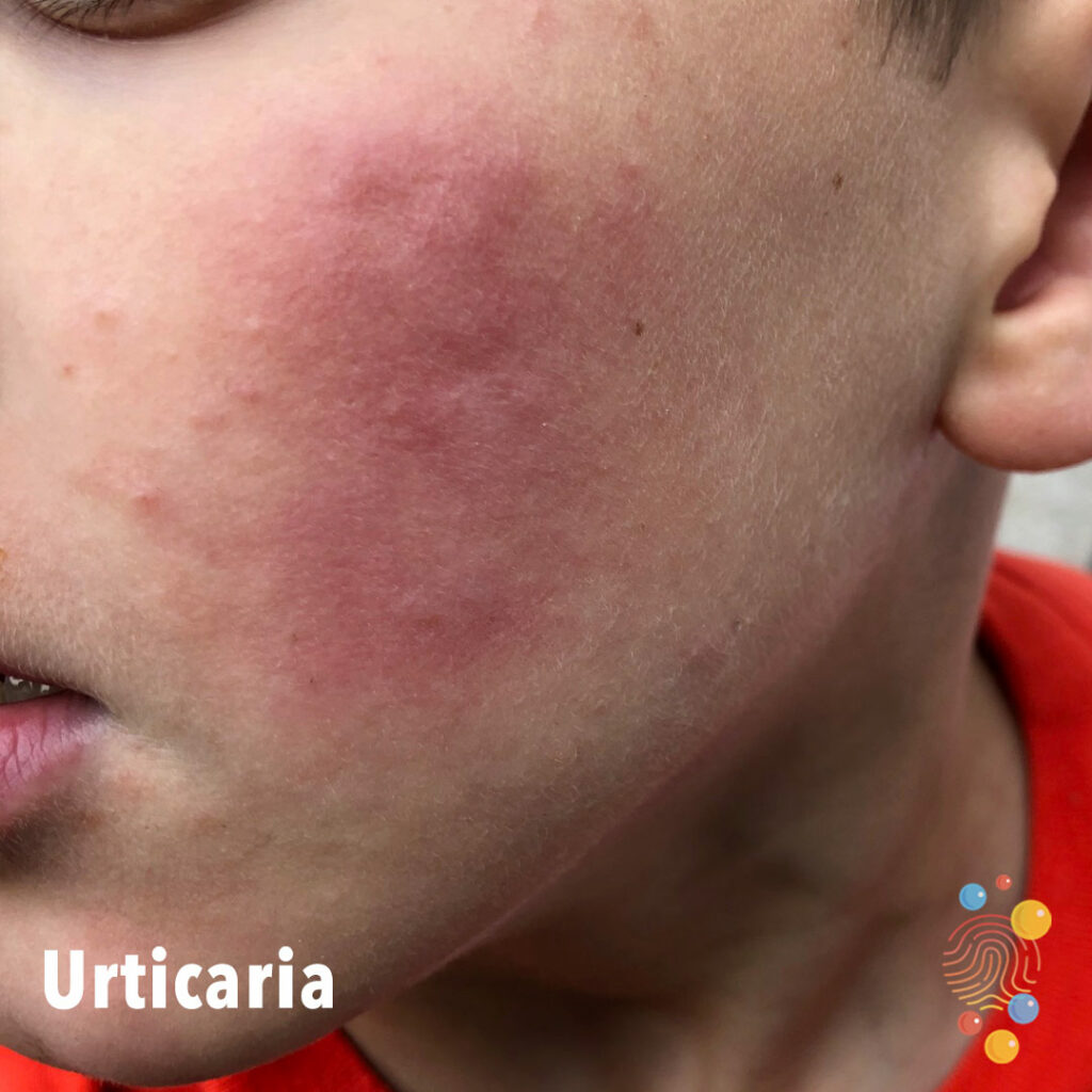
Wheal on cheek (with incidental papules suggesting mild acne).
Learn more about urticaria
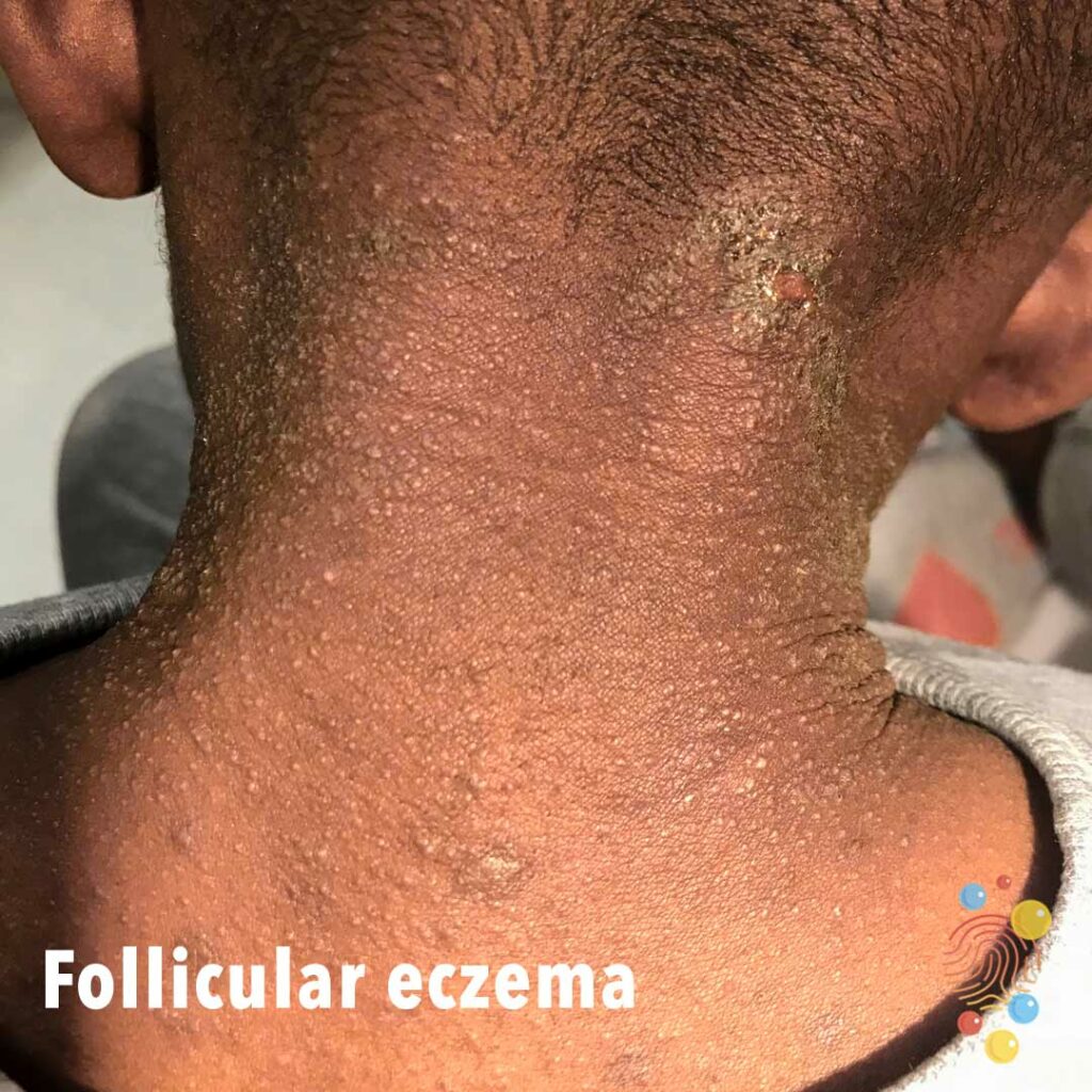
Erythematous papules with surrounding hazy erythema and follicular hyperkeratosis.

Extensive desquamation on back post scarlet fever.

Erythematous maculopapular eruption on trunk.
Learn more about viral exanthem

Skin-coloured protrusion emerging from the umbilicus.
Learn more about umbilical hernias
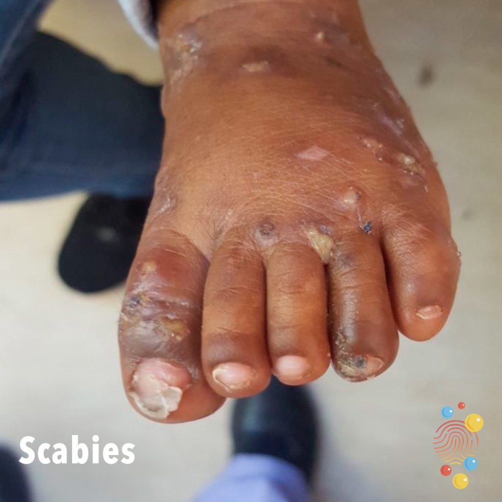
Excoriated papules on dorsal foot, with some pustules.
Learn more about scabies
Generalised erythematous scaly rash on the back.
Learn more about eczema

2 week old with paronychia
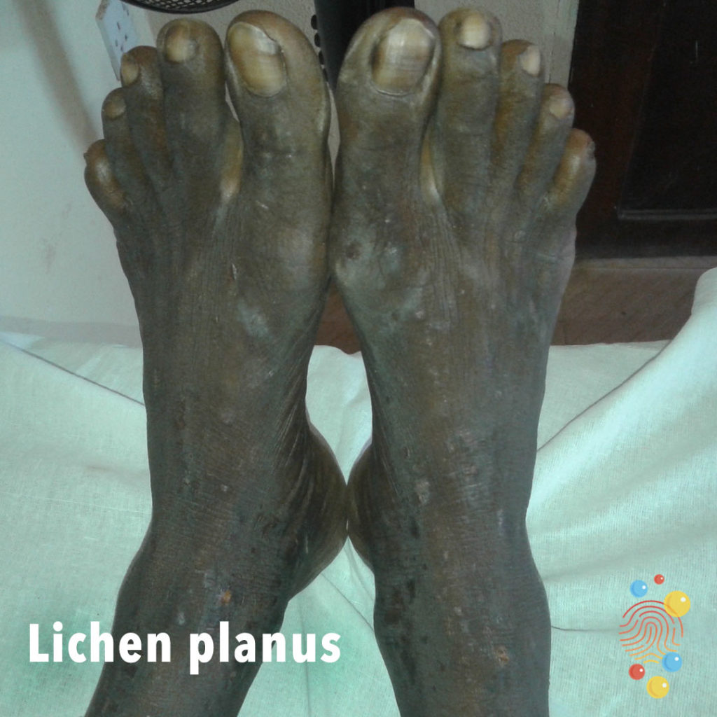
Multiple polygonal yellow papules and plaques on his dorsal area of the feet.
Learn more about lichen planus
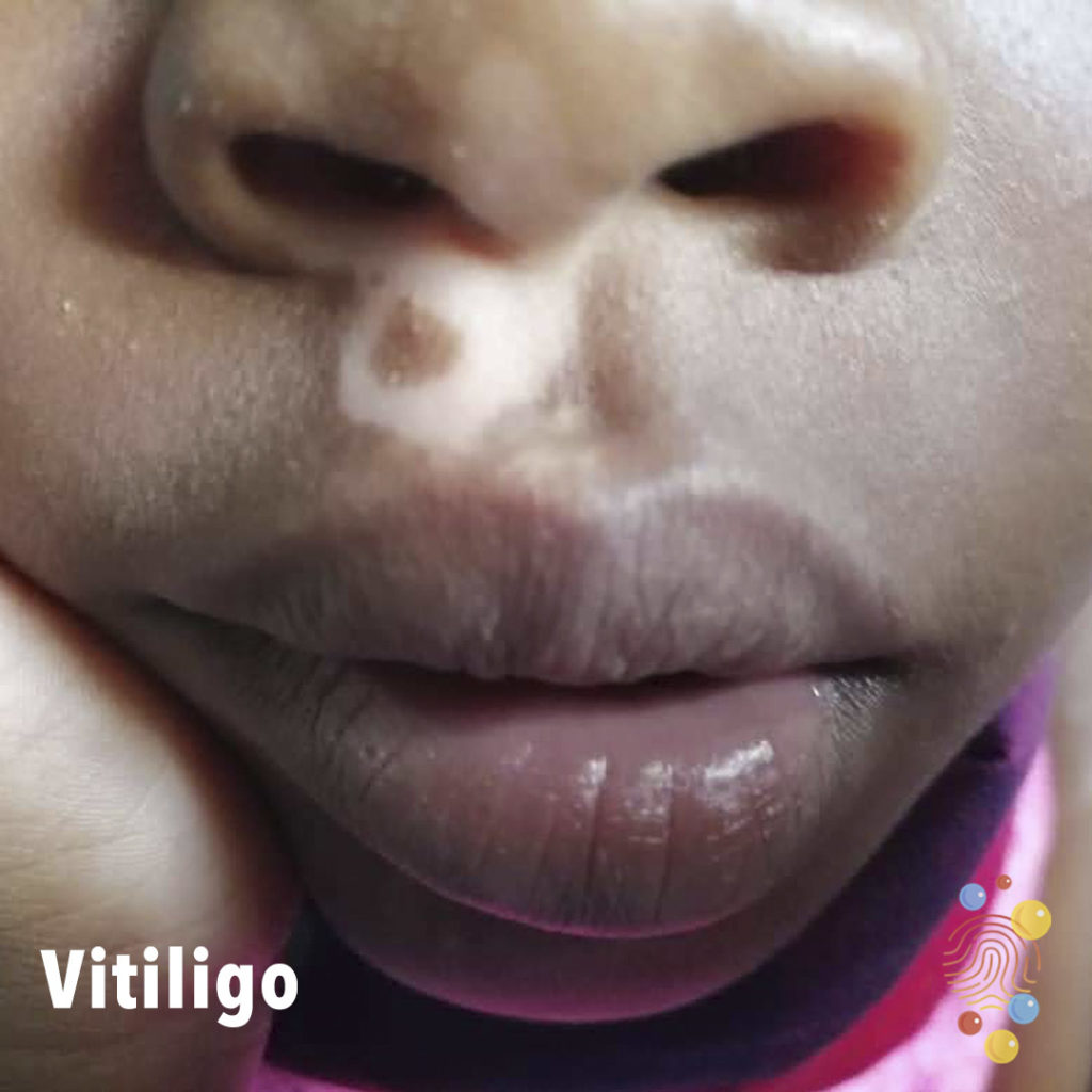
White patch of depigmentation in annular shape with island of sparing centrally on right upper lip.
Learn more about vitiligo

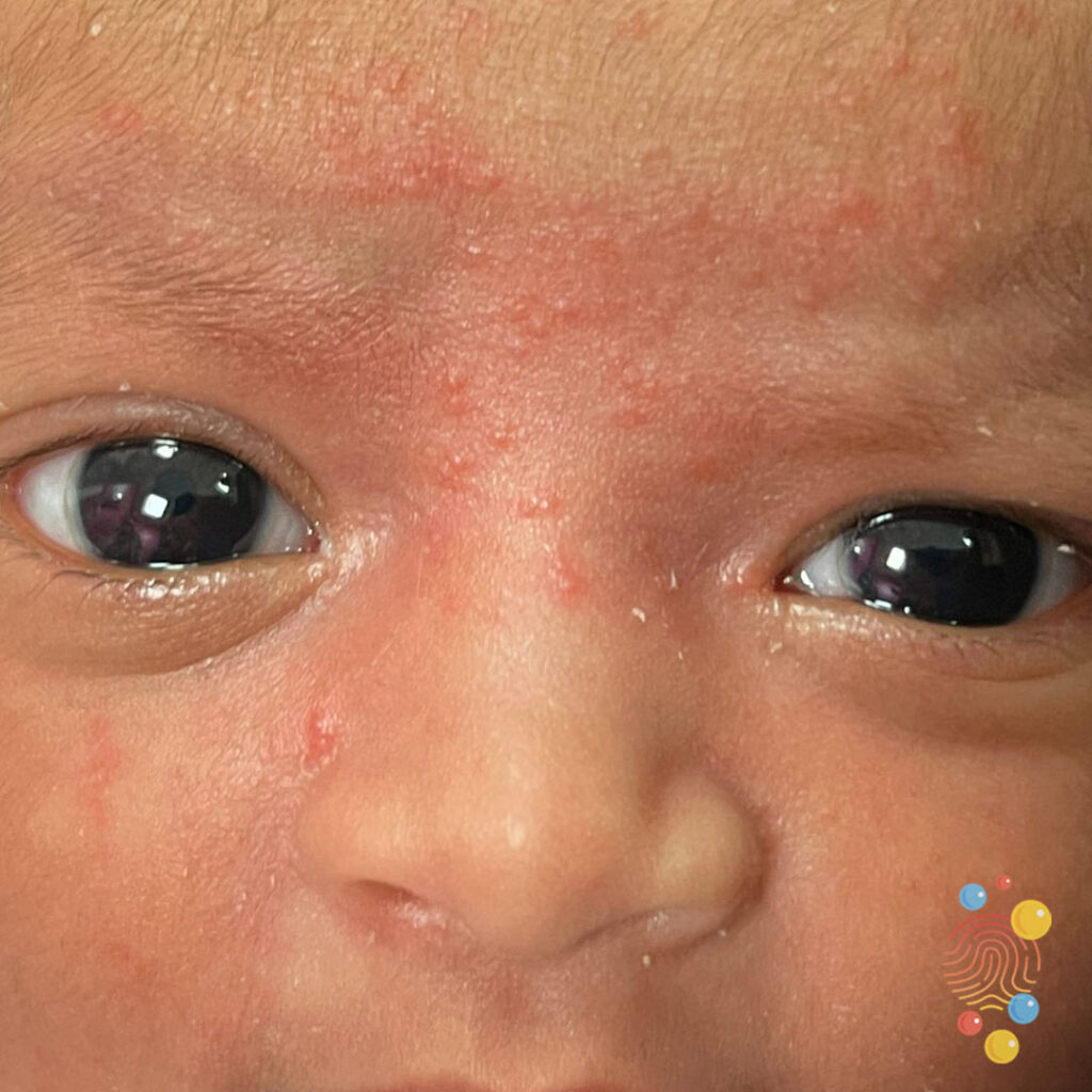
Erythematous rash forehead interspersed with pinpoint papules in a young infant
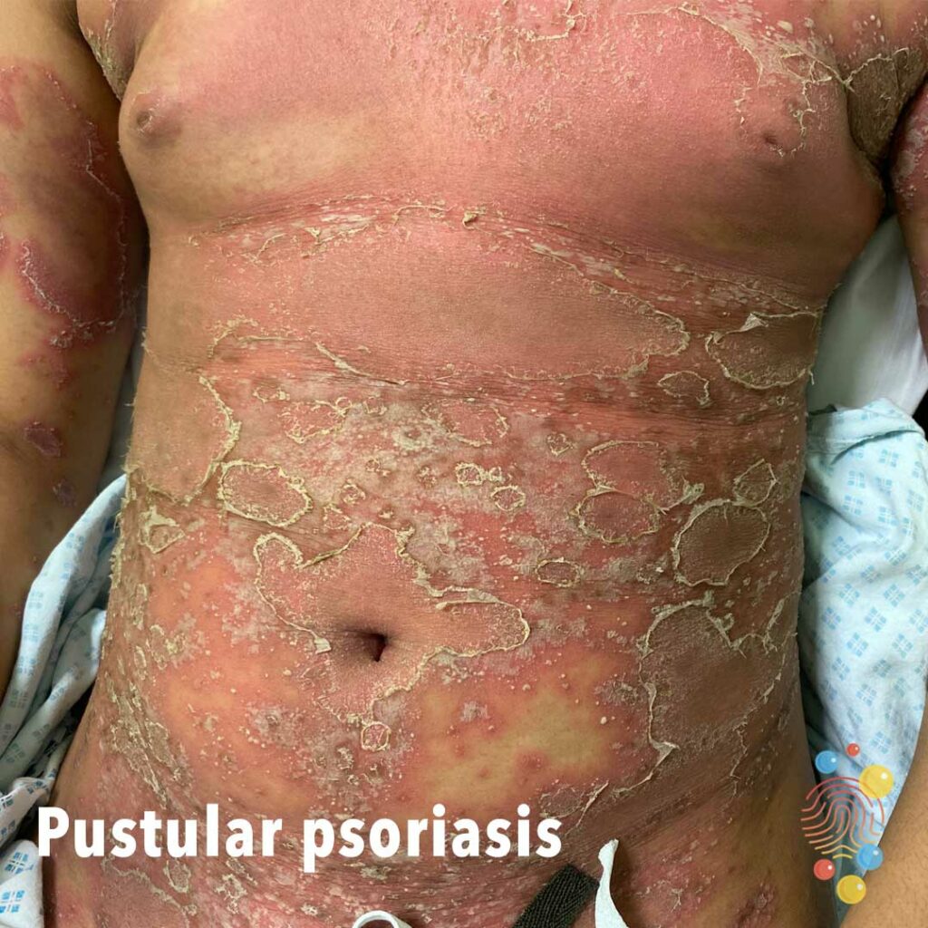
Sheets of pustules on abdomen with generalised erythema and desquamation of skin.
Learn more about psoriasis

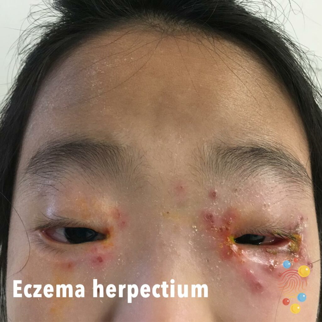
Crops of peri-ocular pustules across both eyes. Background erythema and some excoriation.
Learn more about eczema herpeticum
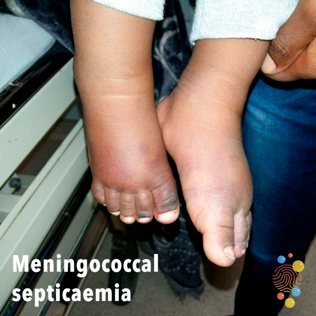
Irregular purpura of distal feet.
Learn more about meningococcal septicaemia
Scarlet Fever
Well circumscribed violaceous umbilical plaque.

Extensive wheals affecting the upper chest and neck. Areas of sparing throughout and irregular borders.
Learn more about urticaria
Follicular based erythematous papules.
Learn more about folliculitis
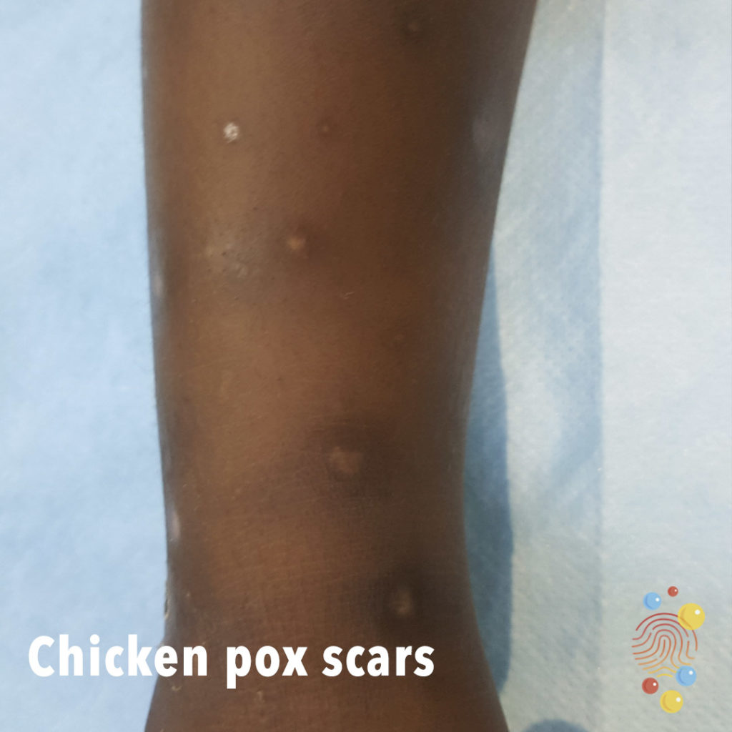
Atrophic and hypo and hyperpigmented round scars.
Learn more about chicken pox

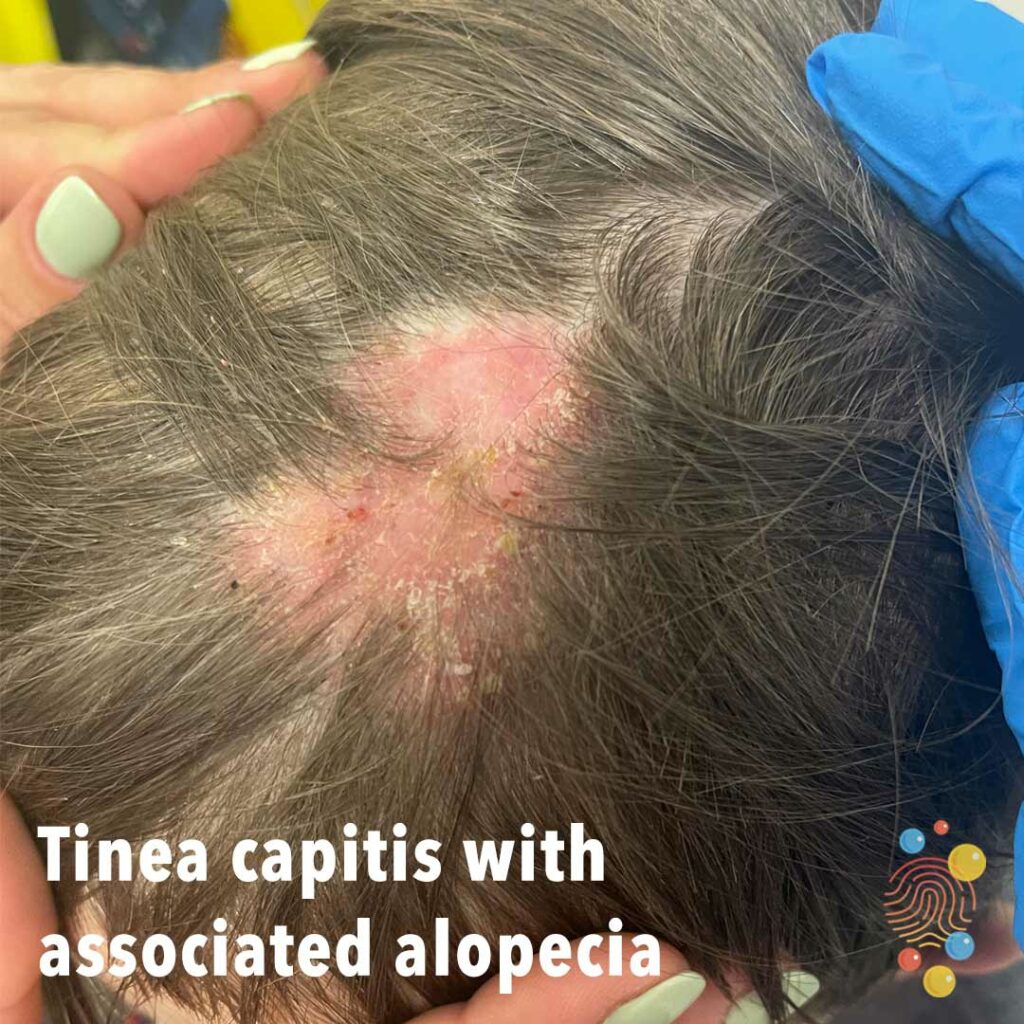
Tinea capitis with associated alopecia
Hyperpigmented annular lesions, featuring erythema and some slight scale.
Learn more about eczema

Annular erythematous wheals.
Learn more about urticaria
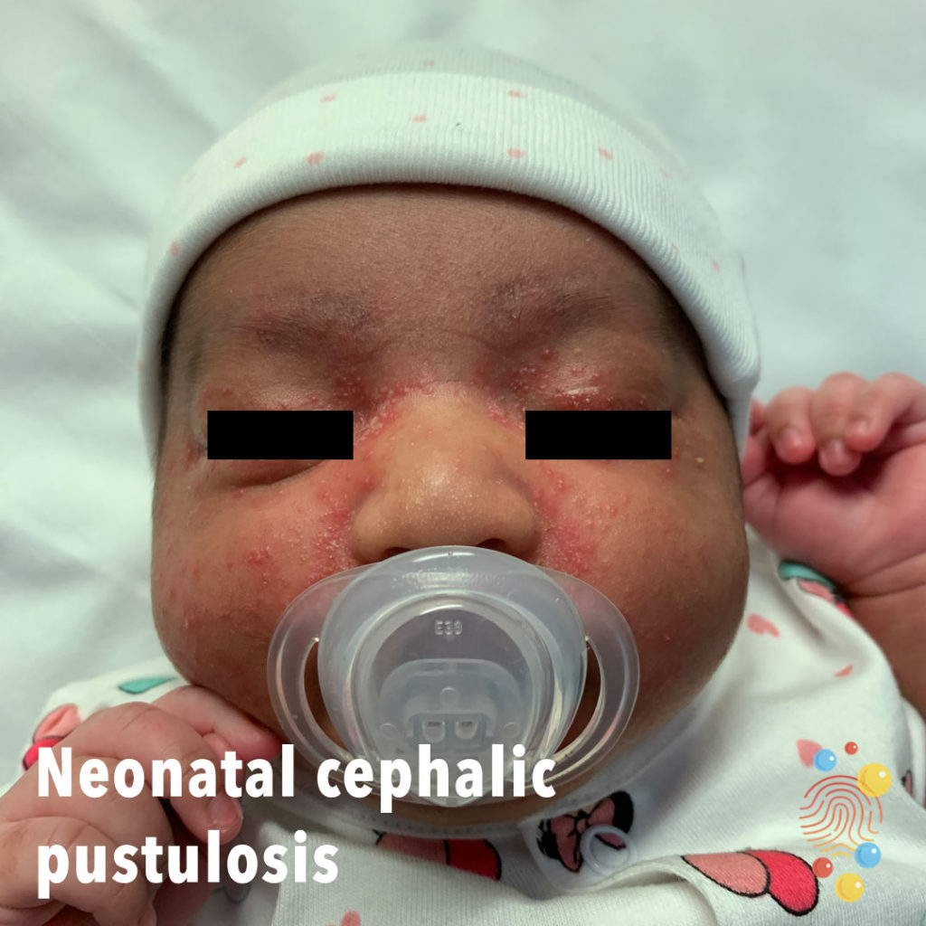
Pustules with erythematous base.
Learn more about neonatal cephalic pustulosis
Multiple little pus-filled pustules with erythematous halo.
Learn more about folliculitis

Mouth Injury
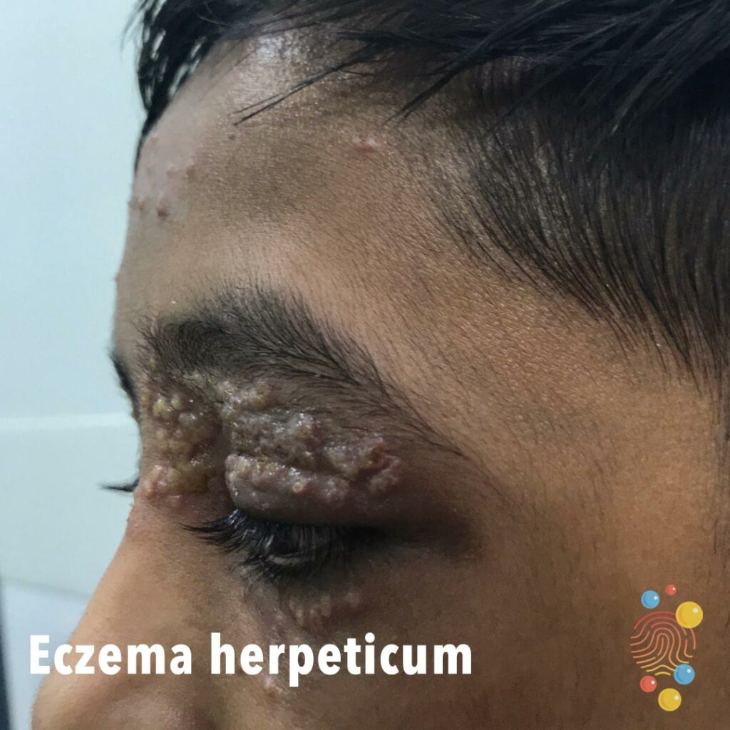
Periorbital vesiculation with hyperpigmentation left side of face.
Learn more about eczema herpeticum
Widespread erythema with generalised lichenification of the flanks generalised. No evidence of secondary infection.
Learn more about eczema
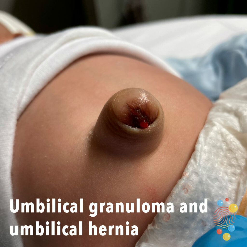
Bulge at the site of the umbilicus with a soft red mass on top of the skin.
Learn more about umbilical hernias

Eruption of dark red macules, vesicles, and erosions distributed across areas previously affected by atopic dermatitis, with relative sparing of the trunk

Impetiginized Eczema
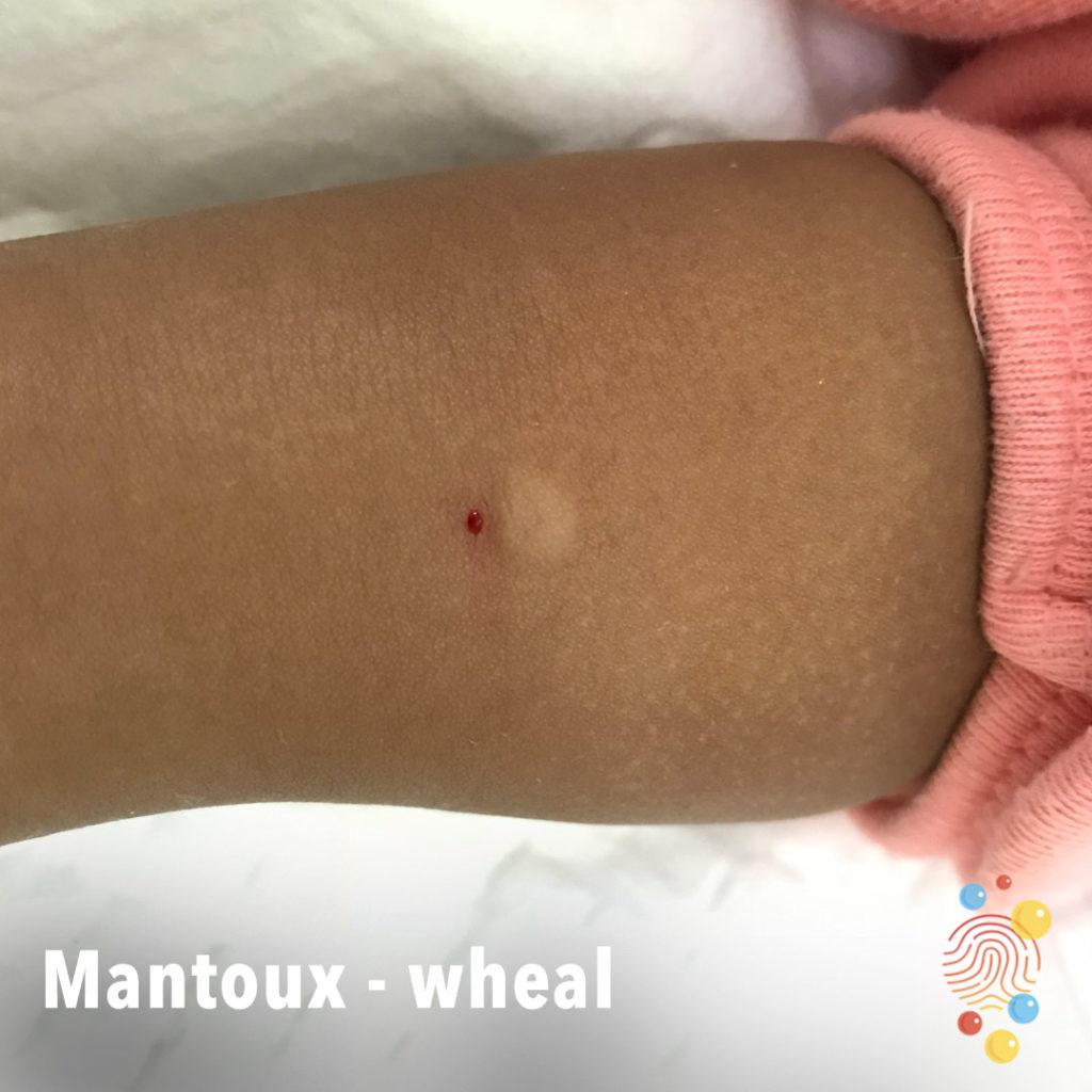
Wheal demonstrated next to intradermal injection site.
Learn more about the Mantoux test

Extensive scale and eczematous patches affecting the upper chest and face with dyspigmentation.
Learn more about eczema
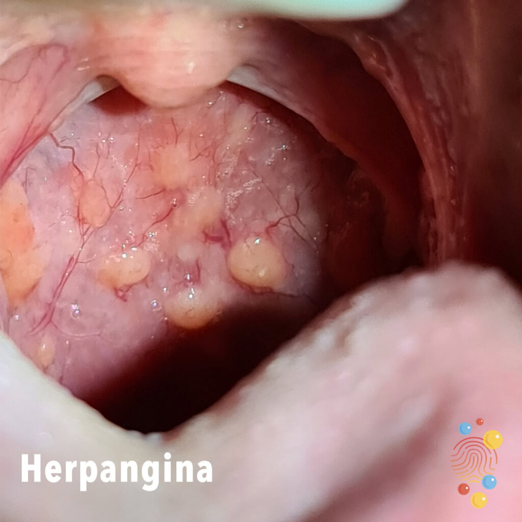
Multiple blisters, yellowish coloured, on mouth mucosa.
Learn more about herpangina

Single erythematous lesion. Well defined, symmetrical and uniform in colour. Likely vascular given colour with ‘strawberry-like’ appearance.
Learn more about haemangiomas
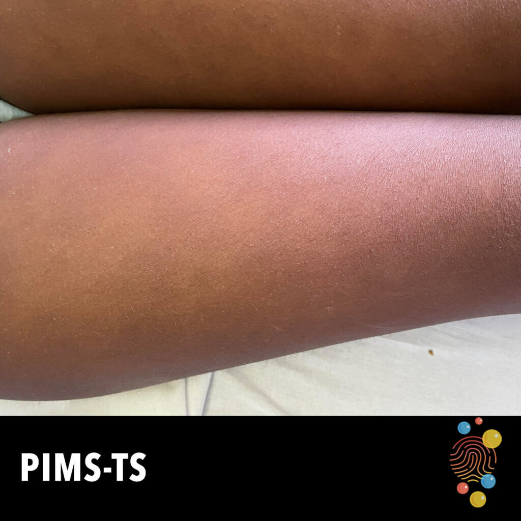
Ill-defined symmetrically distributed erythema on the lower limbs.
Learn more about PIMS-TS
Erythematous macules and papules with some crusting and scale.
Learn more about eczema
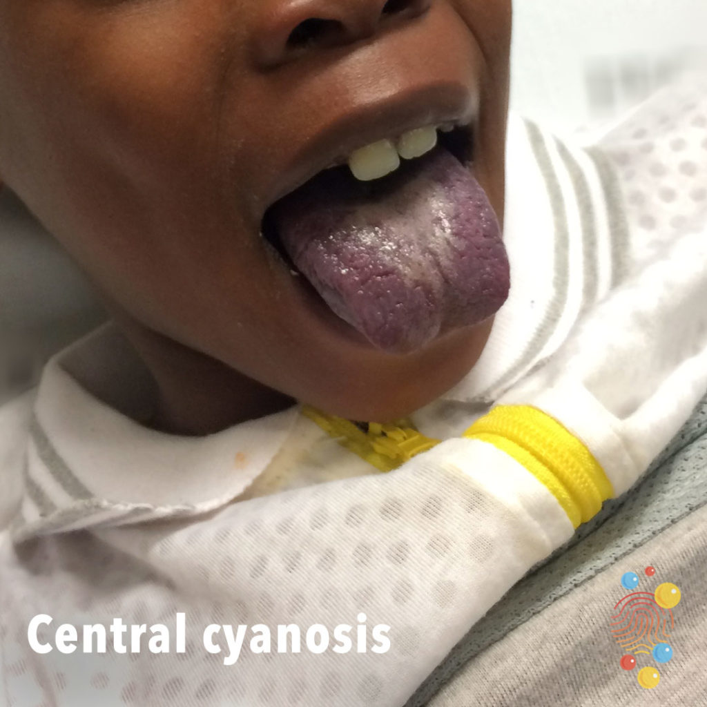
Blue coloured tongue and lips.
Learn more about central cyanosis

Well defined, erythematous, scaling rash, with peeling over the second left toe extending to the first interdigital space.
Learn more about staphylococcal infection

Erythematous area of leg.
Learn more about cellulitis
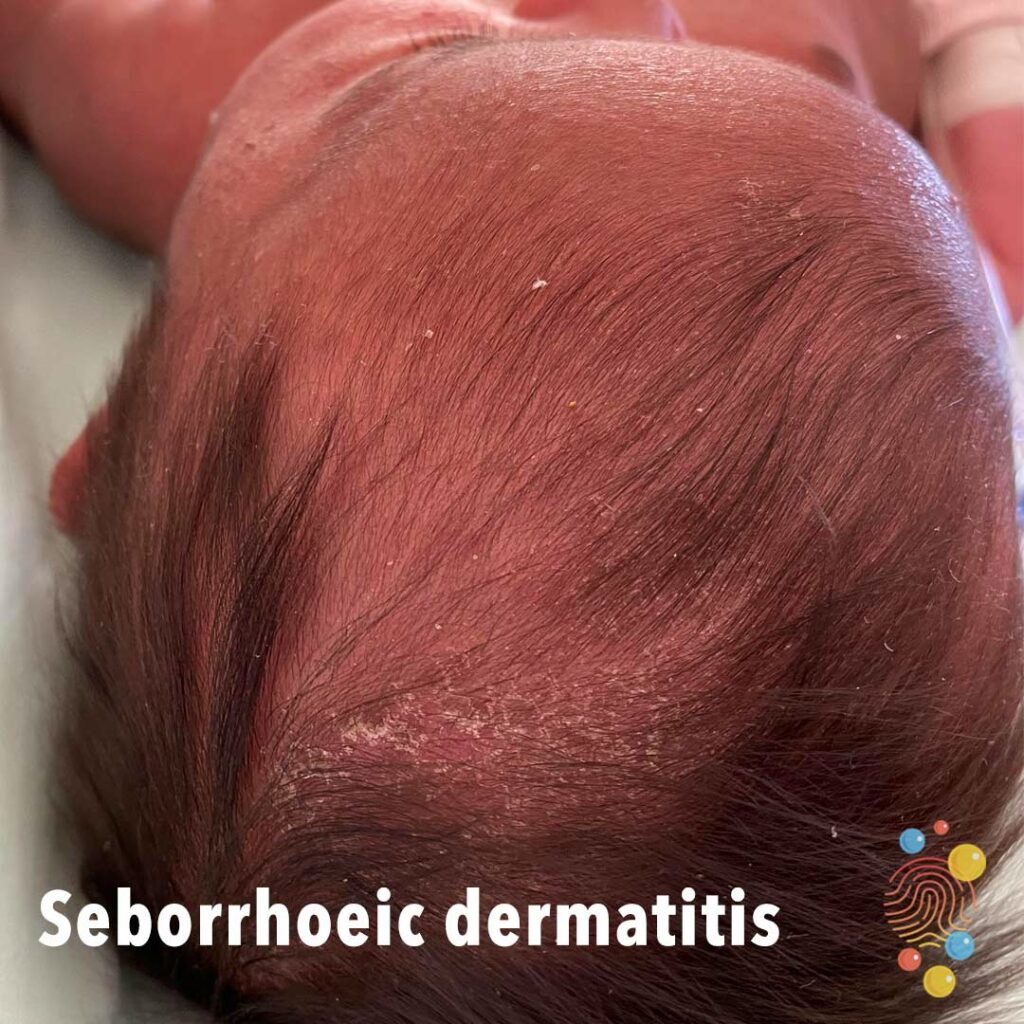
Erythema and adherent scale over scalp.
Learn more about seborrhoeic dermatitis
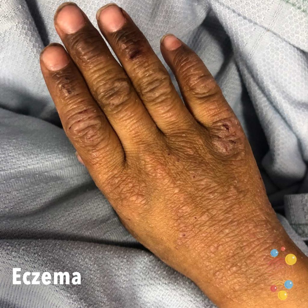
Extensive lichenification of the dorsal fingers, hand, and wrist, with excoriations and erosion.
Learn more about eczema

Post-immunisations (12 month imms)

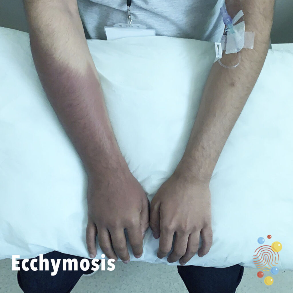
Unilateral, well-defined, purplish discolouration of skin extending from elbow to dorsum of right hand.
Learn more about ecchymosis

Excoriated erythematous micro papules on a background of diffuse erythema.
Learn more about scabies
Raised scaly patches and plaques.
Learn more about psoriasis
Papular change on the cheeks, eyebrows and forehead with no pustule formation.
Learn more about eczema
Erythema and scaling of the cheeks and forehead.
Learn more about eczema
Erythema with lichenification, scale, and excoriations.
Learn more about eczema
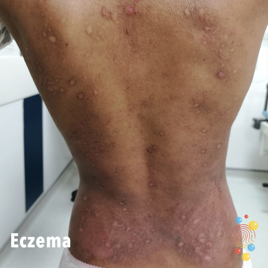
Scaly inflamed patches on the back with excoriations and post-inflammatory hyperpigmentation.
Learn more about eczema
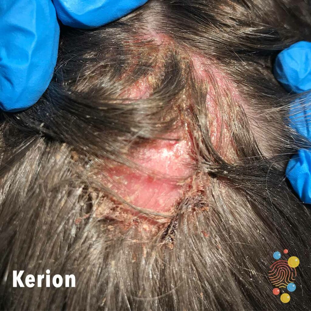
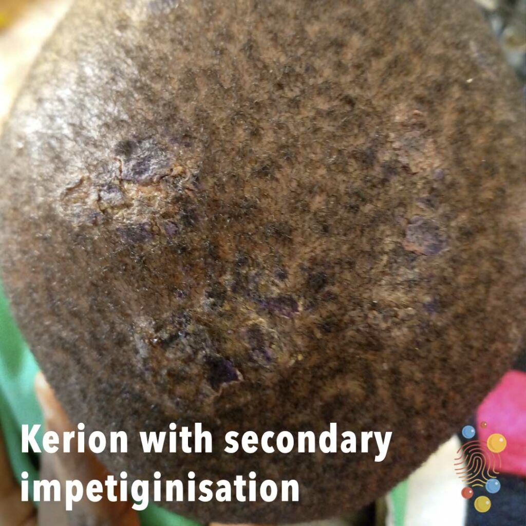
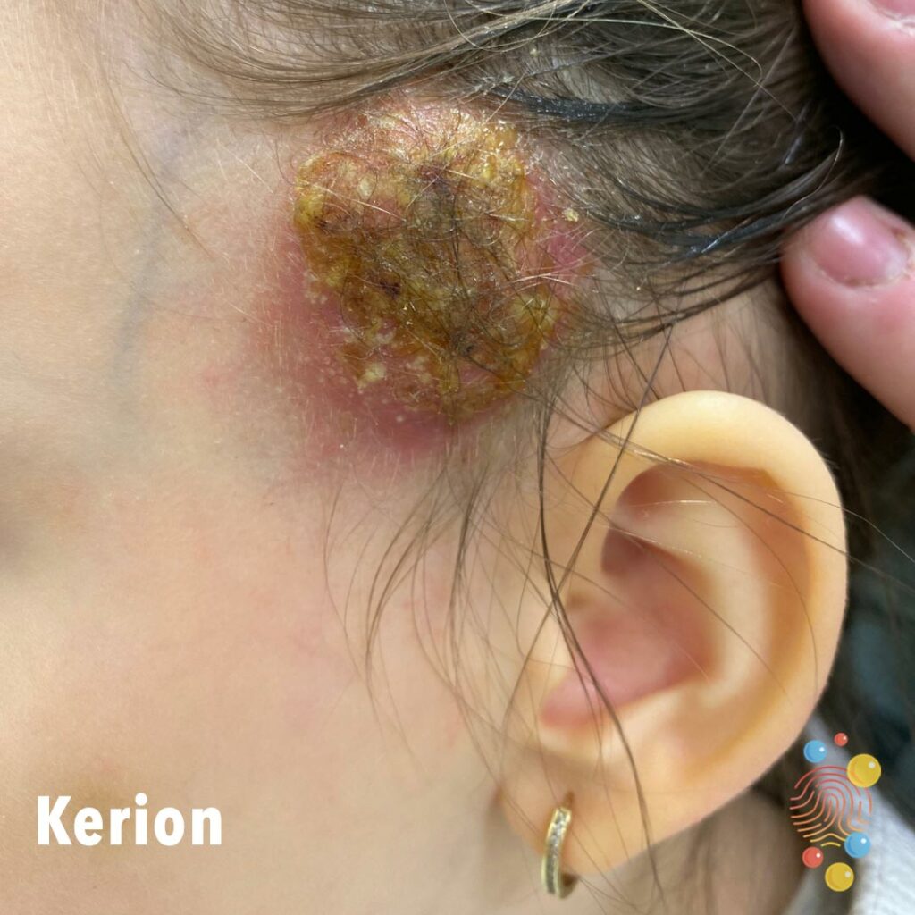
Inflamed boggy mass on scalp with associated hair loss and crusting.
Learn more about kerions
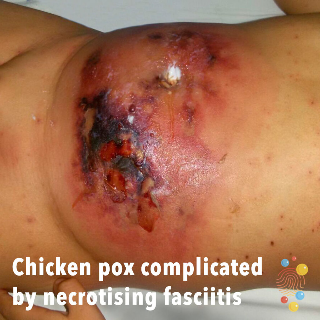
Extensive ulceration and eschar affecting lower abdomen with surrounding erythema.
Learn more about chicken pox

Areas of sloughing of superficial skin and pale, white, leathery appearance suggest possible third degree burn with surrounding blister formation.
Learn more about bullous impetigo
Widespread red, blotchy rash on abdomen.
Learn more about urticaria
Dusky atypical target lesions with blisters and erosions.
Learn more about toxic epidermal necrylosis

The gastrostomy tract is healing. The scar at the medial side of the tract and the vertical lines suggest that this may have been a surgically inserted gastrostomy rather than a true PEG (percutaneous, endoscopic gastrostomy).
There is some redness around the tract and a small amount of discolouration on the dressing which suggests there might be some leakage or maybe infection.
The tube appears to be a ‘PEG tube’ (e.g. a Freak PEG tube) which is held in place by a plastic disc in the stomach and a plastic retainer on the outside which can be seen under the dressing. It is important that once the tract has healed these tubes are ‘exercised’ every week – the outside retainer is released, the tube is pushed in several (4-5) cm and then drawn back until snug and the retainer is re-secured. This helps prevent a buried bumper – where the stomach lining grows over the plastic disc on the inside and often needs to be retrieved with an open operation.
Learn more about gastrostomies

Non-blanching palpable purpura of upper limbs.
Learn more about Henoch-Schonlein purpura
Scaly adherent plaques on scalp.
Learn more about seborrhoeic dermatitis
Flat blue grey birthmark on the lower spine.
Learn more about dermal melanocytosis

Erythematous maculopapular eruption with sparing of vermillion.
Learn more about eczema
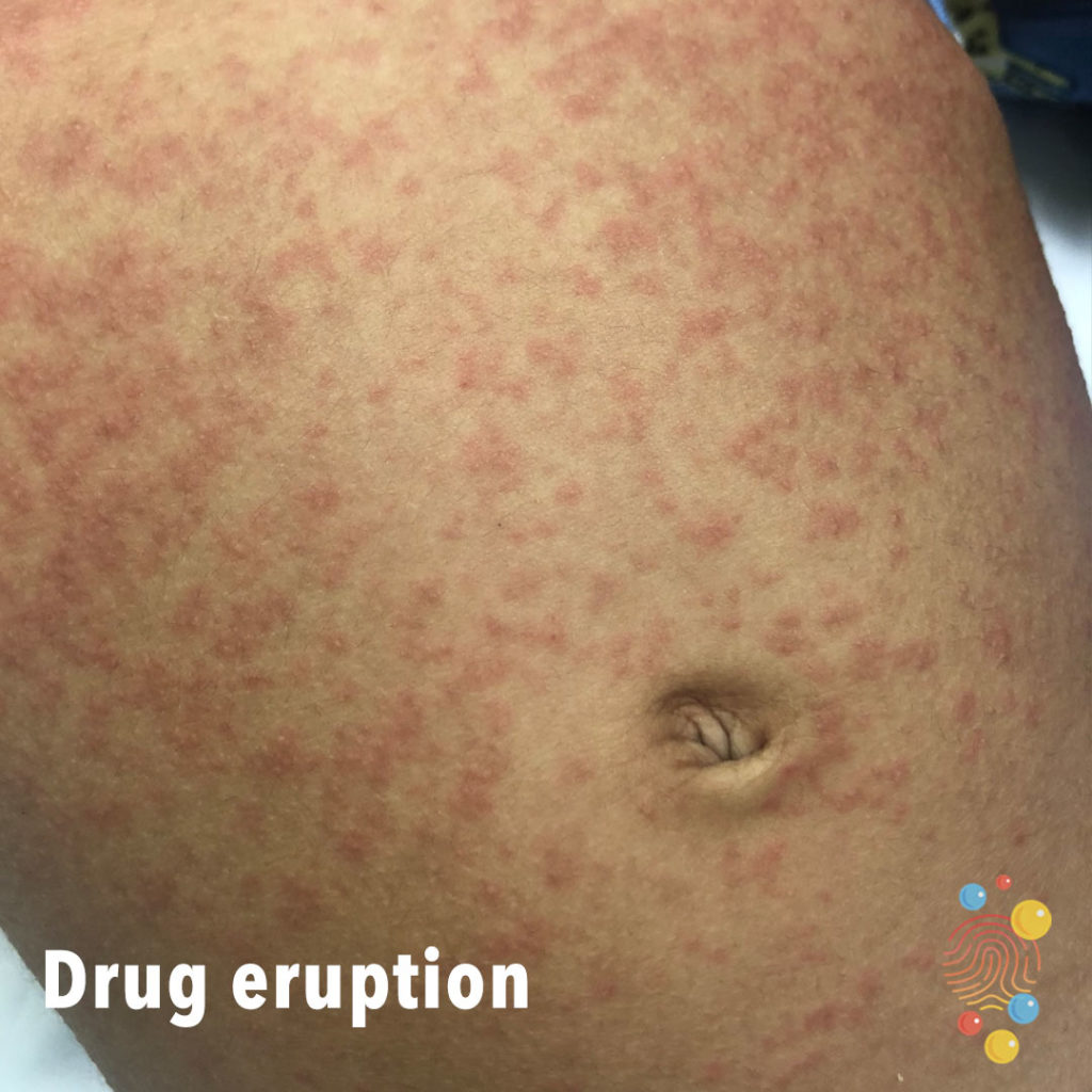
Diffuse erythematous maculopapular eruption on trunk.
Learn more about drug eruptions

Ill-defined hyperpigmentation with follicular prominence over neck. Solitary area of excoriation/ulceration noted on the right side.
Learn more about eczema


Wide spread ill defined erythema over back.
Learn more about PIMS-TS
Swelling of lower limb with hyperpigmentation.
Learn more about lymphoedema

Symmetric swelling of lower limbs associated with hyperkeratosis, plantar keratoderma, and dystrophic toenails
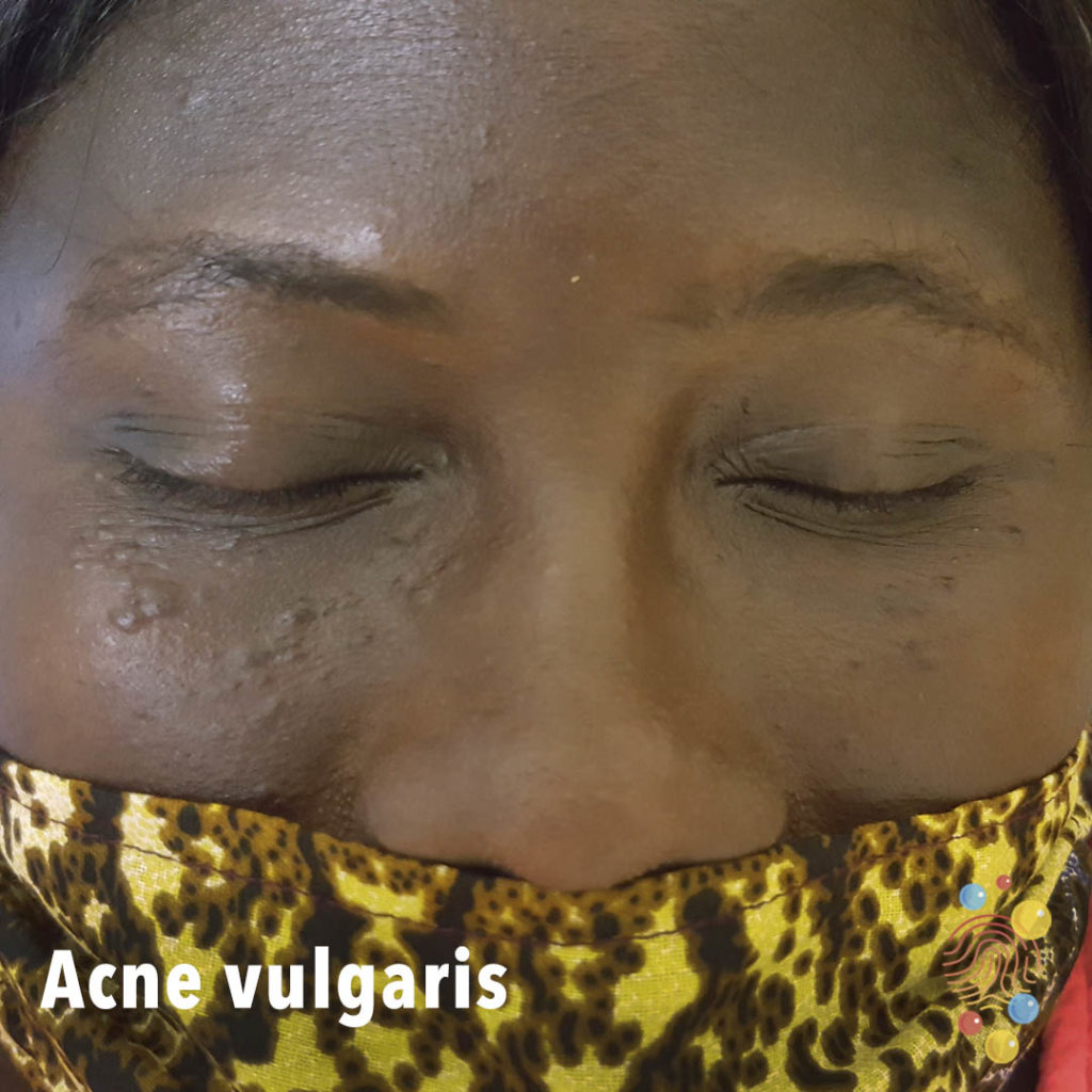
Few comedones (blackheads), papules + cysts with inflammation.
Learn more about acne vulgaris
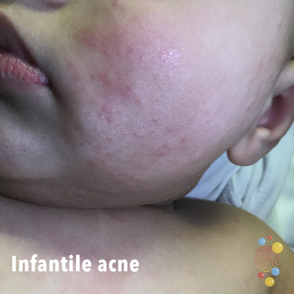
Background erythema with papular component on cheek.
Learn more about infantile acne
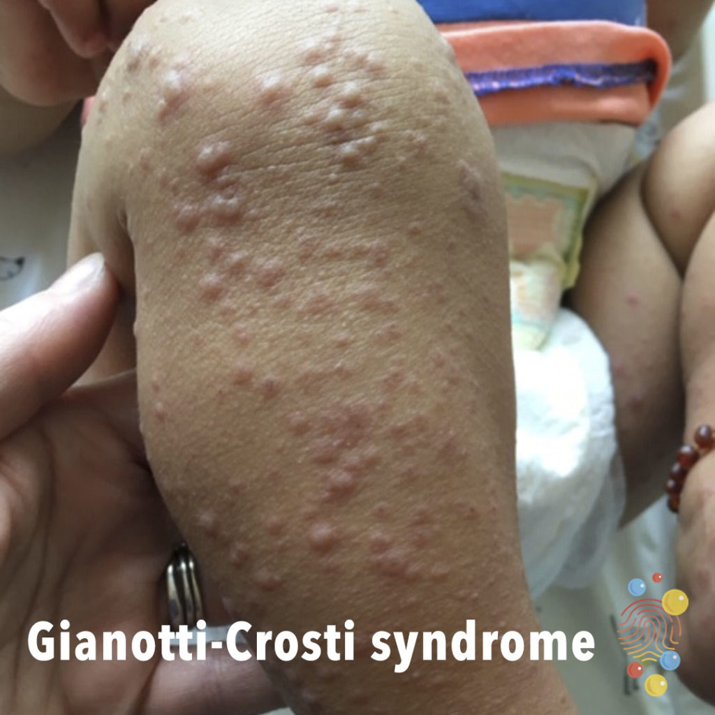
Erythematous papules and pseudo vesicles over shins.
Learn more about Gianotti-Crosti syndrome
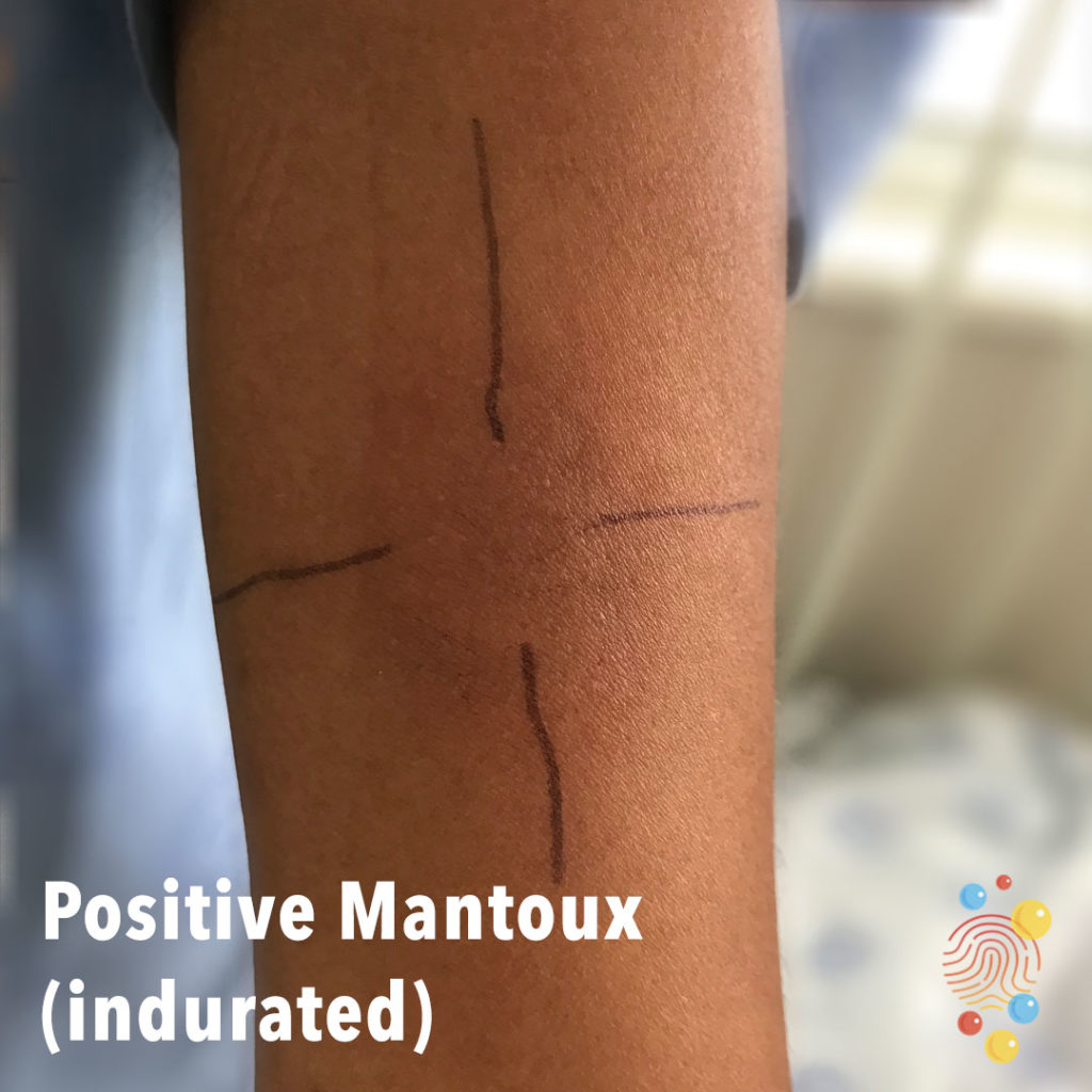
Indurated skin at site of injection.
Learn more about the Mantoux test

Multiple small papules on the leg.
Learn more about scabies
Scarlet Fever
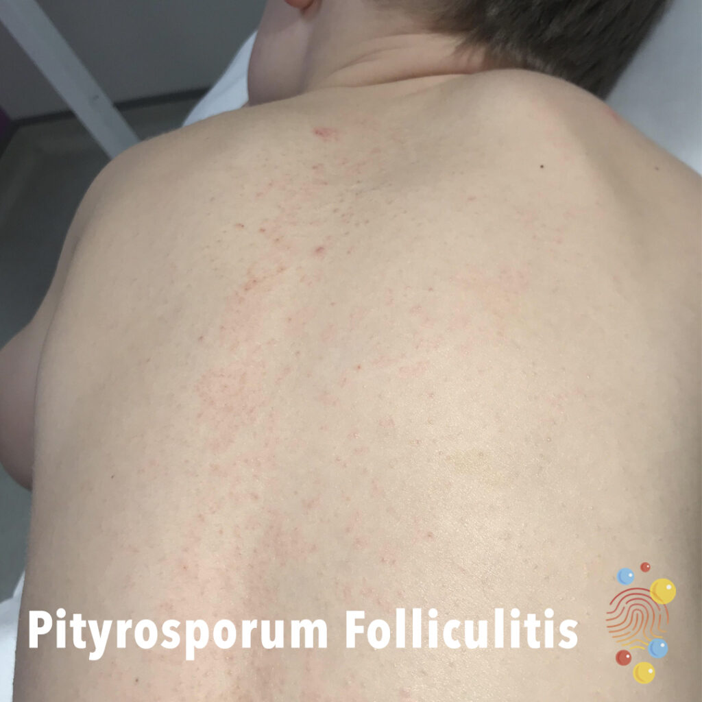
Pityrosporum Folliculitis
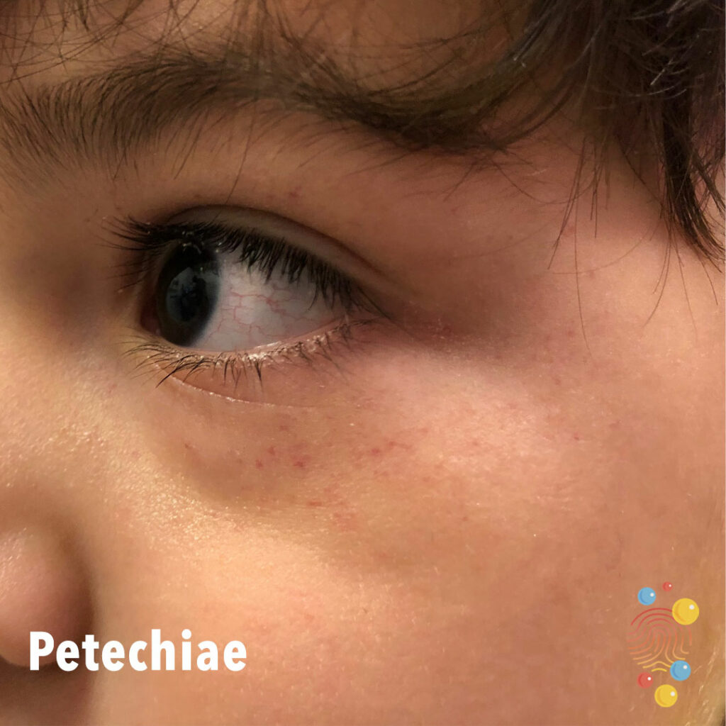
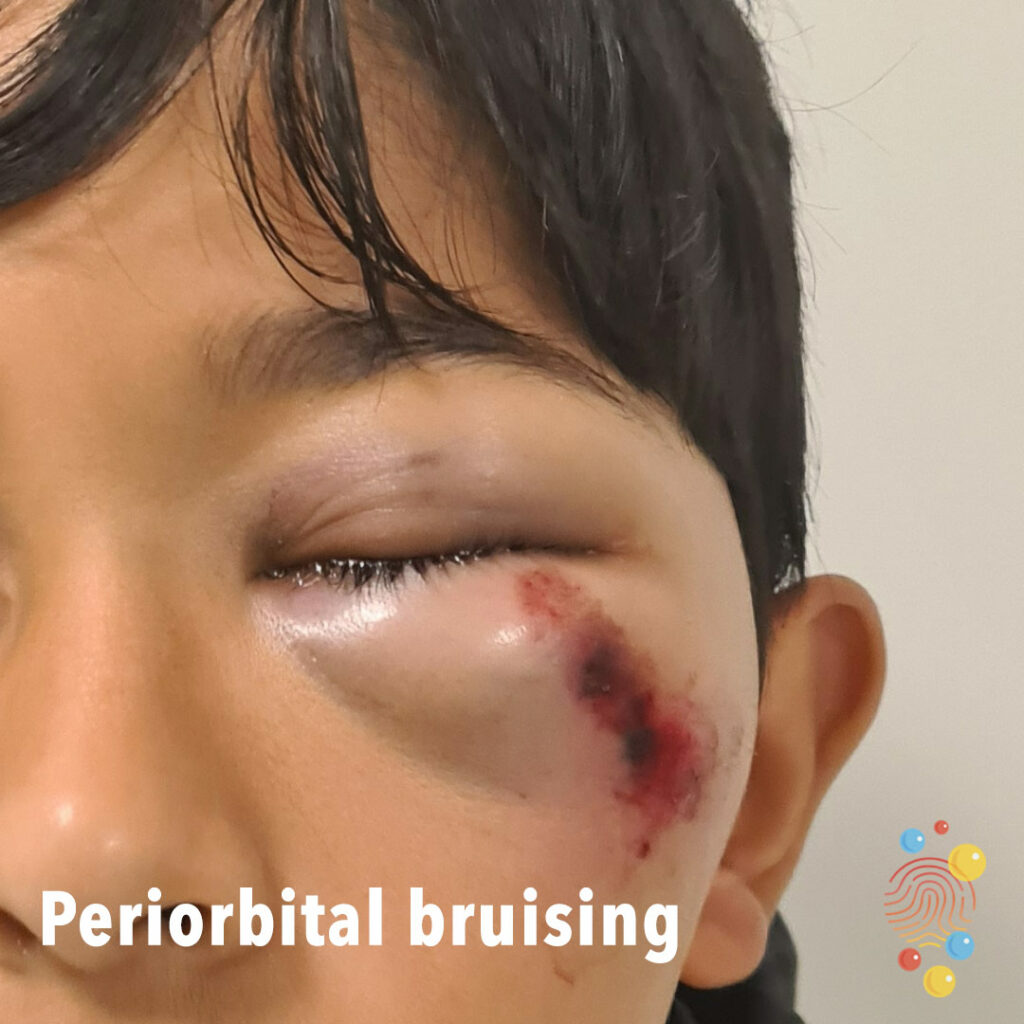
Petechiae around eyes – 4 year old male

Freckle with surrounding lighter area on the abdomen vertical line downward from the nipple.
Learn more about accessory nipples
Round patches of erythema with desquamation on the neckline.
Learn more about PIMS-TS
Superficially eroded patch with golden crust at periphery.
Learn more about bullous impetigo
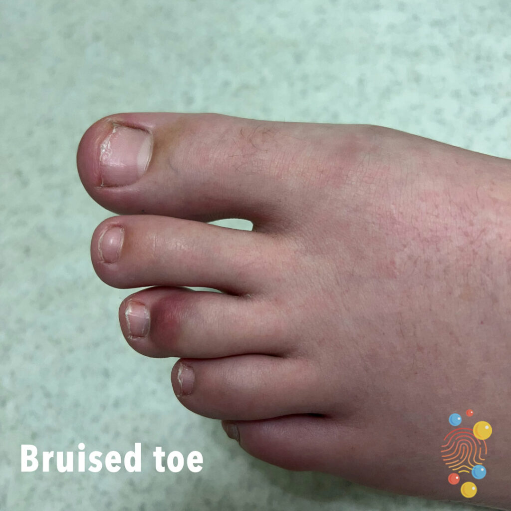
Bruised Toe

Hypopigmented round patches localized on the scalp, as post-inflammatory marks on the skin of atopic patient.
Learn more about pityriasis alba
Non blanching symmetrical purpuric lesions on the lower legs. Dusky erythema on the feet.
Learn more about Henoch-Schonlein purpura
Scaly and erythematous patches localized on the neck.
Learn more about eczema
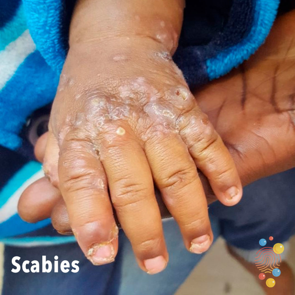
Excoriated papules on dorsal hand with lichenification and pustules.
Learn more about scabies
Abscess with scaling and yellow crust.
Learn more about kerions
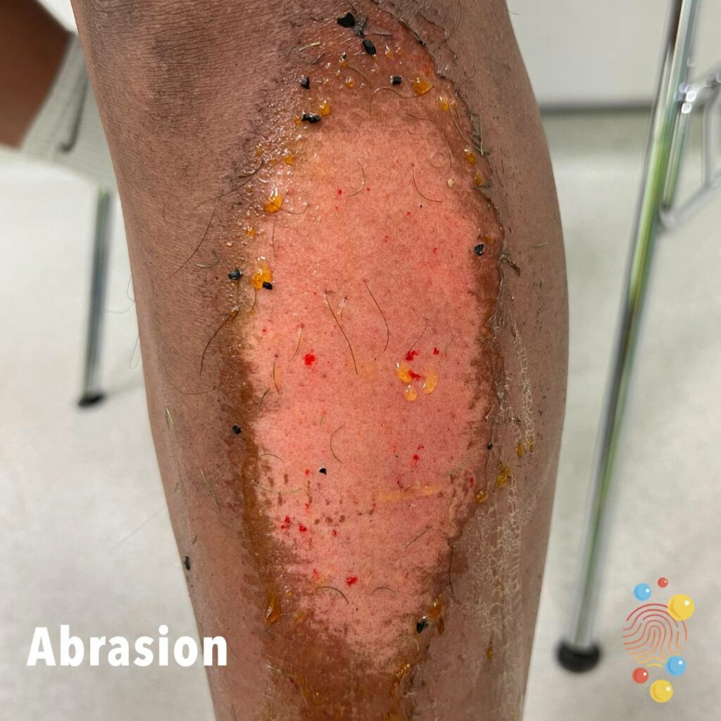
Abrasion to lower leg from AstroTurf – 17 year old male
Lichenified eczematous patch in the distribution of the beltline.
Learn more about eczema
Rough verrucous flat-topped lesions on the dorsal fingers.
Learn more about warts
Widespread multiple greyish blue patches over back of a child with skin type 4.
Learn more about dermal melanocytosis


Multiple urticated bruises, some of which have a targetoid appearance
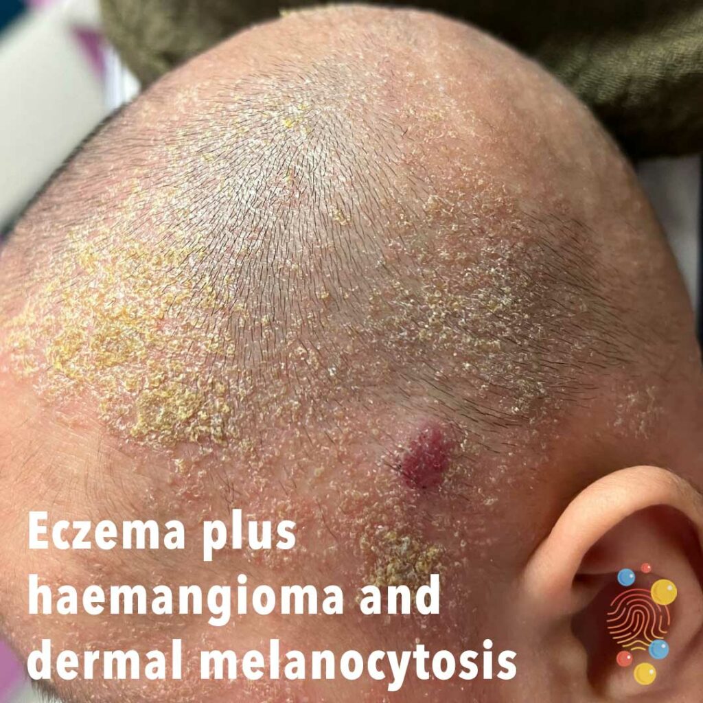
Eczema plus haemangioma and dermal melanocytosis

Yellow scaling of scalp.
Learn more about seborrhoeic dermatitis
Lichenifed patch of eczema.
Learn more about eczema

Widespread xerosis and fissuring, with ichthyotic appearance to skin. No obvious sparing and there are widespread eczematous changes.
Learn more about ichthyosis
Infection extending 2 days later

Lichenified excoriated patch with erosions and weeping.
Learn more about eczema
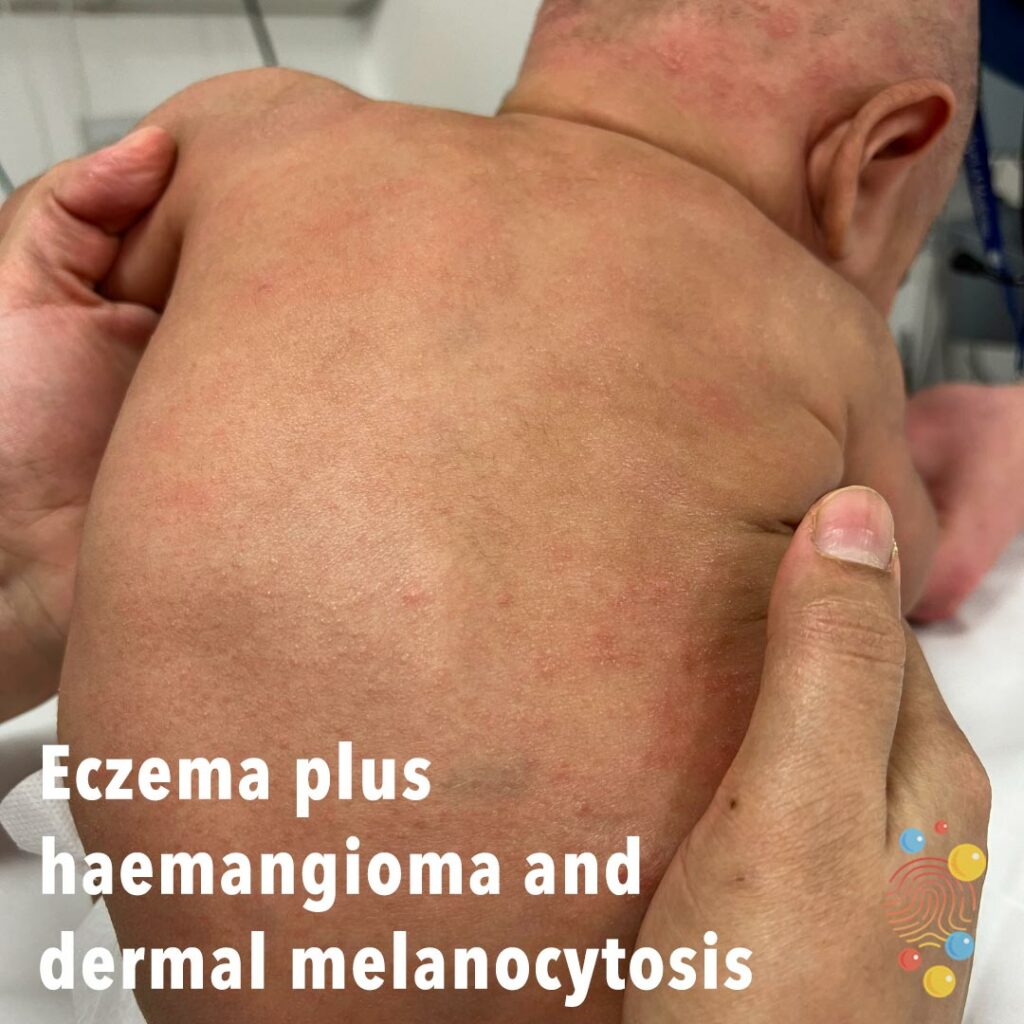
Eczema plus haemangioma and dermal melanocytosis
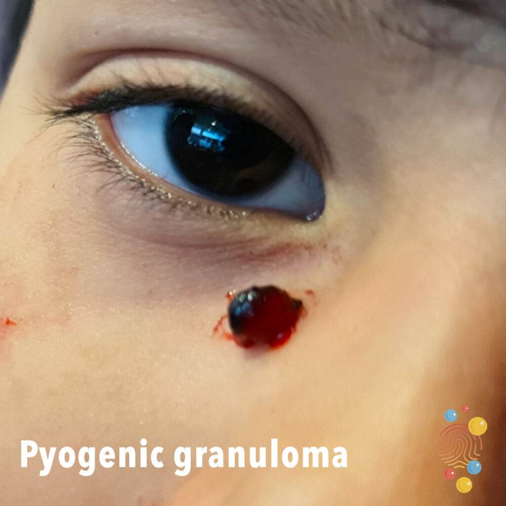
Erythematous vascular-looking nodule inferior to right eye.
Learn more about pyogenic granulomas
Non blanching patch of erythema.

Learn more about corneal abrasions

Nodules over right edge. Dystrophic scarring seen on left from recent abscesses. Old scars from previous abscesses also seen.
Learn more about hidradenitis suppurativa

Blue subcutaneous swelling.
Learn more about cephalhaematoma


Red swollen lesion with yellowish spots on the lower lid.
Learn more about styes
Oval shaped ecchymosis with central sparing.
Learn more about bites

Annular scaly shiny plaque on the dorsal foot.
Learn more about tinea corporis
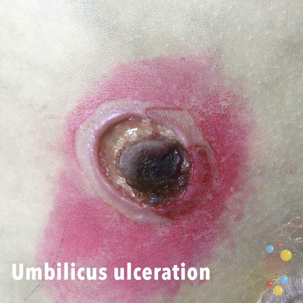
Slightly necrotic central umbilicus with periumbilical ulceration with slightly red raised rim.
Learn more about ulcers
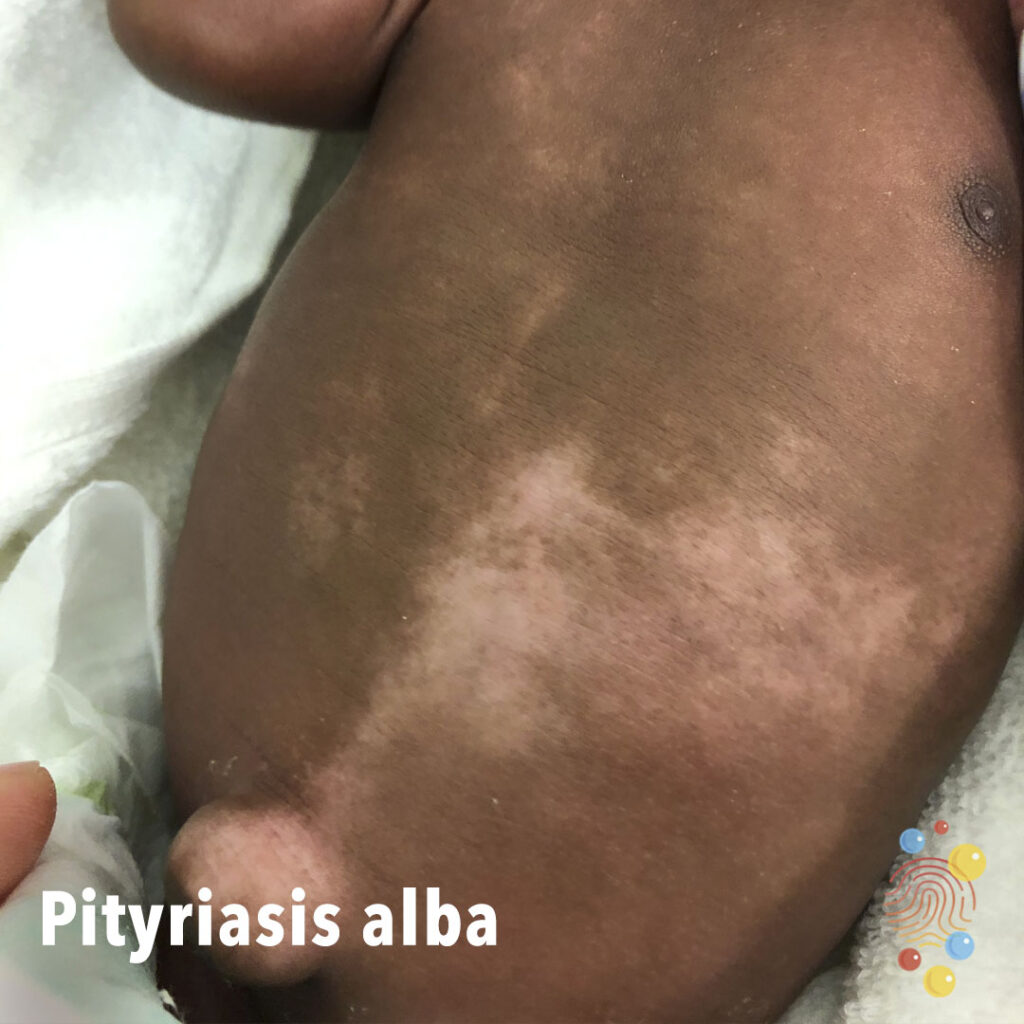
Scattered patches of hypopigmentation with indistinct margins on abdomen.
Learn more about pityriasis alba
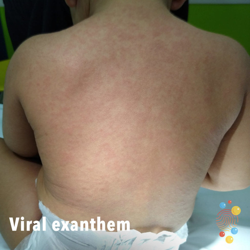
Generalised macular papular rash on the back.
Learn more about viral exanthem
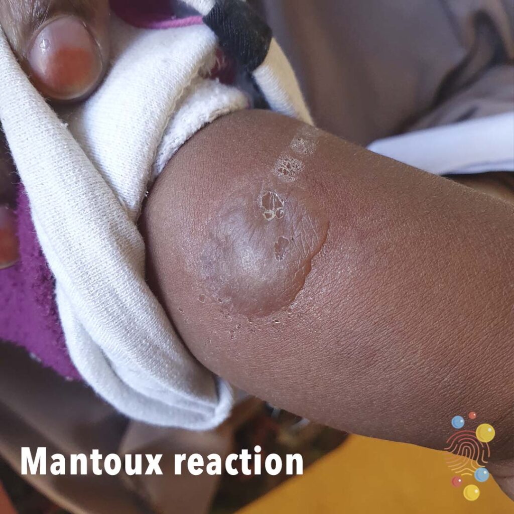
Induration at site of intradermal tuberculin injection.
Learn more about the Mantoux test

Head Injury
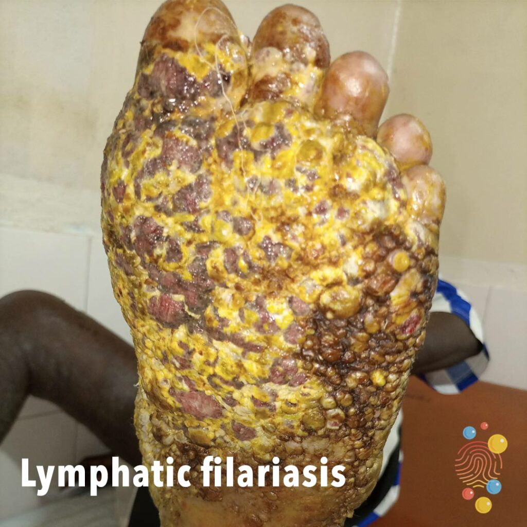
Multiple nodules covering whole of the left sole with slough and ulceration.
Learn more about lymphatic filariasis
Monomorphic erythematous papules with central pustule (this pustule will eventually rupture leaving an ulcer).
Learn more about herpes simplex virus
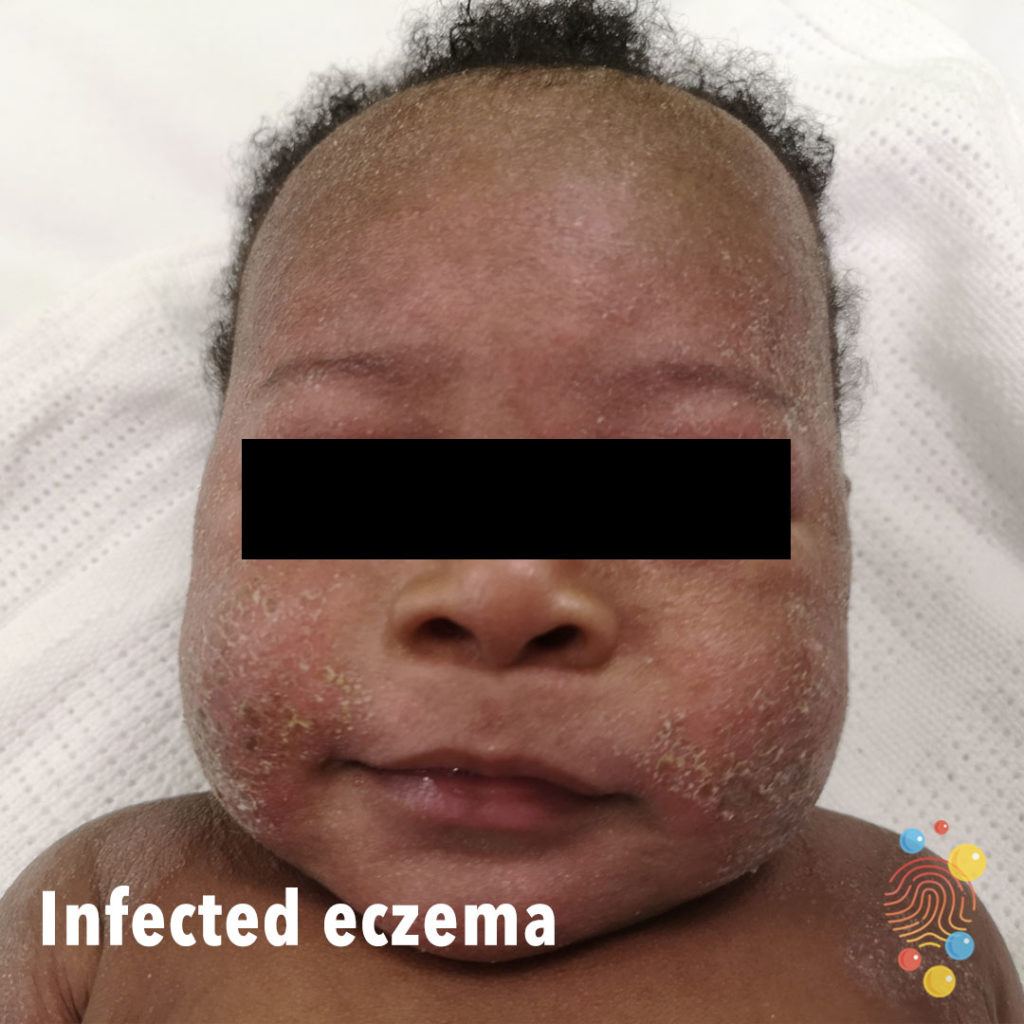
Erythema of cheeks and perioral area with likely secondary bacterial infection (note the honey-coloured crusting).
Learn more about eczema
Swelling and blistering with associated erythema involving the whole finger.
Learn more about bites
Multiple areas of deep blue dermal pigmentation on the back.
Learn more about dermal melanocytosis

Non-blanching palpable purpura of lower limbs.
Learn more about Henoch-Schonlein purpura
4 year old with kerion
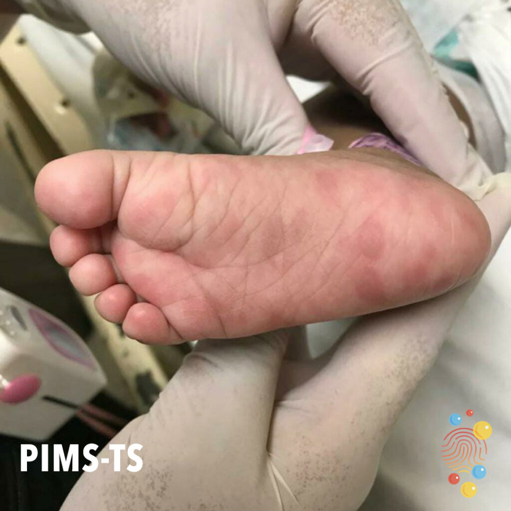
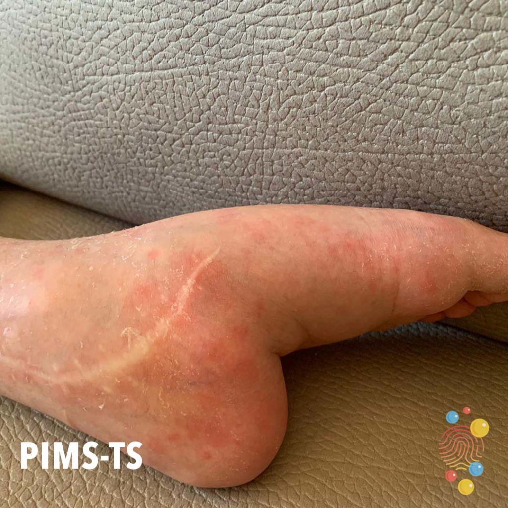
Plantar erythema as manifestation of PIMS-TS.
Learn more about PIMS-TS
Scarlet Fever
Erythema, scale, and excorations on the posterior neck.
Mildly eczematous patch in the antecubital fossa.
Learn more about eczema
Dermatitis over cheeks with a solitary urticated lesion over right upper eyelid. Asteatosis over cheeks bilaterally.
Learn more about eczema
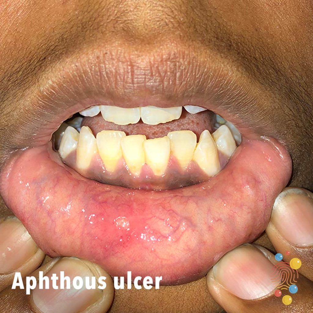
Small superficial erosion of the right lower labial mucosa with surrounding erythema.
Learn more about aphthous ulcers
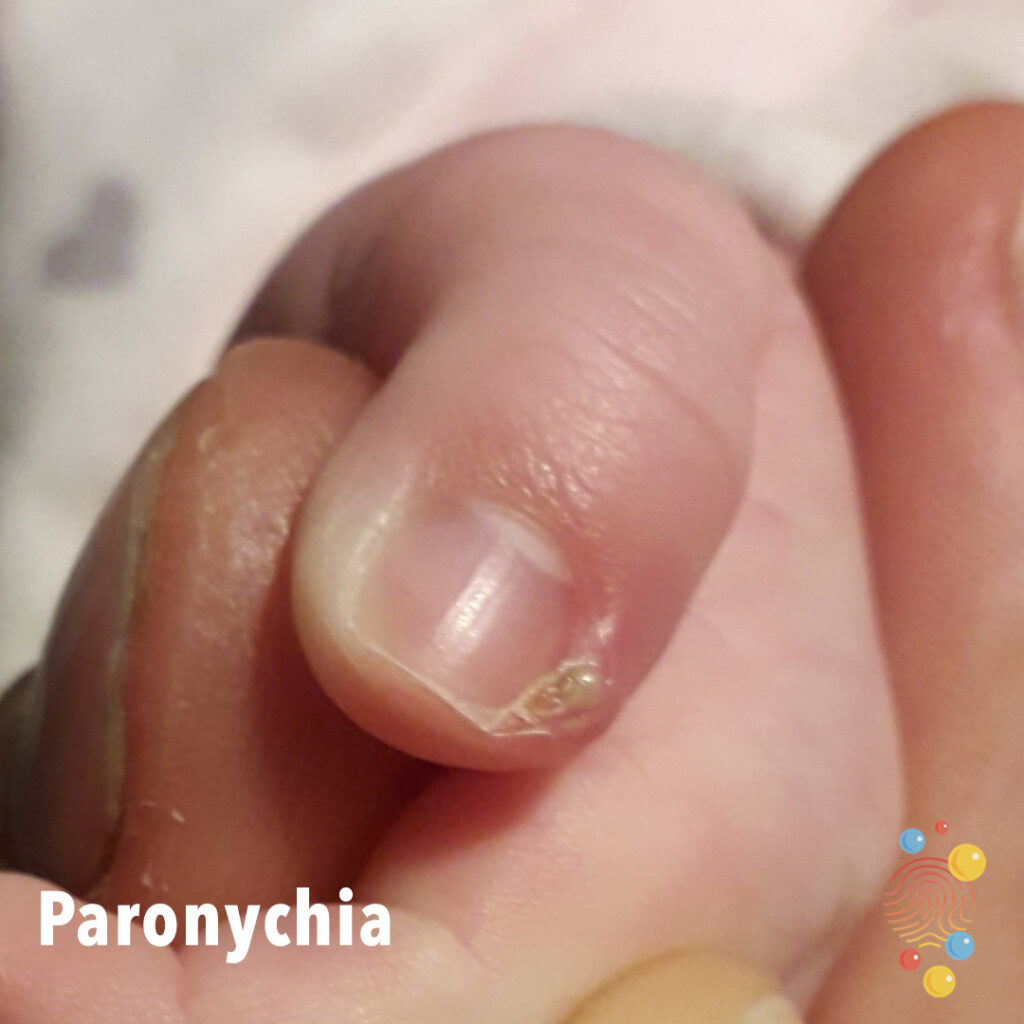
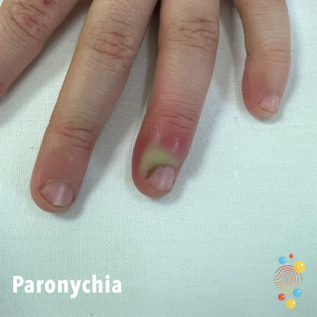
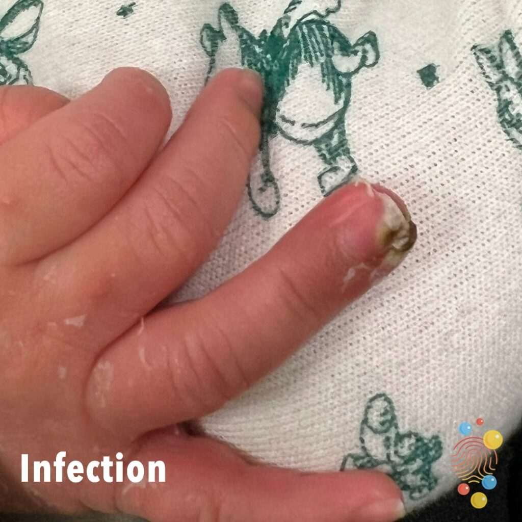
Small area of inflammation with surrounding pus on the skin surrounding the nail.
Learn more about paronychia
Erythematous macules over distal foot, some purpuric. Likely viral exanthem.
Learn more about hand, foot and mouth
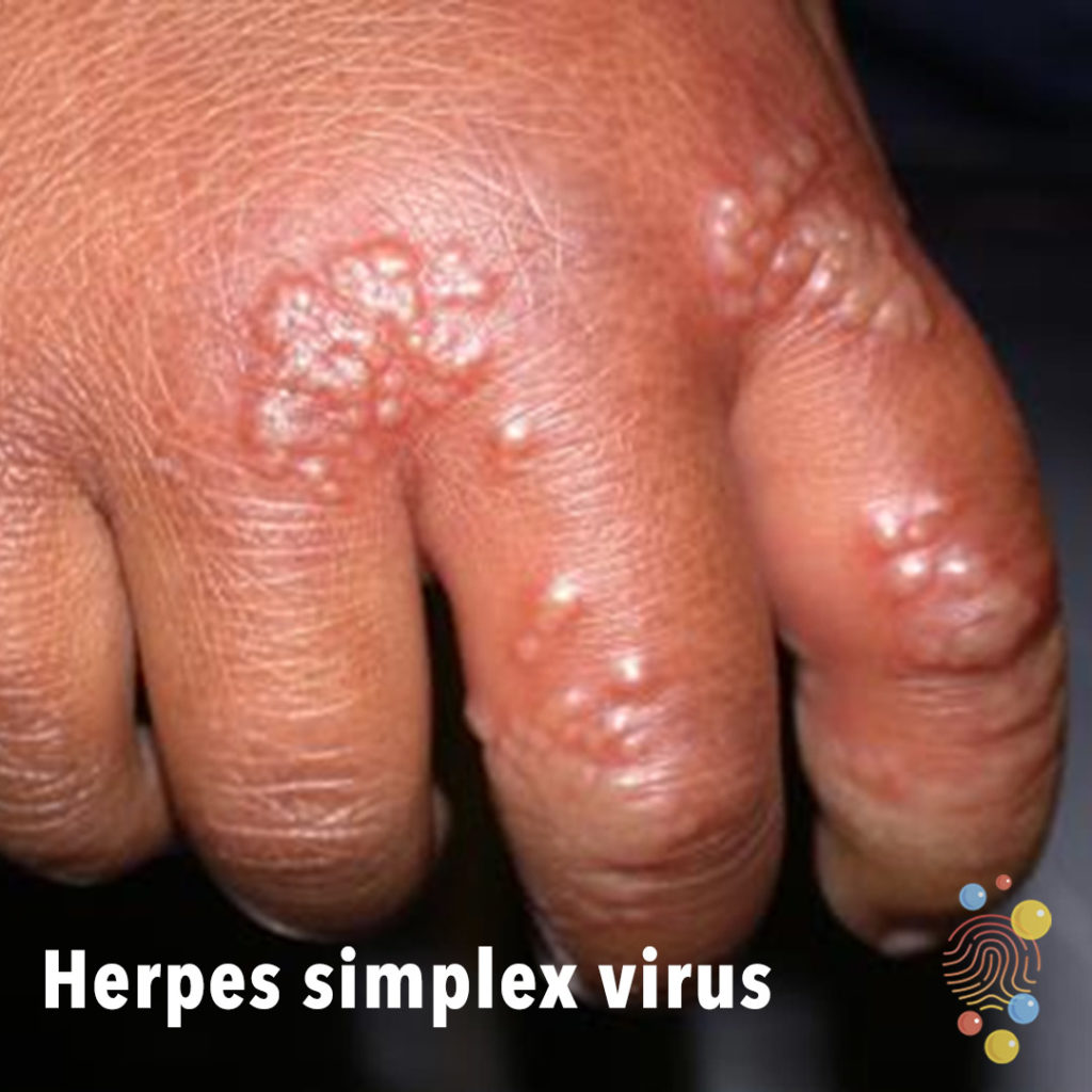
Crops of monomorphic vesicles on dorsal hand and fingers.
Learn more about herpes simplex virus

Bulge at the site of the umbilicus with a soft red mass on top of the skin.
Learn more about umbilical hernias
Central erythema, lichenification, and hyperpigmented edge over flexure. Shiny due to emollient.
Learn more about eczema
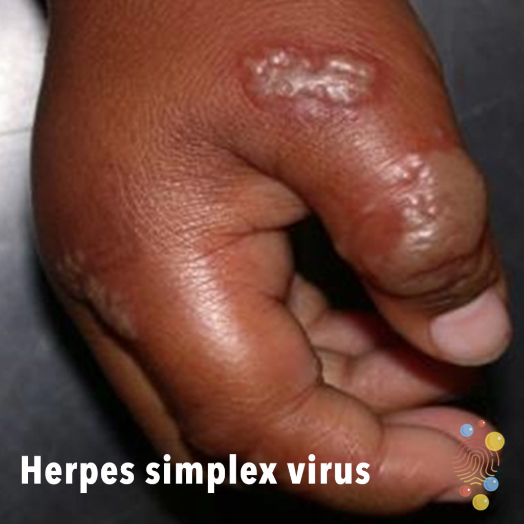
Confluent crops of monomorphic vesicles on dorsal hand and finger.
Learn more about herpes simplex virus
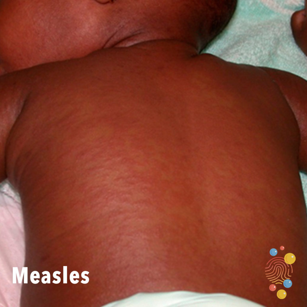
Erythematous maculopapular/morbilliform eruption on trunk.
Learn more about measles
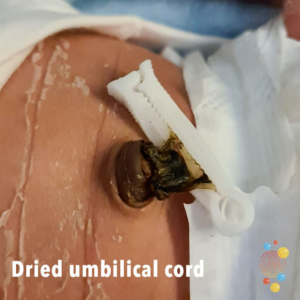
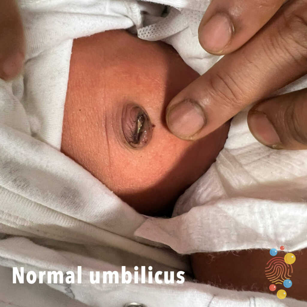
Normal dried umbilical cord
Learn about umbilical hernias
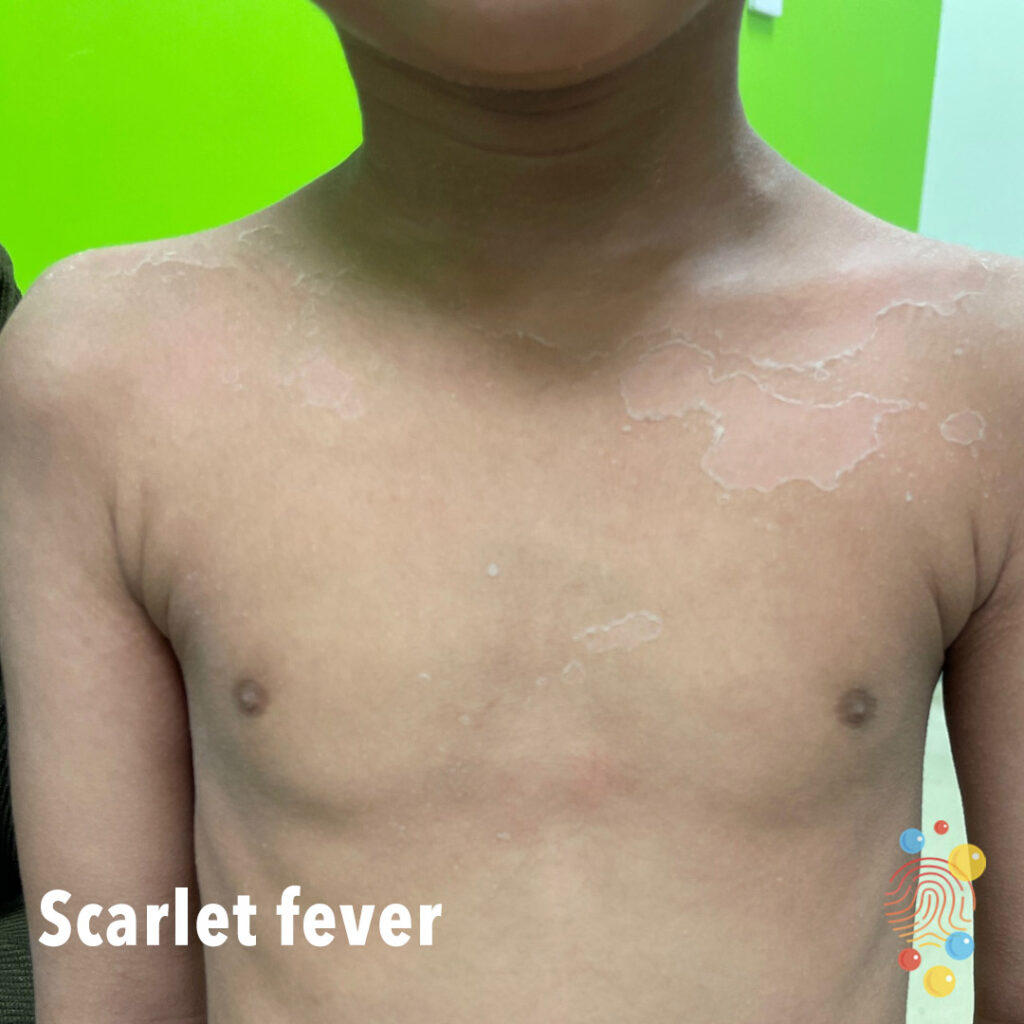
Extensive desquamation on upper chest post scarlet fever.
Dry scaly patch on the scalp.
Learn more about tinea capitits

Multiple eroded areas with surrounding flaccid skin.
Learn more about bullous impetigo

Multiple scaly papules on the skin, with characteristic crusting of the skin.
Learn more about eczema
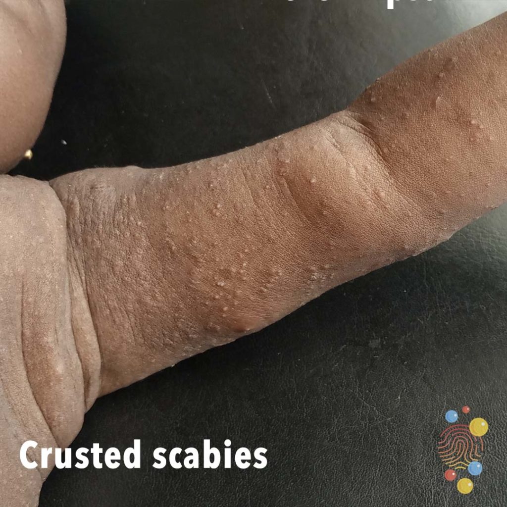
Extensive papular eruption on upper limbs with thick crust affecting trunk.
Learn more about scabies

Generalised widespread scaly plaque rash affecting the torso and arms with areas of lichenification.
Learn more about psoriasis
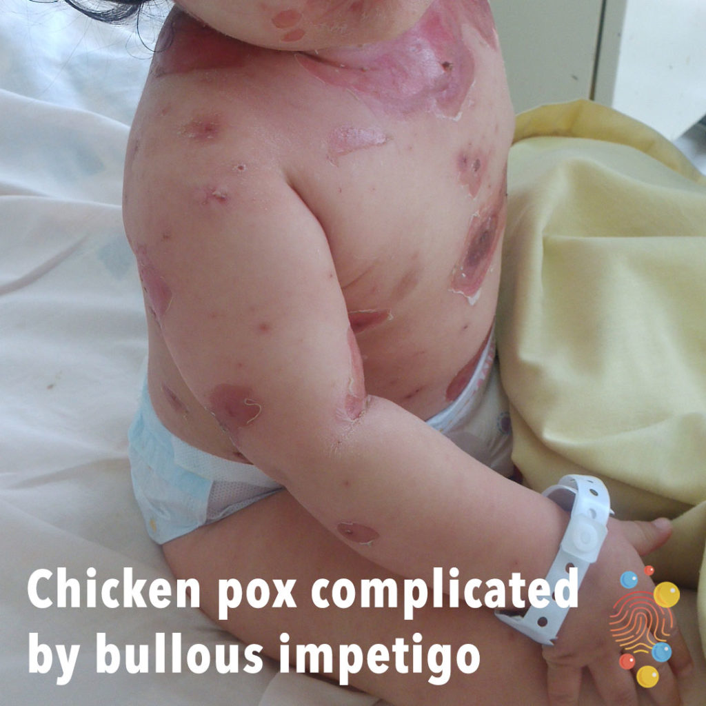
Multiple small crusted erosions with larger erythematous erosions.
Learn more about chicken pox |
Learn more about bullous impetigo
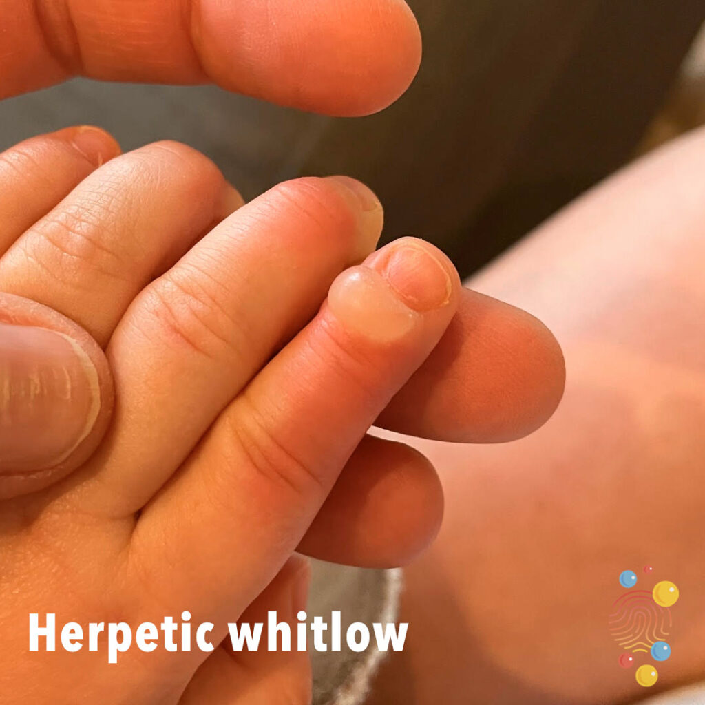
Bullae on fingertip of left second finger.
Learn more about herpes simplex virus


Well defined pink plaque in a flexural site. Some white scaling still evident.
Learn more about psoriasis
Skin coloured blisters coalescing into bullae surrounded by erythema in a multi-dermatomal distribution.
Learn more about chicken pox
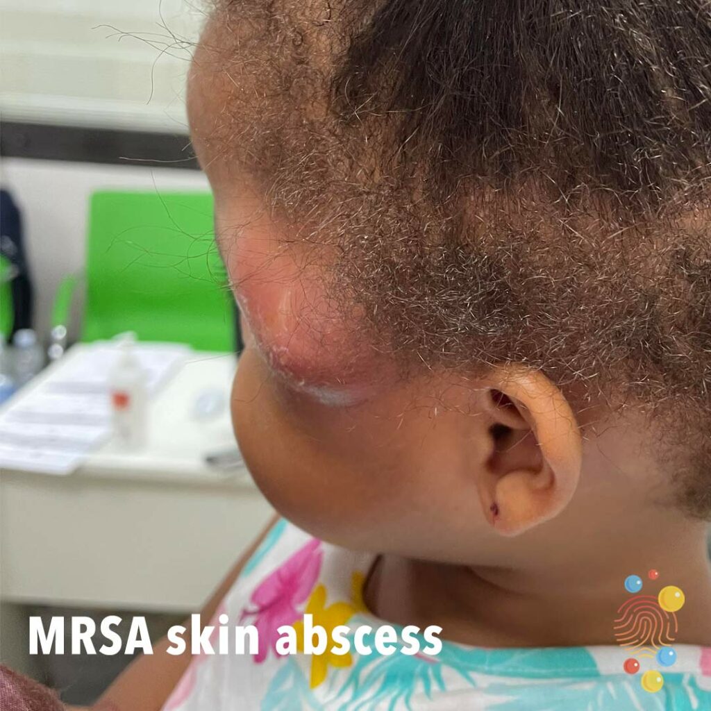
Red tender fluctuant swelling consistent with abscess in this case caused by MRSA.
Scattering of erythematous papules.

Erythematous patches and plaques with surrounding pallor.
Learn more about urticaria

Eczematous patches with lichenification.
Learn more about eczema
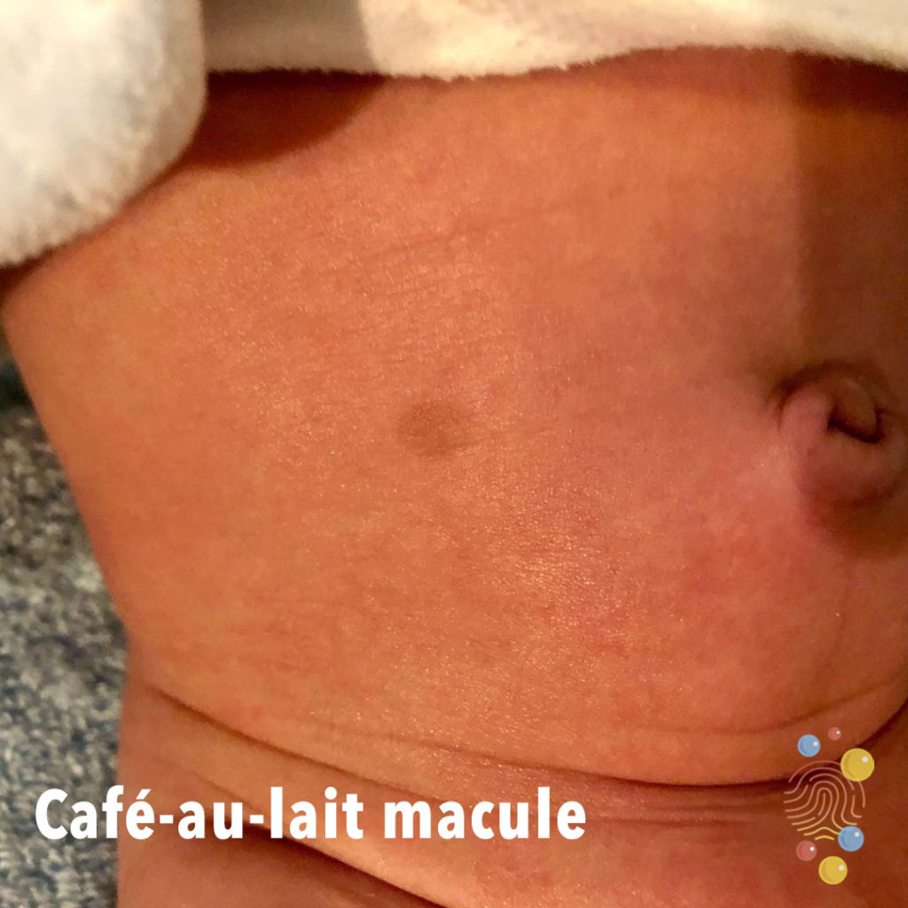
Single well-circumscribed hyperpigmented macular patch on abdomen.
Learn more about café-au-lait macules
Single well-circumscribed hyperpigmented macular patch on chest.
Learn more about café-au-lait macules
Silvery scaly plaques predominantly on extensor arms.
Learn more about psoriasis
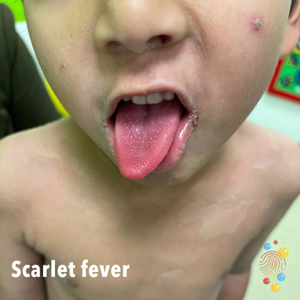
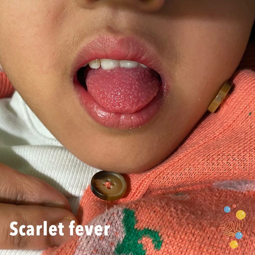
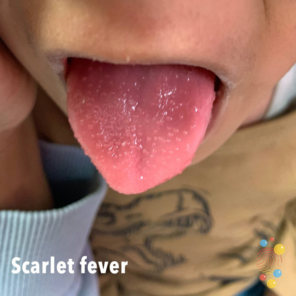
Strawberry tongue (due to reduced filiform papillae with retained fungiform papillae), crusted nodule on left cheek, and desquamation on trunk.

Widespread hyperpigmented, irregular patches across back of patient. Patches have poorly defined borders.
Learn more about dermal melanocytosis

Normal umbilical cord
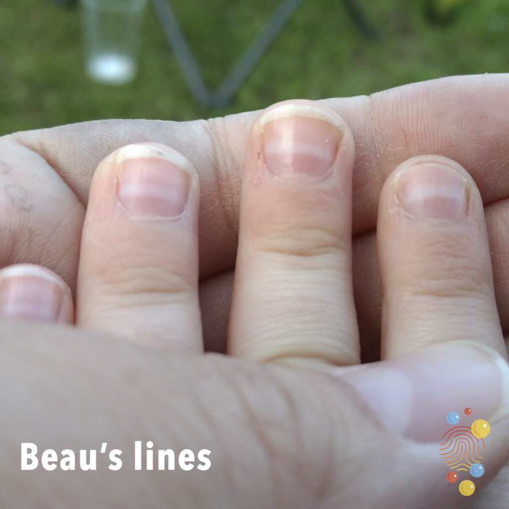
White horizontal lines mid-nails consistent with chemotherapy.
Learn more about Beau’s lines
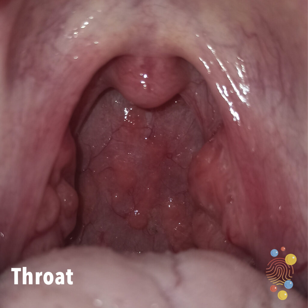
Throat burning with bubbles at the back of the mouth.
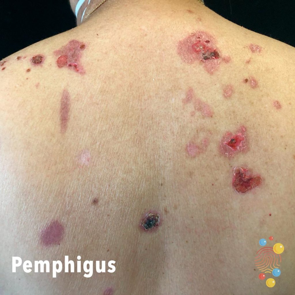
Multiple erythematous irregular eroded areas with areas of scarring.
Learn more about pemphigus
Well defined erythema in the scalp.
Learn more about tick bites
Abrasion
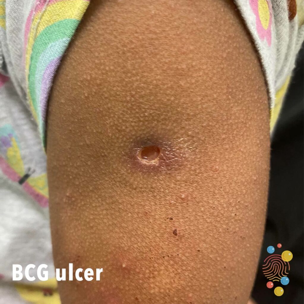
Round ulcer with undermined edges,
Learn more about BCG
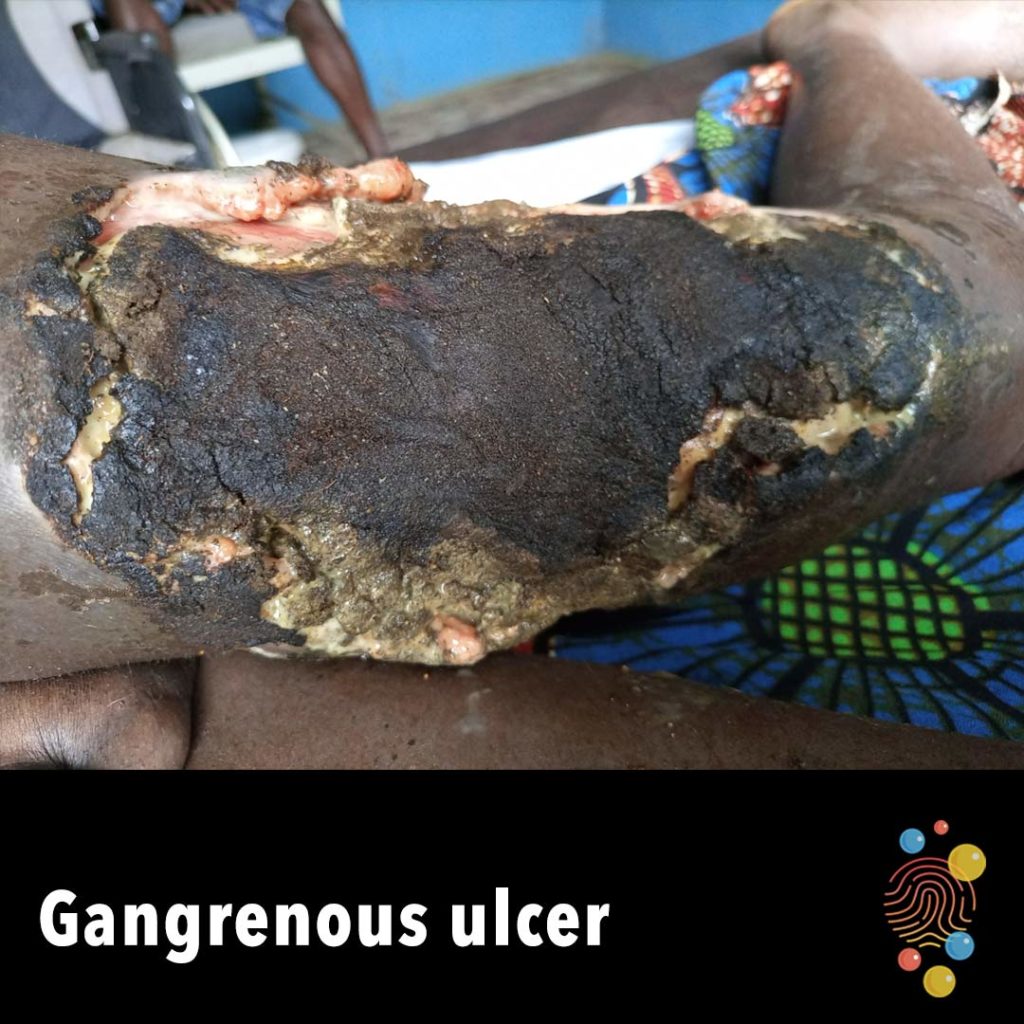
Deep ulceration of the thigh with necrotic tissue and eschar.

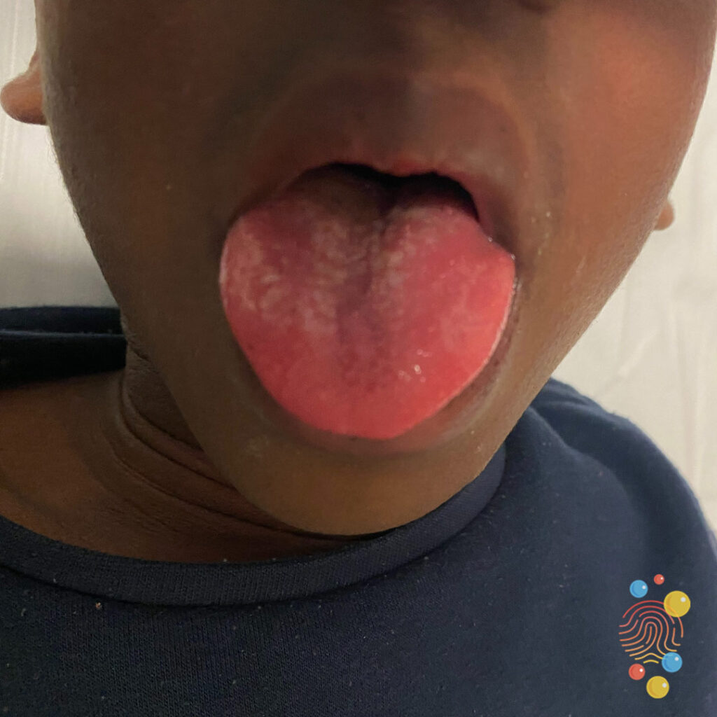
Tiny red spots on the tongue.
Learn more about strawberry tongues
Partially detached papule with bleeding and peripheral scale.
Learn more about molluscum contagiosum